


Indexed In
- Open J Gate
- Genamics JournalSeek
- JournalTOCs
- Ulrich's Periodicals Directory
- RefSeek
- Hamdard University
- EBSCO A-Z
- OCLC- WorldCat
- Publons
- Geneva Foundation for Medical Education and Research
- Euro Pub
- Google Scholar
Useful Links
Share This Page
Journal Flyer

Open Access Journals
- Agri and Aquaculture
- Biochemistry
- Bioinformatics & Systems Biology
- Business & Management
- Chemistry
- Clinical Sciences
- Engineering
- Food & Nutrition
- General Science
- Genetics & Molecular Biology
- Immunology & Microbiology
- Medical Sciences
- Neuroscience & Psychology
- Nursing & Health Care
- Pharmaceutical Sciences
Research Article - (2026) Volume 17, Issue 3
In vitro growth-inhibitory effects of DMTAP-containing cationic hybrid liposomes (DMTAP-cHL) on cholangiocarcinoma cells
Masaki Okumura, Tomoya Hashimoto, Tomoaki Sakoda, Yoko Matsumoto and Hideaki Ichihara*Received: 10-Jul-2026, Manuscript No. JCM-26-32103; Editor assigned: 13-Jul-2026, Pre QC No. JCM-26-32103 (PQ); Reviewed: 27-Jul-2026, QC No. JCM-26-32103; Revised: 03-Aug-2026, Manuscript No. JCM-26-32103 (R); Published: 10-Aug-2026, DOI: 10.35248/2157-2518.26.17.505
Abstract
Cholangiocarcinoma is difficult to detect at an early stage and is one of the most intractable cancers with a poor prognosis. Because current treatments are associated with problems such as adverse effects and therapeutic resistance, the development of new, highly selective therapeutic approaches is required. In this study, we prepared 1,2-Dimyristoyl-3-Trimethylammonium-Propane (DMTAP)-containing cationic Hybrid Liposomes, designated DMTAP-cHL, and investigated their in vitro anticancer effects on Human Cholangiocarcinoma (HuCCT-1) cells.
DMTAP-cHL were prepared by sonication using DMPC, the Polyethylene Glycol (PEG)-based surfactant C12(EO)21, and the cationic lipid DMTAP. Dynamic light scattering analysis revealed that DMTAP-cHL had a hydrodynamic diameter of approximately 30 nm and remained stable for 35 days. Fluorescence polarization analysis showed that DMTAP-cHL exhibited membrane fluidity comparable to that of L-α-Dimyristoylphosphatidylcholine (DMPC) liposomes and HL21. In the WST-8 assay, the IC50 value of DMTAP-cHL was 262 μM, indicating a stronger antiproliferative effect than DMPC liposomes and HL21. Furthermore, Propidium Iodide (PI) staining demonstrated an increase in the DNA fragmentation rate, and activation of caspase-3, caspase-8, and caspase-9 was observed, suggesting that DMTAP-cHL induced caspase-dependent apoptosis.
After treatment with DMTAP-cHL, HuCCT-1 cells showed a rapid increase in plasma membrane fluidity. Confocal laser microscopy revealed that DMTAP-cHL/1-palmitoyl-2-{12-[(7-Nitro-2-1,3-Benzoxadiazol-4-yl)amino]Dodecanoyl}- sn-glycero-3-Phosphocholine (NBD-PC) rapidly fused with and accumulated in HuCCT-1 cells. In contrast, no clear fusion or accumulation was observed in normal bile duct cells. These results indicate that DMTAP-cHL selectively fuse with and accumulate in cholangiocarcinoma cells and induce apoptosis through alteration of cellular membrane fluidity. Therefore, DMTAP-cHL may be useful as a novel cancer-regulating nanomaterial for cholangiocarcinoma.
Keywords
Cationic hybrid liposomes; Chemotherapy; Cholangiocarcinoma; Apoptosis; DMTAP
Abbreviations
C12(EO)21: Polyoxyethylene(21) dodecyl ether; DMPC: L-α-Dimyristoylphosphatidylcholine; HL: Hybrid Liposomes; DMTAP: 1,2-Dimyristoyl-3-Trimethylammonium-Propane; DMTAP-cHL: DMTAP-containing cationic Hybrid Liposomes; IC50: 50% Inhibitory Concentration; DEVD: Aspartic acid-Glutamic acid-Valine- Aspartic acid (Asp-Glu-Val-Asp); IETD: Isoleucine-Glutamic acid-Threonine-Aspartic acid (Ile-Glu-Thr-Asp); LEHD: Leucine-Glutamic acid-Histidine-Aspartic acid (Leu-Glu-His-Asp)
Introduction
Cancer is one of the leading causes of death in Japan and affects a large proportion of the population. At present, approximately one in two Japanese individuals is expected to develop cancer during their lifetime, and the establishment of effective methods for cancer prevention and treatment has become an important social issue. Lifestyle factors, including smoking, alcohol consumption, and dietary habits, are closely associated with cancer development. For example, smoking is associated with an increased risk of several cancers, including lung cancer, while alcohol consumption has been linked to cancers of the liver and digestive system. Cancer incidence also increases with aging because of a decline in Deoxyribonucleic Acid (DNA) damage repair capacity and immune surveillance. As Japan faces a super-aging society, the number of cancer patients and cancer-related deaths is expected to continue increasing [1-2].
Current cancer treatments mainly include surgery, chemotherapy, and radiation therapy. However, each treatment modality has limitations and adverse effects. Surgical treatment can be curative when tumors are completely resectable; however, recurrence may occur when tumors are unresectable or when micrometastases are present. Surgical invasion also carries risks of infection and complications. Chemotherapy has the advantage of acting systemically on both primary and metastatic tumors, but anticancer drugs can affect not only tumor cells but also normal cells, causing serious adverse effects such as nausea, vomiting, bone marrow suppression, and alopecia. Radiation therapy is useful as a local treatment that does not require surgical resection; however, treatment periods may be prolonged, and radiation-induced damage to normal tissues remains a major concern. Therefore, new cancer therapies that are less invasive and more selective are urgently needed.
The bile duct is a tubular tissue that transports bile produced in the liver to the duodenum. Bile is temporarily stored in the gallbladder and secreted into the duodenum through the bile duct to aid in fat digestion and absorption. Cholangiocarcinoma is a malignant tumor arising from the epithelium of the bile ducts, which constitute part of the biliary tract [3-6]. Histologically, most cholangiocarcinomas are adenocarcinomas. Cholangiocarcinoma is often advanced by the time symptoms such as jaundice, abdominal pain, and weight loss appear. In many cases, the tumor is unresectable at diagnosis, and cholangiocarcinoma is therefore regarded as a cancer with a poor prognosis. Risk factors include cholelithiasis, primary sclerosing cholangitis, congenital pancreaticobiliary maljunction, and hepatolithiasis. Because cholangiocarcinoma is often resistant to conventional treatments, new therapeutic strategies that act efficiently and selectively on cholangiocarcinoma cells are required.
Hybrid Liposomes (HL) are nanoparticles prepared by sonication of vesicle-forming lipids and micelle-forming surfactants in buffer solution [5-8]. Although organic solvents are sometimes used in conventional liposome preparation methods, HL can be prepared without organic solvents, thereby reducing the risk of residual solvent contamination and providing advantages in terms of safety and simplicity. HL composed of the phospholipid L-α-Dimyristoylphosphatidylcholine (DMPC) and PEG-based surfactants have been reported to exert antiproliferative and apoptosis-inducing effects on various cancer cells [9-24].
The anticancer effects of HL are thought to be related to the physical properties of cancer cell membranes. Cancer cells generally exhibit higher membrane fluidity than normal cells, and HL are considered to selectively fuse with and accumulate in cancer cell membranes by exploiting such differences in membrane properties [11]. Fusion of HL with cancer cell membranes alters membrane fluidity and structure, which may induce apoptotic signaling. Thus, HL have attracted attention as novel nanomedical materials that exhibit intrinsic anticancer activity without encapsulated drugs.
In addition, negatively charged lipids such as Phosphatidylserine (PS) have been reported to be more frequently exposed on the outer leaflet of cancer cell membranes than on normal cell membranes [26]. Therefore, the surface of cancer cell membranes is considered to be relatively more negatively charged than that of normal cells. By introducing positively charged cationic lipids into HL, electrostatic interactions with cancer cell membranes may be enhanced, promoting fusion and accumulation. It has been reported that cationic hybrid liposomes containing the cationic lipid O,O′-ditetradecanoyl-N-α-trimethylammonioacetyldiethanolamine chloride, DC-6-14, exhibit higher fusogenic and antiproliferative effects on cancer cells than conventional neutral liposomes [27-29].
In this study, we focused on 1,2-dimyristoyl-3-trimethylammoniumpropane chloride, as a new cationic lipid. DMTAP is widely used as a component of cationic liposomes and as a transfection reagent. In addition, DMTAP is readily available in both powder and solution forms, making it convenient for examining experimental conditions and conducting reproducible experiments. DMTAP may also offer advantages in terms of procurement and cost compared with other cationic lipids. Therefore, DMTAP may be a useful constituent lipid for the preparation of cationic hybrid liposomes.
In the present study, we prepared DMTAP-containing cationic hybrid liposomes, DMTAP-cHL, composed of DMPC, a PEG-based surfactant, and DMTAP, and investigated their physicochemical properties and anticancer effects on cholangiocarcinoma cells. Specifically, we evaluated the particle size stability and membrane fluidity of DMTAP-cHL and analyzed their growth-inhibitory effects on HuCCT-1 human cholangiocarcinoma cells, Deoxyribonucleic Acid (DNA) fragmentation, caspase activation, changes in plasma membrane fluidity, and cellular fusion and accumulation. Through these analyses, we aimed to clarify whether DMTAP-cHL could serve as a novel cancer-regulating nanomaterial that selectively acts on cholangiocarcinoma cell membranes and induces apoptosis.
Materials and Methods
Preparation of DMTAP-containing cationic hybrid liposomes
DMTAP-cHL were prepared using 87 mol% of the zwitterionic phospholipid L-α-dimyristoylphosphatidylcholine (DMPC, purity>99%, Nippon oil and fat, Tokyo, Japan), 5 mol% of the PEG-based surfactant polyoxyethylene(21) lauryl ether, (C12(EO)21, purity>99%, Nikko chemicals, Tokyo, Japan) and 8 mol% of the cationic lipid 1,2-dimyristoyl-3-trimethylammonium-propane chloride (DMTAP, purity>99%, Avanti Polar Lipids, AL, USA). Each lipid and surfactant was weighed to obtain the predetermined molar ratio and suspended in physiological saline. The suspension was then sonicated using a desktop ultrasonic cleaner (ASU CLEANER ASU-10, AS ONE, Tokyo, Japan), 240 W, at 45°C under a nitrogen atmosphere for 1 min/mL. The resulting suspension was sterilized by filtration through a 0.20 μm membrane filter and used as the DMTAP-cHL sample solution. The prepared samples were stored at 25°C.
As a comparative sample, HL21 was prepared using 95 mol% DMPC and 5 mol% C12(EO)21 in physiological saline by the same sonication procedure used for DMTAP-cHL. The HL21 sample solution was sterilized by filtration through a 0.20 μm membrane filter and stored at 25°C.
Measurement of hydrodynamic diameter by dynamic light scattering
The hydrodynamic diameters, dhy, of HL21 and DMTAP-cHL were measured by dynamic light scattering using a nanoSAQLA light scattering photometer, Otsuka Electronics, Osaka, Japan. A highpower semiconductor laser with a wavelength of 660 nm was used as the light source at an output power of 70 mW.
The hydrodynamic diameter, dhy, was calculated from the diffusion coefficient, D, using the Stokes-Einste in equation 1:
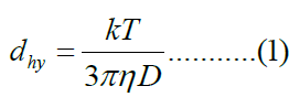
Where k is the Boltzmann constant, T is the absolute temperature, η is the viscosity of the solvent, and D is the diffusion coefficient.
Fluorescence polarization assay for liposome membrane fluidity
The membrane fluidity of liposomes was evaluated by fluorescence polarization analysis using the fluorescent probe 1,6-Diphenyl-1,3,5-Hexatriene (DPH). DPH is incorporated into the hydrophobic region of lipid bilayers, and membrane fluidity can be evaluated by measuring the rotational mobility of DPH within the membrane. In general, when membrane fluidity is low, the rotational mobility of DPH is restricted and the fluorescence polarization value, P, increases. Conversely, when membrane fluidity is high, the rotational mobility of DPH increases and the P value decreases.
The fluorescence polarization value, P, was calculated using the following equation 2:
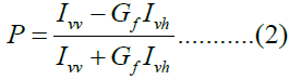
The correction factor, Gf, was calculated using the following equation 3:
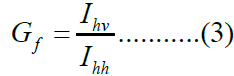
Where Ivv and Ivh represent fluorescence intensities detected in the vertical and horizontal directions, respectively, when vertically polarized excitation light was used. Ihv and Ihh represent fluorescence intensities detected in the vertical and horizontal directions, respectively, when horizontally polarized excitation light was used.
Each liposome sample was adjusted to a DMPC concentration of 0.1 mM and transferred to a quartz cell. DPH was added at a final concentration of 2 μM, and the samples were incubated at 37°C for 15 min. Fluorescence intensity was then measured for 5 min using a spectrofluorometer (F-7100, Hitachi High-Tech Science, Tokyo, Japan), and the P value was calculated from the obtained fluorescence intensities.
Determination of the 50% inhibitory concentration by WST-8 assay
The growth-inhibitory effect of DMTAP-cHL on cholangiocarcinoma cells was evaluated using the WST-8 assay. WST-8 is reduced by mitochondrial dehydrogenases in viable cells via the coenzyme Reduced Nicotinamide Adenine Dinucleotide (NADH) and the electron mediator 1-methoxy PMS to form Water-Soluble Tetrazolium-8 (WST-8) formazan. Because the amount of WST-8 formazan produced is proportional to the number of viable cells, absorbance was measured as an indicator of cell proliferation.
HuCCT-1 human cholangiocarcinoma cells were prepared as a cell suspension at 5.0 × 10⁴ cells/mL, and 100 μL of the suspension was seeded into each well of a 96-well plate. The cells were incubated at 37°C under 5% CO2 for 24 h. After incubation, samples at various concentrations were added, and the cells were incubated for an additional 48 h. After treatment, WST-8 reagent was added to each well, and the color reaction was allowed to proceed for 3 h. Absorbance was measured using a microplate reader, Thermo Fisher Scientific, MA, USA.
Cell viability relative to the control was calculated from the obtained absorbance values, and the 50% inhibitory concentration, IC50, was determined.
Measurement of DNA content by flow cytometry
To evaluate apoptosis induction in HuCCT-1 cells by DMTAP-cHL, DNA content was measured using propidium iodide, Propidium Iodide (PI). PI is a fluorescent dye that emits red fluorescence upon intercalation into DNA. Because PI does not readily penetrate intact plasma membranes, it is commonly used to measure DNA content and detect DNA-fragmented cells after membrane permeabilization.
HuCCT-1 cells were prepared at 5.0 × 10⁴ cells/mL and seeded in 60 mm dishes at 5 mL per dish, corresponding to 2.5 × 10⁵ cells. The cells were cultured at 37 °C under 5% CO2 for 24 h. After incubation, samples were added at final concentrations of 0.16, 0.26, 0.36, and 0.46 mM, and the cells were treated for 48 h. After treatment, the cells were collected and centrifuged at 3,000 rpm for 5 min, and the supernatant was removed. The cell pellet was resuspended in 1 mL of Phosphate-Buffered Saline [PBS(-)] and transferred to a microtube. The cells were centrifuged again at 3,000 rpm for 5 min, and the supernatant was removed. Then, 150 μL of 0.1% Triton X-100 and an equal volume of RNase solution were added for membrane permeabilization and RNA degradation. Subsequently, 300 μL of PBS(-) was added to re-suspend the cells. A 60 μL aliquot of PI solution was added to each measurement tube, and 540 μL of the 600 μL cell suspension was transferred to the tube through a nylon mesh. PI fluorescence was then measured using a CytoFLEX Flow Cytometer (Beckman Coulter, Brea, CA, USA), and the DNA fragmentation rate was analyzed.
Measurement of caspase activation using fluorescent substrates
To investigate the apoptosis induction pathway activated by DMTAP-cHL, the activation rates of caspase-3, caspase-8, and caspase-9 were measured. Fluorescein Isothiocyanate (FITC)-labeled fluorescent substrates with caspase-specific recognition sequences were used, including a cleaved caspase-3 staining kit, FITC, ab65613; caspase-8 active FITC staining kit, ab65614; and caspase-9 active FITC staining kit, ab65615; all from Abcam, Cambridge, United Kingdom.
Substrates containing the DEVD, IETD, and LEHD sequences were used for caspase-3, caspase-8, and caspase-9, respectively. These substrates react with activated caspases to generate fluorescent signals, allowing caspase-activated cells to be detected by flow cytometry.
HuCCT-1 cells were seeded at 5.0 × 10⁴ cells/mL in 60 mm dishes and cultured at 37°C under 5% CO2 for 24 h. After incubation, DMTAP-cHL was added at a final concentration of 360 μM, and the cells were treated for 48 h. After treatment, the cells were collected, and 1 μL of the fluorescent substrate corresponding to each caspase was added, followed by incubation at 37°C for 45 min. After staining, the cells were washed, and the cell suspension was passed through a nylon mesh. The activation rates of caspase-3, caspase-8, and caspase-9 were then measured using a CytoFLEX flow cytometer (Beckman Coulter, CA, USA).
Measurement of cellular membrane fluidity by fluorescence polarization
Changes in the membrane fluidity of HuCCT-1 cells after treatment with DMTAP-cHL were evaluated by fluorescence polarization analysis using DPH. HuCCT-1 cells were prepared at 1.0 × 10⁶ cells/mL, and DPH was added at a final concentration of 2 μM. The cells were then stained at 37°C for 35 min.
After staining, the cells were washed with Hanks' Balanced Salt Solution (HBSS), and a HuCCT-1 cell suspension was prepared at 5.0 × 10⁵ cells/mL. Fluorescence polarization values after sample addition were measured using an F-7100 spectrofluorometer, Hitachi High-Tech Science. Changes in the membrane fluidity of HuCCT-1 cells induced by DMTAP-cHL treatment were evaluated based on the obtained P values.
Confocal laser microscopic observation of fusion and accumulation of DMTAP-cHL in cholangiocarcinoma cells
To evaluate the fusion and accumulation of DMTAP-cHL in HuCCT-1 cells, HL21/NBD-PC and DMTAP-cHL/NBD-PC containing the fluorescent lipid NBD-PC were prepared and observed by confocal laser microscopy.
DMTAP-cHL/NBD-PC was prepared using 83 mol% DMPC, 5 mol% C12(EO)21, 8 mol% DMTAP, and 4 mol% fluorescent lipid 1-palmitoyl-2-{6-[(7-nitro-2,1,3-benzoxadiazol-4-yl)amino]hexanoyl}-sn-glycero-3-phosphocholine (16:0 NBD-PC, Avanti Polar Lipids, AL, USA). The sample solution was prepared by sonication at 45°C for 1 min/mL in physiological saline under a nitrogen atmosphere using a desktop ultrasonic cleaner, ASU CLEANER ASU-10, AS ONE, 240 W. The solution was sterilized by filtration through a 0.20 μm membrane filter and stored at 4°C to prevent photobleaching of the fluorescent lipid.
HuCCT-1 cells were prepared at 5.0 × 10⁴ cells/mL and seeded at 2 mL per 35 mm glass-bottom dish. The cells were cultured at 37°C under 5% CO2 for 24 h. After incubation, DMTAP-cHL/NBD-PC was added, and the cells were incubated in a 37°C, 5% CO2 incubator or in a 4°C humidified chamber for 1, 2, 3, 24, or 48 h. After incubation, the cells were washed with PBS(-) and fixed with 10% neutral buffered formalin for 10 min. After fixation, the cells were washed again with PBS(-), and the fusion and accumulation of DMTAP-cHL/NBD-PC in HuCCT-1 cells were observed using a TCS SP confocal laser microscope (TCS-SP; Leica Microsystems, Berlin, Germany).
Statistical analysis
Data were analyzed using a Student’s t-test. All results are presented as the mean ± Standard Error (SE). A p-value of less than 0.05 was considered statistically significant.
Results and Discussion
Particle size stability of DMTAP-cHL
Changes in the hydrodynamic diameters of DMPC liposomes, HL21, and DMTAP-cHL during storage at 25°C were measured by dynamic light scattering. The results are shown in Figure 1.
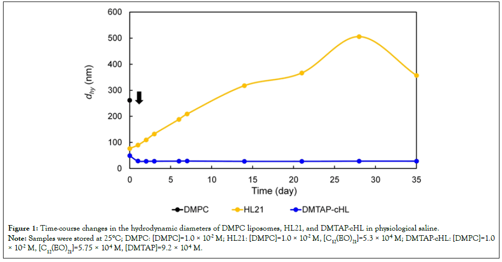
Figure 1: Time-course changes in the hydrodynamic diameters of DMPC liposomes, HL21, and DMTAP-cHL in physiological saline. Note: Samples were stored at 25°C; DMPC: [DMPC]=1.0 × 10⁻² M; HL21: [DMPC]=1.0 × 10⁻² M, [C12(EO)21]=5.3 × 10⁻⁴ M; DMTAP-cHL: [DMPC]=1.0 × 10⁻² M, [C12(EO)21]=5.75 × 10⁻⁴ M, [DMTAP]=9.2 × 10⁻⁴ M.
For DMPC liposomes, precipitation was observed on the day after preparation, indicating low stability as a particle dispersion system. HL21 showed a hydrodynamic diameter of approximately 70 nm immediately after preparation; however, the diameter increased during storage and changed markedly by 35 days. In contrast, DMTAP-cHL maintained a hydrodynamic diameter of approximately 30 nm from immediately after preparation to 35 days, indicating stable formation of nanosized particles over a prolonged period.
The smaller diameter and long-term stability of DMTAP-cHL compared with HL21 may be attributable to the positive charge introduced by DMTAP, which increased electrostatic repulsion between liposome particles. In general, the particle diameter of liposome suspensions tends to increase as a result of aggregation and fusion among particles. However, incorporation of cationic lipids may suppress aggregation through electrostatic repulsion. In addition, the PEG-based surfactant C12(EO)21 may contribute to the dispersion stability of DMTAP-cHL.
The stable hydrodynamic diameter of approximately 30 nm is important for evaluating the fusion and accumulation of DMTAP-cHL in cancer cells. Stable nanosized liposomes are expected to exhibit uniform behavior when interacting with cell membranes. Therefore, DMTAP-cHL were considered suitable for subsequent in vitro evaluation as physicochemically stable cationic hybrid liposomes.
Membrane fluidity of DMTAP-cHL
To evaluate the membrane properties of DMTAP-cHL, membrane fluidity was measured by fluorescence polarization analysis using DPH. The results are shown in Figure 2.
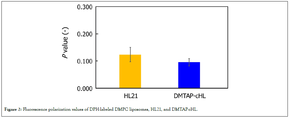
Figure 2: Fluorescence polarization values of DPH-labeled DMPC liposomes, HL21, and DMTAP-cHL.
No significant differences were observed in the P values of DMPC liposomes, HL21, and DMTAP-cHL, indicating that DMTAP-cHL had membrane fluidity comparable to that of conventional liposomes. The P value reflects the rotational mobility of DPH incorporated into the membrane; a lower P value indicates higher membrane fluidity. These results suggest that the incorporation of 8 mol% DMTAP did not significantly alter the overall fluidity of the liposome membrane.
This finding is important for understanding the function of DMTAP-cHL. Although the surface charge of DMTAP-cHL was altered by the introduction of a cationic lipid, membrane fluidity was maintained at a level comparable to that of HL21. Excessive reduction in membrane fluidity could potentially decrease fusogenicity with cell membranes. However, DMTAP-cHL prepared in this study appeared to retain the flexible membrane properties characteristic of hybrid liposomes. Thus, DMTAP-cHL acquired cationic properties while maintaining membrane fluidity comparable to that of conventional HL.
Growth-inhibitory effect of DMTAP-cHL on cholangiocarcinoma cells
To evaluate the growth-inhibitory effect of DMTAP-cHL on HuCCT-1 human cholangiocarcinoma cells, the 50% inhibitory concentration, IC50, was determined using the WST-8 assay. The results are shown in Figure 3.
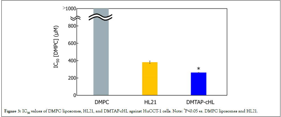
Figure 3: IC50 values of DMPC liposomes, HL21, and DMTAP-cHL against HuCCT-1 cells. Note: *P<0.05 vs. DMPC liposomes and HL21.
The IC50 values of DMPC liposomes and HL21 against HuCCT-1 cells were>1000 μM and 382 μM, respectively. In contrast, the IC50 value of DMTAP-cHL was 262 μM, which was lower than those of DMPC liposomes and HL21. These results indicate that DMTAP-cHL exerted a strong antiproliferative effect on HuCCT-1 cells.
The enhanced antiproliferative effect of DMTAP-cHL compared with HL21 may be due to the positive charge derived from DMTAP. Negatively charged lipids such as phosphatidylserine are reported to be more frequently exposed on the outer leaflet of cancer cell membranes than on normal cell membranes. Therefore, positively charged DMTAP-cHL may interact electrostatically with the negatively charged HuCCT-1 cell membrane, thereby promoting adhesion, fusion, and accumulation.
In addition, the small and stable particle size of DMTAP-cHL, approximately 30 nm, suggests that DMTAP-cHL acted as uniform nanoparticles on the cell membrane. Because particle size stability can affect cell-contact efficiency and experimental reproducibility, the physicochemical stability of DMTAP-cHL may contribute to its antiproliferative activity.
These findings suggest that introduction of the cationic lipid DMTAP enhanced the interaction of DMTAP-cHL with HuCCT-1 cell membranes and resulted in a stronger antiproliferative effect than that of HL21.
Induction of apoptosis in cholangiocarcinoma cells by DMTAP-cHL
To investigate apoptosis induction in HuCCT-1 cells by DMTAP-cHL, DNA fragmentation was measured using PI staining. The results are shown in Figure 4.
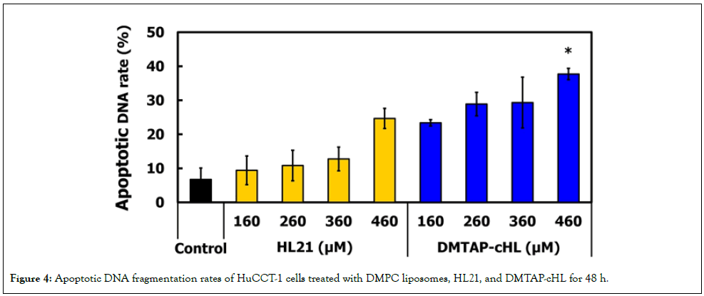
Figure 4: Apoptotic DNA fragmentation rates of HuCCT-1 cells treated with DMPC liposomes, HL21, and DMTAP-cHL for 48 h.
DNA fragmentation was also observed in the DMPC-treated group. However, because precipitation was observed in the DMPC samples at the time of addition, physical stimulation by precipitates or non-specific cell damage may have influenced DNA fragmentation. Therefore, DNA fragmentation in the DMPC group may include non-specific cell damage caused by dispersion instability rather than apoptosis specifically induced by the liposome membrane itself.
In contrast, HL21 and DMTAP-cHL induced concentration-dependent increases in DNA fragmentation in the range of 160-460 μM. In particular, DMTAP-cHL treatment increased DNA fragmentation in association with its antiproliferative effect, suggesting that apoptosis induction is involved in the growth inhibition of HuCCT-1 cells.
DNA fragmentation is a representative marker of apoptosis and suggests that cell death occurred through a regulated cell death pathway rather than simple necrosis. In this study, DMTAP-cHL treatment increased DNA fragmentation in a concentration-dependent manner, indicating that DMTAP-cHL exerted its antiproliferative effect on HuCCT-1 cells by inducing apoptosis. However, because PI staining alone cannot completely distinguish apoptosis from necrosis, these findings should be evaluated together with caspase activation analysis.
Apoptosis induction pathway activated by DMTAP-cHL
To investigate the apoptosis induction pathway in HuCCT-1 cells treated with DMTAP-cHL, the activation rates of caspase-3, caspase-8, and caspase-9 were measured by flow cytometry. The results are shown in Figure 5.
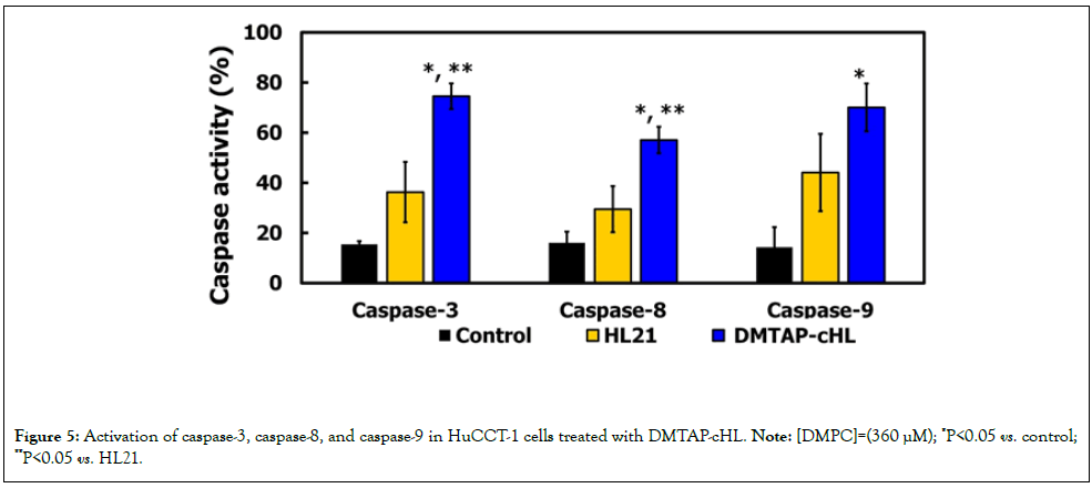
Figure 5: Activation of caspase-3, caspase-8, and caspase-9 in HuCCT-1 cells treated with DMTAP-cHL. Note: [DMPC]=(360 µM); *P<0.05 vs. control;
**P<0.05 vs. HL21.
DMTAP-cHL treatment significantly increased the activation of caspase-3, caspase-8, and caspase-9 compared with the control group. Caspase-3, an executioner caspase in apoptosis, was also significantly activated compared with the HL21-treated group.
Caspase-8 is mainly involved in the death receptor-mediated extrinsic apoptosis pathway, whereas caspase-9 is involved in the mitochondria-mediated intrinsic apoptosis pathway. Caspase-3 is activated by these upstream caspases and plays a central role in the execution phase of apoptosis, including DNA fragmentation and morphological changes. Therefore, these results indicate that DMTAP-cHL may activate the caspase cascade in HuCCT-1 cells through both extrinsic and intrinsic pathways, ultimately inducing caspase-3-mediated apoptosis.
The stronger activation of caspase-3 by DMTAP-cHL than by HL21 suggests that incorporation of the cationic lipid DMTAP enhances interaction with the cell membrane and promotes apoptotic signaling. Consistent with the increase in DNA fragmentation described above, the growth-inhibitory effect of DMTAP-cHL appears to be closely associated with caspase-dependent apoptosis. These results indicate that DMTAP-cHL induces caspase-8-, caspase-9-, and caspase-3-mediated apoptosis in HuCCT-1 cells.
Changes in membrane fluidity of cholangiocarcinoma cells induced by DMTAP-cHL
Changes in the membrane fluidity of HuCCT-1 cells after addition of DMTAP-cHL were evaluated by fluorescence polarization analysis using DPH. The results are shown in Figure 6.
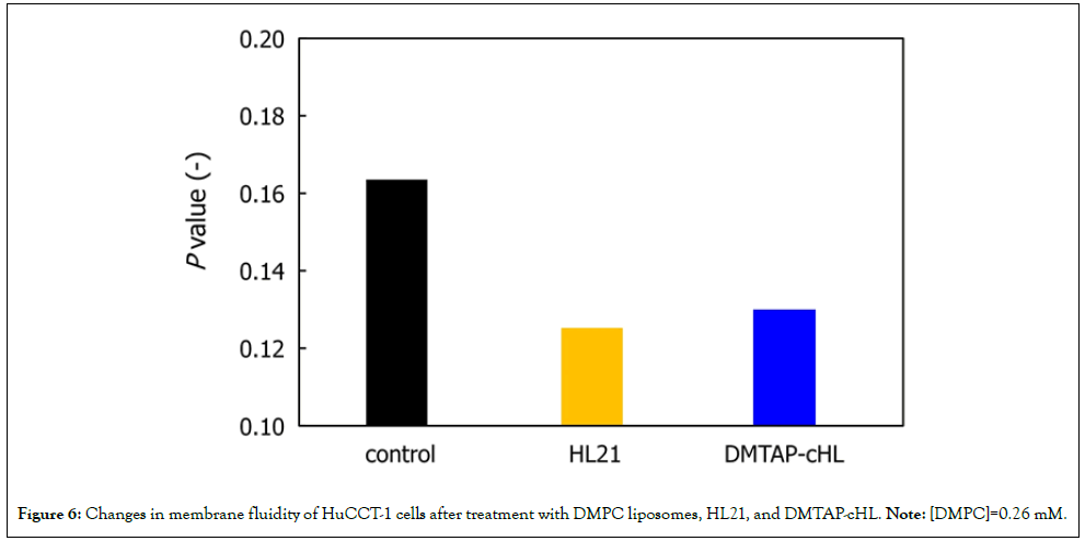
Figure 6: Changes in membrane fluidity of HuCCT-1 cells after treatment with DMPC liposomes, HL21, and DMTAP-cHL. Note: [DMPC]=0.26 mM.
After addition of DMPC liposomes, HL21, or DMTAP-cHL, the P value of HuCCT-1 cells decreased within 5 min, indicating an increase in membrane fluidity. DMTAP-cHL increased membrane fluidity to a degree comparable to that of the other liposome samples.
The increase in plasma membrane fluidity may reflect contact and fusion of liposomes with the plasma membrane, resulting in changes in lipid arrangement and membrane structure. In the case of DMTAP-cHL, the cationic lipid DMTAP may enhance electrostatic interactions with the HuCCT-1 plasma membrane, allowing DMTAP-cHL to act rapidly on the membrane surface.
Changes in membrane fluidity may also affect intracellular signal transduction, the arrangement of membrane proteins, and mitochondrial function. Therefore, the increase in plasma membrane fluidity induced by DMTAP-cHL may represent not only a physical alteration but also an early event leading to caspase activation and apoptosis induction.
These results suggest that DMTAP-cHL rapidly acts on the plasma membrane of HuCCT-1 cells and increases membrane fluidity, thereby inducing changes in the membrane environment that are involved in cell death.
Fusion and accumulation of DMTAP-cHL in cholangiocarcinoma cells
To observe the fusion and accumulation of DMTAP-cHL in HuCCT-1 cells, DMTAP-cHL/NBD-PC containing the fluorescent lipid NBD-PC was prepared and examined by confocal laser microscopy. The results are shown in Figure 7.
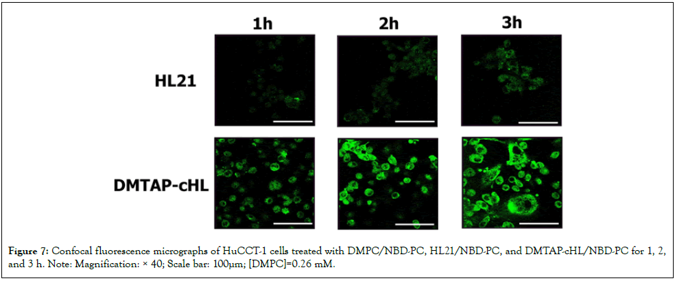
Figure 7: Confocal fluorescence micrographs of HuCCT-1 cells treated with DMPC/NBD-PC, HL21/NBD-PC, and DMTAP-cHL/NBD-PC for 1, 2, and 3 h. Note: Magnification: × 40; Scale bar: 100µm; [DMPC]=0.26 mM.
Green fluorescence was observed from 1 h after addition of DMTAP-cHL/NBD-PC to HuCCT-1 cells, indicating that DMTAP-cHL fused with and accumulated in HuCCT-1 cells within a short period. Compared with DMPC/NBD-PC and HL21/NBD-PC, the fluorescence signal of DMTAP-cHL/NBD-PC was detected at an earlier stage, suggesting that introduction of DMTAP improved the affinity of the liposomes for HuCCT-1 cells.
These results support the possibility that the cationic surface charge of DMTAP-cHL electrostatically interacts with negatively charged components on the HuCCT-1 cell membrane, thereby promoting adhesion, fusion, and accumulation. In addition, the fluorescence signal was detected within a relatively short period of 1 h, suggesting that the action of DMTAP-cHL on the cell membrane progresses rapidly.
In contrast, accumulation of DMTAP-cHL/NBD-PC in normal cholangiocytes was not clearly observed up to 3 h after addition. These results suggest that DMTAP-cHL do not readily fuse with or accumulate in normal cholangiocytes and may act selectively on HuCCT-1 cells. However, additional studies, including evaluation of cell number, observation time, and quantitative fluorescence intensity, are necessary to further assess the effects on normal cells.
These results suggest that DMTAP-cHL selectively and rapidly fuse with and accumulate in HuCCT-1 cells and are involved in subsequent changes in membrane fluidity and apoptosis induction.
Conclusion
In this study, we investigated the anticancer effects of DMTAP-cHL on HuCCT-1 human cholangiocarcinoma cells.
- DMPC liposomes precipitated after preparation and were unstable. HL21 showed a hydrodynamic diameter of approximately 70 nm immediately after preparation, but the diameter increased during storage. In contrast, DMTAP-cHL maintained a hydrodynamic diameter of approximately 30 nm and remained stable for a prolonged period.
- DMTAP-cHL exhibited membrane fluidity comparable to that of DMPC liposomes and HL21, indicating that the introduction of DMTAP did not significantly alter membrane fluidity.
- DMTAP-cHL showed a lower IC50 value than DMPC liposomes and HL21, indicating a stronger antiproliferative effect on HuCCT-1 cells.
- DMTAP-cHL induced DNA fragmentation and apoptosis in HuCCT-1 cells.
- DMTAP-cHL activated caspase-3, caspase-8, and caspase-9, suggesting that DMTAP-cHL induces cell death in HuCCT-1 cells via both extrinsic and intrinsic apoptotic pathways.
- DMTAP-cHL rapidly increased the membrane fluidity of HuCCT-1 cells, suggesting that DMTAP-cHL alters the membrane environment by fusing with the cell membrane.
- DMTAP-cHL rapidly fused with and accumulated in HuCCT-1 cells but did not clearly accumulate in normal cholangiocytes, suggesting that DMTAP-cHL selectively act on cholangiocarcinoma cells.
These results indicate that DMTAP-cHL selectively fuse with and accumulate in HuCCT-1 cells, increase membrane fluidity, and induce caspase-dependent apoptosis, thereby exhibiting strong growth-inhibitory activity. Further evaluation of the safety of DMTAP-cHL in normal cells, as well as in vivo assessment of their antitumor effects and toxicity, will be necessary to verify the usefulness of DMTAP-cHL for clinical application.
Conflicts of Interest
No potential conflict of interest was reported by the authors.
Acknowledgment
We thank Takumi Tabata for technical assistance. We would like to thank Editage (www.editage.jp) for English language editing.
References
- Nagashima F, Furuse J. Treatments for elderly cancer patients and reforms to social security systems in Japan. Int J Clin Oncol. 2022;27(2):310-315.
[Crossref] [Google Scholar] [PubMed]
- Shiratori Y, Hutfless S, Rateb G, Fukuda K. The burden of gastrointestinal diseases in Japan, 1990-2019, and projections for 2035. JGH Open. 2023;7(3):221-227.
[Crossref] [Google Scholar] [PubMed]
- Razumilava N, Gores GJ. Cholangiocarcinoma. Lancet. 2014;383(9935):2168-2179.
[Crossref] [Google Scholar] [PubMed]
- Vithayathil M, Khan SA. Current epidemiology of cholangiocarcinoma in Western countries. J Hepatol. 2022;77(6):1690-1698.
[Crossref] [Google Scholar] [PubMed]
- Patel T. Cholangiocarcinoma. Nat Clin Pract Gastroenterol Hepatol. 2006;3(1):33-42.
[Crossref] [Google Scholar] [PubMed]
- Mantripragada S, Chawla A. Cholangiocarcinoma: Part 1, pathological and morphological subtypes, spectrum of imaging appearances, prognostic factors and staging. Curr Probl Diagn Radiol. 2022;51(3):351-361.
[Crossref] [Google Scholar] [PubMed]
- Matsumoto Y, Iwamoto Y, Matsushita T, Ueoka R. Novel mechanism of hybrid liposomes-induced apoptosis in human tumor cells. Int J Cancer. 2005;115(3):377-382.
[Crossref] [Google Scholar] [PubMed]
- Nagami H, Nakano K, Ichihara H, Matsumoto Y, Ueoka R. Two methylene groups in phospholipids distinguish between apoptosis and necrosis for tumor cells. Bioorg Med Chem Lett. 2006;16(4):782-785.
[Crossref] [Google Scholar] [PubMed]
- Ichihara H, Nagami H, Kiyokawa T, Matsumoto Y, Ueoka R. Chemotherapy using hybrid liposomes along with induction of apoptosis. Anticancer Res. 2008;28(2B):1187-1195.
[Google Scholar] [PubMed]
- Shimoda S, Ichihara H, Matsumoto Y, Ueoka R. Chemotherapy with hybrid liposomes for human breast tumors along with apoptosis in vivo. Int J Pharm. 2009;372(1-2):162-168.
[Crossref] [Google Scholar] [PubMed]
- Komizu Y, Nakata S, Goto K, Matsumoto Y, Ueoka R. Membrane-targeted nanotherapy with hybrid liposomes for tumor cells leading to apoptosis. ACS Med Chem Lett. 2011;2(4):275-279.
[Crossref] [Google Scholar] [PubMed]
- Ichihara H, Hino M, Umebayashi M, Matsumoto Y, Ueoka R. Intravenous injection of hybrid liposomes suppresses the liver metastases in xenograft mouse models of colorectal cancer in vivo. Eur J Med Chem. 2012;57:143-148.
[Crossref] [Google Scholar] [PubMed]
- Ichihara H, Yamasaki S, Hino M, Ueoka R, Matsumoto Y. Hybrid liposomes inhibit the growth and angiogenesis in human breast cancer model. J Carcinog Mutagen. 2015;6(4):1-7.
[Crossref]
- Ichihara H, Komizu Y, Ueoka R, Matsumoto Y. Inhibitory effects of hybrid liposomes on the growth of non-small cell lung carcinoma cells and anti-invasive activity by ceramide generation without any drugs. J Carcinog Mutagen. 2015;6(4): 1-5.
[Crossref]
- Ichihara H, Muneaki M, Matsumoto Y. Apoptotic and anti-invasive effects of hybrid liposomes without drugs against human breast cancer cells in vitro. J Nanomedicine Biotherapeutic Discov. 2017;7:1-4.
- Kuwabara K, Ichihara H, Matsumoto Y. Inhibitory effects and anti-invasive activities of trehalose liposomes on the proliferation of lung carcinoma cells. J Carcinog Mutagen. 2017;8(1):1-5.
[Crossref]
- Okumura M, Ichihara H, Matsumoto Y, Ueoka R. Hybrid liposomes showing enhanced accumulation in tumors as theranostic agents in the orthotopic graft model mouse of colorectal cancer. Drug Deliv. 2018;25(1):1192-1199.
[Crossref] [Google Scholar] [PubMed]
- Kuwabara K, Ichihara H, Matsumoto Y. Inhibitory effect of hybrid liposomes on the growth of NP2 glioma cell. J Carcinog Mutagen. 2020;11(1):1-5.
- Kuwabara K, Ichihara H, Matsumoto Y. Novel therapy with hybrid liposomes for orthotopic graft mouse. J Carcinog Mutagen. 2021;12(2):1-5.
- Kuwabara K, Ichihara H, Matsumoto Y. Trehalose liposomes inhibit the growth of glioblastoma cell in vitro and in vivo. J Carcinog Mutagen. 2021;12:1-5.
- Ichihara H, Okumura M, Matsumoto Y. Photodynamic therapy effects of hybrid liposomes including indocyanine green against a xenograft mouse model mice of colorectal cancer in vivo. J Carcinog Mutagen. 2022;13(5):1-5.
- Ichihara H, Miyamoto S, Takai J, Okumura M, Otsuka H, Takai J, et al. Therapeutic effects of hybrid liposomes against triple negative mouse breast cancer in vitro and in vivo. J Carcinog Mutagen. 2025;16(2):1-5.
- Okumura M, Otsuka H, Takai J, Goto K, Matsumoto Y, Ichihara H. Growth suppression of triple-negative breast cancer cells by the photodynamic therapy effect of hybrid liposomes containing indocyanine green. J Carcinog Mutagen. 2025;16(2):1000471-1-5.
- Okumura M, Otsuka H, Takai J, Goto K, Matsumoto Y, Ichihara H. Enhanced therapeutic efficacy of photodynamic therapy using hybrid liposomes containing indocyanine green in a triple-negative breast cancer mouse model. Biol Pharm Bull. 2026;49(2):310-315.
[Crossref] [Google Scholar] [PubMed]
- Utsugi T, Schroit AJ, Connor J, Bucana CD, Fidler IJ. Elevated expression of phosphatidylserine in the outer membrane leaflet of human tumor cells and recognition by activated human blood monocytes. Cancer Res. 1991;51(11):3062-3066.
[Google Scholar] [PubMed]
- Ran S, Downes A, Thorpe PE. Increased exposure of anionic phospholipids on the surface of tumor blood vessels. Cancer Res. 2002;62(21):6132-6140.
[Google Scholar] [PubMed]
- Ichihara H, Motomura M, Matsumoto Y. Negatively charged cell membranes-targeted highly selective chemotherapy with cationic hybrid liposomes against colorectal cancer in vitro and in vivo. J Carcinog Mutagen. 2016;7(3):1-8.
[Crossref]
- Ichihara H, Matsumoto Y. Inhibitory effects of cationic hybrid liposomes against metastasis of bile duct cancer in vitro and in vivo. J Carcinog Mutagen. 2024;15(2):1-6.
- Ichihara H, Takaki H, Motomura M, Okumura M, Goto K, Matsumoto Y. Efficacy of cationic hybrid liposomes composed of L-α-dimyristoylphosphatidylcholine in targeting negatively charged cholangiocarcinoma cell membranes without any drug in vitro and in vivo. J Carcinog Mutagen. 2024;15(6):1-5.
Citation: Okumura M, Hashimoto T, Sakoda T, Goto K, Matsumoto Y, Ichihara H. (2026). In Vitro Growth-Inhibitory Effects of DMTAP-Containing Cationic Hybrid Liposomes (DMTAP-cHL) on Cholangiocarcinoma Cells. J Carcinog Mutagene. 17:505.
Copyright: © Okumura M. This is an open-access article distributed under the terms of the Creative Commons Attribution License, which permits unrestricted use, distribution, and reproduction in any medium, provided the original author and source are credited.