Indexed In
- Open J Gate
- Academic Keys
- JournalTOCs
- ResearchBible
- RefSeek
- Hamdard University
- EBSCO A-Z
- OCLC- WorldCat
- Publons
- Geneva Foundation for Medical Education and Research
- Euro Pub
- Google Scholar
Useful Links
Share This Page
Journal Flyer

Open Access Journals
- Agri and Aquaculture
- Biochemistry
- Bioinformatics & Systems Biology
- Business & Management
- Chemistry
- Clinical Sciences
- Engineering
- Food & Nutrition
- General Science
- Genetics & Molecular Biology
- Immunology & Microbiology
- Medical Sciences
- Neuroscience & Psychology
- Nursing & Health Care
- Pharmaceutical Sciences
Research Article - (2020) Volume 8, Issue 2
TGF-Beta Signaling Pathways in the Study of Expression in Patients with Atrial Fibrillation Atrial Tissue
Zhiliang Huang*, Yu Gao and Dianchen Hua, DOI: 10.35248/2329-8847.20.08.228
Abstract
Objective: To explore the relationship between TGF-beta signaling pathway and atrial fibrillation in patients with mitral valve disease, and reveal the mechanism of TGF-beta signaling pathway in development of atrial fibrillation.
Methods: Firstly, the fibrosis extent of right atrial appendage tissues were tested by Masson’s trichrome staining. Secondly, the protein expression of TGF-beta1, TGF-beta RI, RII, RIII, P-Smad2/3, Smad2/3, Smad4, Smad7, TAK1, p38, ATF-2 were tested by Western blot.
Results: There were significant difference between atrial fibrillation group and control group in fibrosis extent and the diameter of left atria (p0.05). The patients with atrial fibrillation are associated with the more serious fibrosis and larger diameter of left atria. For further study, the Western bolt results reveal that protein expression of TGF-beta1 and Smad4 were significant higher in atrial fibrillation and sever fibrosis group, other factors were no difference between trial and control group.
Conclusion: There are severing atrial fibrosis in atrial fibrillation patients with mitral valve diseases. The over expression of TGF-beta1 and Smad4 maybe is an important reason that induces atrial fibrosis in atrial fibrillation, and the atrial fibrosis appears to play a role in the development of atrial fibrillation. Fibrosis response by TGF-beta1 initiating maybe only depends on mutations of the TGF-beta Smad signaling pathway.
Keywords
Atrial fibrillation; Fibrosis; TGF-beta; Smad
Introduction
Atrial fibrillation (AF) is the most common arrhythmia and can occur in three different clinical situations: no organic heart disease can be identified; atrial fibrillation is a primary arrhythmia secondary to other organs; non cardiac lesions, such as hyperthyroidism; patients with organic heart disease, atrial fibrillation secondary arrhythmia. TGF-beta, as a powerful regulatory cell and signal pathway of intercellular substance, plays an important role in the interstitial fibrosis and apoptosis of [1-3] this study was to investigate the role of TGF-beta signaling pathway in the development of atrial fibrillation in the most common organic heart disease of cardiovascular surgery.
Materials and Methods
Experimental material
Right atrial tissues were taken from the Inner Mongolia Forestry General Hospital, cardiovascular surgery department patients from January 2014 to June 2016 (approved by both the Inner Mongolia Forestry General Hospital Ethics Committee and the patient). The mitral valvular disease with atrial fibrillation of the heart ear specimens fibrillation were used as the experimental group; coronary artery bypass and other specimens without atrial fibrillation were used as the control group, 10 cases in each group. Each sample was taken for paraffin embedding and the remaining liquid nitrogen was kept in reserve. The experiment included 10 cases of male patients, 10 cases of female patients; 10 cases younger than 60 years, more than 60 years; 10 patients with atrial fibrillation, 10 patients with no atrial fibrillation; left atrial diameter less than 50 mm in 13 cases, more than 50 mm in 7 cases; left ventricular diameter less than 55 mm for 12 cases, left ventricular diameter greater than 55 mm in 8 cases; there were 4 cases of left atrial thrombosis, without left atrial thrombus in 16 cases; ejection fraction less than 60 in 11 cases, ejection fraction more than 60 in 9 cases; heart function grade, in 8 cases, cardiac function grade , in 12 cases; 12 patients with mitral valve disease, there were 8 cases of other primary disease patients.
TGF-beta1, TGF-beta receptor I, receptor II, receptor III, phosphorylated Smad2/3, Smad2/3, Smad4, Smad7, phosphorylase inhibitor, phosphatase inhibitor, TAK1, p38, ATF-2 were purchased from Santa Cruz Corporation; the second antibody kit was purchased from Beijing Zhongyu Jinqiao Biotechnology Co., Ltd.; PVDF film was purchased from Millipore Company; ECL test kit was purchased from Amersham Biosciences; Fixing solution and X-ray film purchased from Kodak company.
Masson staining method: paraffin sections of formalin fixed tissue, conventional dew axing to water; Masson composite dyeing liquid 5 min, 0.2% acetic acid aqueous solution, 5% phosphotungstic acid 5-10 min, 0.2% acetic acid aqueous solution , Aniline blue dye solution 5 min, 0.2% acetic acid washed 2 times, anhydrous ethanol dehydration, xylene transparent, neutral gum seal.
Results: Arranged according to the different content of collagen fibers in the heart, respectively, with +, ++, +++, ++++. The fibrous area<25% is +, the fibrotic area between 25% and 50% is ++, the fibrous area between 50% -75% is +++, and >75% is + +++.
Western blot method: extracted total protein lysate, measure the protein concentration by Bradford method, glue, different samples of total protein of each of the 50 tested, SDS-PAGE electrophoresis; assembly of wet rotary device, anode and cathode - fiber membrane -1 Whatman 3 MM filter - gel -PVDF film -1 Whatman 3 MM filter - fiber membrane, fully discharged bubbles closed after transfer film device, transfer film; TBS-T solution containing 5% skim milk powder added closed; dilution of 1:2000 anti dilution; adding 1:2000 two anti ECL; fluorescence imaging, membrane cleaning, scanning, analysis. 1:3000 dilution of beta-actin and two dilutions of 1:2000 dilutions were used as internal controls.
Statistical analysis method: SPSS21.0 statistical software used for statistical analysis. Comparison of the two groups of data using ANOVA test with p<0.05, the difference was significant.
Results
Masson’s trichrome staining showed that the atrial fibrosis of atrial fibrillation group was obviously fibrosis. The staining showed that the collagen fibers were blue and the muscle fibers were orange red. The 7 cases of fibrosis +, 4 cases of ++, 6 cases of +++ and 3 cases of + + + +. The fibrosis + and + + for mild fibrosis group, a total of 11 cases, and + + + and + + + + as severe fibrosis group, a total of 9 cases. Of these, 9 patients with severe fibrosis were from the atrial fibrillation group, and the atrial fibrosis in the atrial fibrillation group was markedly reduced and myocardial cells were markedly reduced (Figure 1).
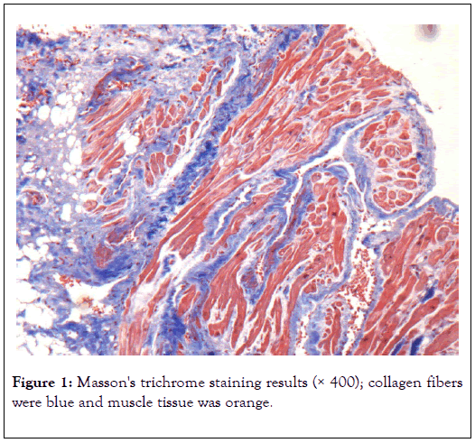
Figure 1: Masson's trichrome staining results (× 400); collagen fibers were blue and muscle tissue was orange.
Comparison of clinical parameters between atrial fibrillation group and control group; SPSS21.0 software x2 and ANOVA test method for statistical treatment, in the left atrial diameter there are significant differences (Figure 2).
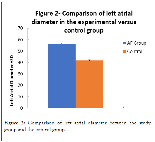
Figure 2: Comparison of left atrial diameter between the study group and the control group.
There was no significant difference in gender, age, left ventricular diameter, EF value and cardiac function grade.
Groups according to the degree of fibrosis; fibrosis + and ++ were set to mild fibrosis, +++ and ++++ were severe fibrosis. The expression of each signal factor in TGF-beta signaling pathway was analyzed between the two groups. The results showed TGFBeta 1 with p<0.05. The expression of Smad4 and protein was significantly increased in the two groups (p<0.05). TGF-beta receptor I, receptor II, receptor III, phosphorylated Smad2/3, Smad2/3, Smad7, TAK1, p38 and ATF-2 showed no significant change (Figure 3: Left four strips for atrial fibrillation group, right four bands for control group; Figure 4: Histograms of signal factors expressed between the two groups).
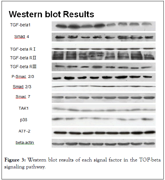
Figure 3: Western blot results of each signal factor in the TGF-beta signaling pathway.
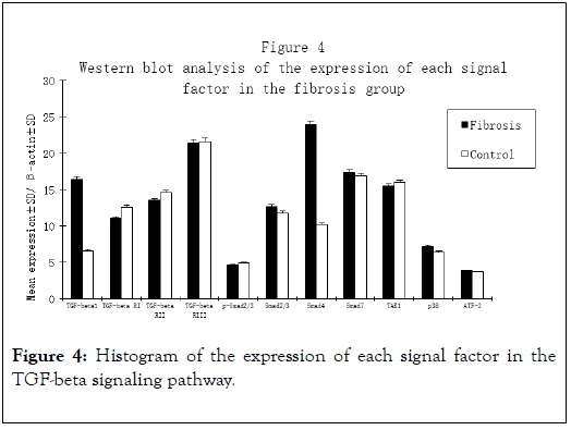
Figure 4: Histogram of the expression of each signal factor in the TGF-beta signaling pathway.
Discussion
Atrial fibrillation is the most common arrhythmia. Atrial fibrillation caused by cerebrovascular accident, limb embolism and heart failure has become the main lethal, disabling disease. Atrial fibrillation is characterized by a significant atrial fibrosis, which plays an important role in structural remodeling in triggering and maintaining atrial fibrillation. The results of this study are in line with the research direction at home and abroad: there is a significant difference between the degree of fibrosis between the AF group and the control group. Masson's trichrome staining showed that there was significant interstitial fibrosis in the atrial myocardium of the atrial fibrillation group and the normal cardiomyocytes were significantly reduced. The study found that [4]: atrial fibrillation secondary to heart disease is significantly different from pathological changes in atrial fibrillation in other cases. Organic heart disease associated with atrial fibrillation, like common atrial dilatation and fibrosis of uneven distribution; on the contrary, secondary to systemic diseases such as hyperthyroidism or electrolyte disorder in atrial fibrillation, usually are not associated with pathological abnormalities, or at least have non-specific scattered fibers. Some studies have found that atrial fibrosis may sometimes serve as a pathological basis for atrial fibrillation. [5], such as fibrosis at the pulmonary venous orifice, can alter local electrophysiological activity and trigger atrial fibrillation. Atrial fibrosis changes the electrical activity of the atrium to reduce the conduction velocity, thereby promoting the generation and maintenance of the reentry ring [6]. In conclusion, atrial fibrosis is associated with organic atrial fibrillation [7].
TGF-beta signaling pathway is an important factor in regulating fibrosis progression in vivo [8]. In this study, we further studied the expression of each signal factors in TGF-beta signaling pathway. TGF-beta pathway, as an important signaling pathway in cells, plays an important regulatory role in cell differentiation, proliferation, apoptosis, immune suppression and interstitial fibrosis [9]. The pathological basis of atrial fibrillation is atrial structural remodeling [10], which is characterized by interstitial fibrosis, which is an important aspect of the biological effects of TGF-beta signaling pathway regulation [11]. In this study, the expression of signal transduction factors in TGF-beta signaling pathway in atrial myocardium of patients with atrial fibrillation (AF) was analyzed. Western blot results in the experimental group TGF-beta1, Smad4 expression was significantly increased, other signal factors expression, suggesting that TGF-beta signaling system plays an important role in the evolution of atrial fibrillation. And what role does the signaling system play? For further analysis of the association between TGF-beta signaling pathway and atrial fibrosis, the fibrosis area (50%) were divided into two groups, the signal factors of TGF-beta signaling pathway were analyzed one by one. The expression of TGF-beta1 and Smad4 was also significantly increased, while the other signal factors were not different.
The results show that there is significant fibrosis in the atrial tissue of atrial fibrillation, and the over expression of TGF-beta1 and Smad4 is closely related to atrial fibrosis and atrial fibrillation. From the above results, we can conclude that TGFbeta1 may be involved in atrial fibrillation by means of the following processes[6,12]: atrial enlargement, increased pressure, increased angiotensin II, TGF-beta1 over expression; in atrial fibrois and atrial fibrillation, over expression of TGF-beta1 fibers increased [13,14].
Conclusion
TGF-beta signaling plays an important role in the development of atrial fibrillation by promoting atrial fibrosis, and further understanding of this signaling system is expected to make progress in the treatment of atrial fibrillation, such as through the development of TGF-beta inhibitors to control atrial fibrosis, thereby inhibiting or slowing the evolution of atrial fibrillation. The occurrence and evolution of atrial fibrillation is a complex pathophysiological process, its development process involves a variety of cytokine interactions, its more profound understanding, needs further scientific research.
REFERENCES
- Bennett RL, Steinhaus KA, Uhrich SB, O’Sullivan CK, Resta RG, Lochner-Doyle D, et al. Recommendations for standardized human pedigree nomenclature. pedigree standardization task force of the national society of genetic counselors. Am J Hum Genet. 1995;56(3): 745-752.
- He X, Gao X, PengL,Wang S, Zhu Y, Ma H, et al. Atrial fibrillation induces myocardial fibrosis through angiotensin II type 1 receptor-specific Arkadia-mediated downregulation of Smad7. Circ Res. 2011;108(2): 164-175.
- Li Q, Xu Y, Li X, Guo Y, Liu G. Inhibition of Rho-kinase ameliorates myocardial remodeling and fibrosis in pressure overload and myocardial infarction: role of TGF-beta1-TAK1. ToxicolLett. 2012;211(2): 91-97.
- Schoonderwoerd BA, Van Gelder IC, Van Veldhuisen DJ, Vanden Berg M, Crijns HJGM. Electrical and structural remodeling: role in the genesis and maintenance of atrial fibrillation. ProgCardiovasc Dis. 2005;48(3): 153-168.
- Canpolat U, Oto A, Hazirolan T, Sunman H, Yorgun H, Sahiner L, et al. A prospective DE-MRI study evaluating the role of TGF-beta1 in left atrial fibrosis and implications for outcomes of cryoballoon-based catheter ablation: New insights into primary fibrotic atriocardiomyopathy. J Cardiovasc Electrophysiol. 2015;26(3): 251-259.
- Zhang D, Liu X, Chen X, Gu J, Li F, Zhang W, et al. Role of the MAPKs/TGF-beta1/TRAF6 signaling pathway in atrial fibrosis of patients with chronic atrial fibrillation and rheumatic mitral valve disease. Cardiology. 2014;129(4): 216-223.
- Feng J, Chen X, Wan L, He S, Zhou Y, Wan S. Relationship between intermedin and atrial fibrosis in patients of hypertension combined with atrial fibrillation. Sheng Wu Yi Xue Gong Cheng XueZ aZhi. 2014;31(5): 1097-1101.
- Ghavami S, Cunnington RH, Gupta S, Yeganeh B, Filomeno KL, Freed DH, et al. Autophagy is a regulator of TGF-beta1-induced fibrogenesis in primary human atrial myofibroblasts. Cell Death Dis. 2015;6(3):e1696.
- Sun Y, Huang ZY, Wang ZH, Li CP, Meng XL, Zhang YJ, et al. TGF-beta1 and TIMP-4 regulate atrial fibrosis in atrial fibrillation secondary to rheumatic heart disease. Mol Cell Biochem. 2015;406(1): 131-138.
- Wu H, Xie J, Li GN, Chen QH, Li R, Zhang XL, et al. Possible involvement of TGF-beta/periostin in fibrosis of right atrial appendages in patients with atrial fibrillation. Int J ClinExpPathol. 2015;8(6): 6859-6869.
- Kornej J, Ueberham L, Schmidl J, Husser D, Adams V, Hindricks G, et al. Addition of TGF-beta1 to existing clinical risk scores does not improve prediction for arrhythmia recurrences after catheter ablation of atrial fibrillation. Int J Cardiol. 2016;221: 52-54.
- Polejaeva IA, Ranjan R, Davies CJ, Regouski M, Hall J, Olsen AL, et al. Increased susceptibility to atrial fibrillation secondary to atrial fibrosis in transgenic goats expressing transforming growth factor-beta1. Cardiovasc Electrophysiol. 2016;27(10): 1220-1229.
- Liu LJ, Yao FJ, Lu GH, Xu CG, Xu Z, Tang Z, et al. The role of the Rho/ROCK pathway in Ang II and TGF-beta1-induced atrial remodeling. PLoS One. 2016;11(9): e0161625.
- Chen X, Xu J, Jiang B, Liu D. Bone morphogenetic protein-7 antagonizes myocardial fibrosis induced by atrial fibrillation by restraining transforming growth factor-beta (TGF-beta)/Smads signaling. Med Sci Monit. 2016;22: 3457-3468.
Citation: Huang Z, Gao Y, Hua D (2020) TGF-Beta Signaling Pathways in the Study of Expression in Patients with Atrial Fibrillation Atrial Tissue. J Aging Sci.8:228. Doi:10.35248/2329-8847.20.08.228.
Copyright: © 2020 Huang Z, et al. This is an open-access article distributed under the terms of the Creative Commons Attribution License, which permits unrestricted use, distribution, and reproduction in any medium, provided the original author and source are credited.
