Indexed In
- Genamics JournalSeek
- RefSeek
- Hamdard University
- EBSCO A-Z
- OCLC- WorldCat
- Publons
- Geneva Foundation for Medical Education and Research
- Euro Pub
- Google Scholar
Useful Links
Share This Page
Journal Flyer
Open Access Journals
- Agri and Aquaculture
- Biochemistry
- Bioinformatics & Systems Biology
- Business & Management
- Chemistry
- Clinical Sciences
- Engineering
- Food & Nutrition
- General Science
- Genetics & Molecular Biology
- Immunology & Microbiology
- Medical Sciences
- Neuroscience & Psychology
- Nursing & Health Care
- Pharmaceutical Sciences
Case Report - (2023) Volume 12, Issue 5
Ping Pong Fracture in a Newborn with Maternal Motor Vehicle Accident Exposure during Pregnancy and Underlying Vitamin D Deficiency
Jean Matthes*Received: 24-Sep-2023, Manuscript No. JNB-23-23170; Editor assigned: 28-Sep-2023, Pre QC No. JNB-23-23170(PQ); Reviewed: 13-Oct-2023, QC No. JNB-23-23170; Revised: 19-Oct-2023, Manuscript No. JNB-23-23170(R); Published: 26-Oct-2023, DOI: 10.35248/2167-0897.23.12.426
Abstract
We present a case of a Ping pong fracture, also recognized as a depressed skull fracture, in a late preterm female infant. This type of fracture can manifest spontaneously or as a result of obstetrical or external trauma. In present case, the mother had a history of involvement in a motor vehicle accident, and the newborn displayed low vitamin D levels attributed to maternal Vitamin D deficiency. The management of this type of fracture lacks a universally accepted standard protocol. It should be tailored to factors including clinical presentation, the underlying cause of the fracture, and the response to conservative management.
In the case, the infant was treated with vitamin D and the fracture was managed conservatively. Remarkably, the fracture resolved spontaneously over few months. The case also underscores the significance of evaluating vitamin D levels in newborns with depressed skull fractures, even when other risk factors such as obstetrical or external trauma are present, to ensure appropriate treatment.
Keywords
Ping pong fracture, Depressed skull fracture, Vitamin D deficiency
Introduction
A Ping Pong fracture, also known as a depressed skull fracture, occurs when the calvarial bone buckles inward without a loss of bone continuity. It is a rare type of fracture in newborns, with an incidence ranging from 1 to 2.5 cases per 10,000 live births [1,2]. This type of fracture can occur spontaneously or as a result of obstetrical or external trauma. The management of Ping Pong fractures lacks a universally accepted standard protocol. In this report, we present a case of a Ping Pong fracture in a late preterm female infant. This case is particularly interesting due to the infant's history of in utero exposure to a maternal motor vehicle accident and low vitamin D levels.
Case Presentation
A newborn girl was delivered to a 34-year-old Childbearing 3, Afro-American woman at 36 weeks and 3 days of gestation via an emergent C-section following a motor vehicle accident. The accident occurred while the woman was an unrestrained rear passenger, resulting in non-reassuring fetal heart tones. The mother sustained cervical and pelvic fractures in the accident. The infant had a birth weight of 2360 g. The mother had received regular prenatal care, and her history revealed anemia with Rh-negative blood group status post Rhogam administration. Because of a history of recurrent Herpes, the mother was on Valacyclovir prophylaxis, and had no active lesions during delivery. Prenatal multivitamins and iron were part of the mother's pregnancy regimen. The delivery proceeded without complications, and the APGAR scores were 8 and 9 at 1 and 5 minutes, respectively.
On initial examination, the newborn exhibited soft cystic swellings measuring approximately 2.5 cm × 2.5 cm on the right parietal area, and 1 cm × 1 cm on the left parietal area, with a soft depressed area over the occipital region (Figure 1). The anterior fontanelle was wide and evaluation for congenital hypothyroidism revealed Thyroid Stimulating Hormone (TSH) and free T4 levels at 31.450 uIU/mL and 1.08 ng/dL, respectively, at about 25 hours after birth. A head ultrasound showed no abnormalities. Skull X-rays were performed due to the maternal motor vehicle accident and showed mild contour abnormality in the parietal bone without any definite fractures. A complete metabolic profile on day 4 of life indicated low calcium levels at 7.6 mg/dL, high normal phosphorus levels at 8.1 mg/dL, and high alkaline phosphatase levels at 527 U/L. Parathyroid hormone and 25-hydroxy vitamin D levels were 238.10 pg/mL and 15 ng/ml. Fractional excretion of phosphorus was calculated at 17.53% (reference range<20%). Maternal 25-hydroxy vitamin D level was found to be<7 ng/ml.
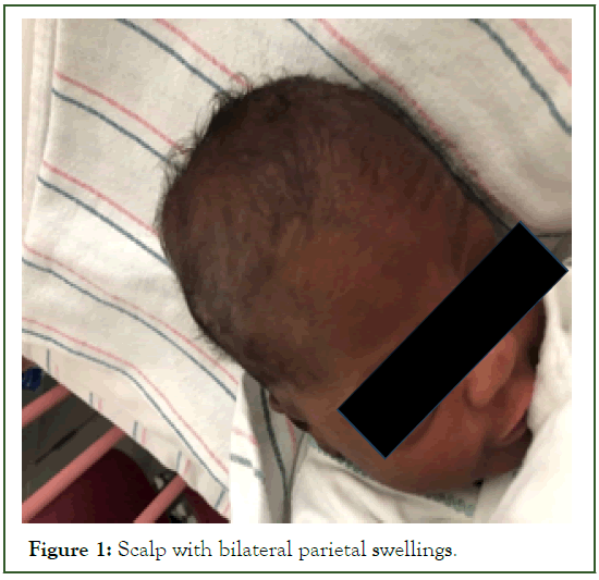
Figure 1: Scalp with bilateral parietal swellings.
A skeletal survey showed no fractures, thinning of bones or rachitic changes. A repeated TSH level indicated an increase to 45.440 uIU/mL. A reassessment ultrasound of the scalp cystic lesion showed a superficial fluid collection with a small step-off of the underlying bone (Figure 2).
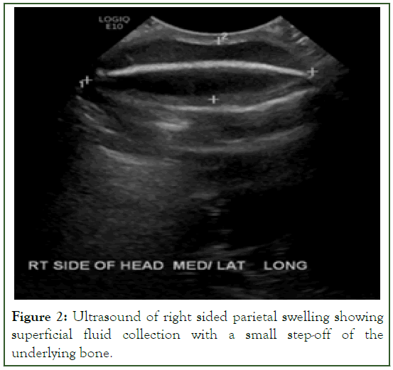
Figure 2: Ultrasound of right sided parietal swelling showing superficial fluid collection with a small step-off of the underlying bone.
A Computed Tomography (CT) of head without contrast revealed fractures of the bilateral posterior parietal calvarium, larger on the right side, along with extra-calvarial soft tissue swelling but no intracranial hemorrhage (Figure 3). The neurosurgery team recommended monitoring the head circumference without neurosurgical intervention. The patient was started on levothyroxine at 25 mcg/day for possible congenital hypothyroidism, ergocalciferol 1000 IU daily, and calcium carbonate with 50 mg of elemental Ca/kg/day for vitamin D deficiency. Repeat parathyroid hormone, calcium, phosphorous, TSH, and free T4 levels on day 11 of life were normalized at 44.47 pg/mL,10.4 mg/dl, 6.3 mg/dl, 0.360 uIU/mL, and 1.33 ng/dL, respectively.
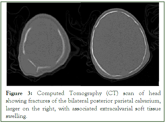
Figure 3: Computed Tomography (CT) scan of head showing fractures of the bilateral posterior parietal calvarium, larger on the right, with associated extracalvarial soft tissue swelling.
At 4 months, the scalp swelling had completely resolved, and the thyroid function tests were normal without supplementation. Repeat serum calcium and phosphorus levels at 2-month visit were normal. Alkaline phosphatase peaked at 760 U/L at 7 weeks of life and had normalized by the 6-month visit. The Osteogenesis Imperfecta Gene panel yielded negative results.
Results and Discussion
A Ping Pong fracture, also known as a depressed skull fracture, results from the inward buckling of the calvarial bone without a loss of bone continuity. The incidence of Ping Pong fractures varies from 1 to 2.5 cases per 10,000 live births [1,2]. This type of fracture may occur due to constant pressure from maternal bony prominences during intrauterine constraint, obstetrical trauma, or external trauma [3,4]. It is considered spontaneous if the pregnancy is uneventful [1-3]. In the case, the mother had a history of a motor vehicle accident resulting in cervical and pelvic fractures. Insufficient fetal Vitamin D levels, stemming from maternal Vitamin D deficiency, may predispose the fetus to such fractures due to softened skull bones resulting from immature mineralization [5-6]. Studies indicate increased risks of Vitamin D deficiency in both pregnant mothers and newborns, particularly in Black populations [7,8]. The mother in our case had a known Vitamin D deficiency with inadequate supplementation. A correlation between low Vitamin D levels in mothers and infants has been observed [9]. Pregnancy-associated Vitamin D deficiency is linked to adverse outcomes for both mothers and children, including preterm birth, low birth weight, respiratory infections, and bone-related abnormalities [10]. Both external trauma and Vitamin D deficiency are considered individual risk factors for depressed skull fractures [9].
In the case, a history of a maternal motor vehicle accident during pregnancy, combined with low Vitamin D levels, may have collectively heightened the risk. To our knowledge, this is the first reported instance of a newborn with depressed skull fractures following maternal motor vehicle accident exposure during pregnancy and documented low vitamin D levels. This underscores the significance of evaluating vitamin D levels in newborns with depressed skull fractures, even when other risk factors such as obstetrical or external trauma are present, to ensure appropriate treatment. While plain skull X-rays and ultrasound were relatively unremarkable, a CT scan of the skull revealed bilateral posterior parietal calvarium fractures without intracranial hemorrhage, highlighting the importance of CT scans to rule out underlying brain injuries.
Our case exhibited low calcium and high phosphorus levels, suggestive of hypoparathyroidism, but elevated Parathyroid Hormone (PTH) levels. PTH levels can be elevated in response to Vitamin D deficiency, normalizing with correction of calcium and Vitamin D levels, as observed in our case. The initial thyroid function test suggested hypothyroidism, which improved after thyroid hormone supplementation and proved transient. Hypothyroidism and Vitamin D deficiency are reported to have a correlation in adults, and in our case, thyroid function improved with treatment [11].
Management of depressed skull fractures is contentious and depends on clinical presentation, the cause of the fracture, and the response to conservative treatment. Options include observation, non-surgical interventions, or surgery [12-14]. In our case, due to the absence of neurological manifestations and the absence of underlying brain hemorrhage, observation was chosen, and the bilateral depressed skull fractures resolved spontaneously during follow-up visits.
Conclusion
Ping Pong or Depressed skull fractures can manifest either spontaneously or as a result of obstetrical or external trauma. The management of this fracture lacks a universally accepted standard protocol and should be tailored to factors including clinical presentation, the underlying cause of the fracture, and the response to conservative treatment. Our case also underscores the significance of evaluating vitamin D levels in newborns with depressed skull fractures, even when other risk factors such as obstetrical or external trauma are present, to ensure appropriate treatment.
References
- Basaldella L, Marton E, Bekelis K, Longatti P. Spontaneous resolution of atraumatic intrauterine ping-pong fractures in newborns delivered by cesarean section. J Child Neurol. 2011;26(11):1449-1451.
[Crossref] [Google Scholar] [PubMed]
- Ilhan O, Bor M, Yukkaldiran P. Spontaneous resolution of a ‘ping-pong’fracture at birth. BMJ Case Rep. 2018; 2018.
[Crossref] [Google Scholar] [PubMed]
- Dupuis O, Silveira R, Dupont C, Mottolese C, Kahn P, Dittmar A, et al. Comparison of “instrument-associated” and “spontaneous” obstetric depressed skull fractures in a cohort of 68 neonates. J Obstet Gynaecol. 2005;192(1):165-170.
[Crossref] [Google Scholar] [PubMed]
- Fantacci F, Capozz D, Romano V, Ferrara P, Chiaretti A, Massimi L. “Spontaneous” ping-pong fracture in newborns: Case report and review of the literature. Signa Vitae. 2015;10(1):103-109.
- Namgung R, Tsang RC, Specker BL, Sierra RI, Ho ML. Low bone mineral content and high serum osteocalcin and 1, 25-dihydroxyvitamin D in summer-versus winter-born newborn infants: An early fetal effect? J Pediatr Gastroenterol Nutr. 1994;19(2):220-227.
[Crossref] [Google Scholar] [PubMed]
- Viljakainen HT, Saarnio E, Hytinantti T, Miettinen M, Surcel H, Makitie O, et al. Maternal vitamin D status determines bone variables in the newborn. J Clin Endocrinol Metab. 2010;95(4):1749-1757.
[Crossref] [Google Scholar] [PubMed]
- Dawodu A, Wagner CL. Mother-child vitamin D deficiency: An international perspective. Arch Dis Child. 2007;92(9):737-740.
[Crossref] [Google Scholar] [PubMed]
- Marshall I, Mehta R, Ayers C, Dhumal S, Petrova A. Prevalence and risk factors for vitamin D insufficiency and deficiency at birth and associated outcome. BMC Pediatr. 2016;16:1-7.
[Crossref] [Google Scholar] [PubMed]
- Dória M, Viveiros C, e Rodrigues LR, Soares F. A rare case of spontaneous intrauterine skull fracture. Acta Med Port. 2020;33(5):344-346.
[Crossref] [Google Scholar] [PubMed]
- Zhang H, Wang S, Tuo L, Zhai Q, Cui J, Chen D, et al. Relationship between maternal Vitamin D levels and adverse outcomes. Nutrients. 2022;14(20):4230.
[Crossref] [Google Scholar] [PubMed]
- Mackawy AM, Al-Ayed BM, Al-Rashidi BM. Vitamin D deficiency and its association with thyroid disease. Int J Health Sci. 2013;7(3):267.
[Crossref] [Google Scholar] [PubMed]
- Zalatimo O, Ranasinghe M, Dias M, Iantosca M. Treatment of depressed skull fractures in neonates using percutaneous microscrew elevation. J Neurosurg Pediatr. 2012;9(6):676-679.
[Crossref] [Google Scholar] [PubMed]
- Hung KL, Liao HT, Huang JS. Rational management of simple depressed skull fractures in infants. J Neurosurg. 2005;103(1):69-72.
[Crossref] [Google Scholar] [PubMed]
- Altieri R, Grasso E, Cammarata G, Garozzo M, Marchese G, Certo F, et al. Decision-Making Challenge of Ping-Pong Fractures in Children: Case Exemplification and Systematic Review of Literature. World Neurosurg. 2022;165:69-80.
[Crossref] [Google Scholar] [PubMed]
Citation: Matthes J (2023) Ping Pong Fracture in a Newborn with Maternal Motor Vehicle Accident Exposure during Pregnancy and Underlying Vitamin D Deficiency. J Neonatal Biol. 12:426.
Copyright: © 2023 Matthes J. This is an open access article distributed under the terms of the Creative Commons Attribution License, which permits unrestricted use, distribution, and reproduction in any medium, provided the original author and source are credited.
