Indexed In
- Genamics JournalSeek
- RefSeek
- Hamdard University
- EBSCO A-Z
- OCLC- WorldCat
- Publons
- Geneva Foundation for Medical Education and Research
- Euro Pub
- Google Scholar
Useful Links
Share This Page
Journal Flyer

Open Access Journals
- Agri and Aquaculture
- Biochemistry
- Bioinformatics & Systems Biology
- Business & Management
- Chemistry
- Clinical Sciences
- Engineering
- Food & Nutrition
- General Science
- Genetics & Molecular Biology
- Immunology & Microbiology
- Medical Sciences
- Neuroscience & Psychology
- Nursing & Health Care
- Pharmaceutical Sciences
Case Report - (2023) Volume 12, Issue 1
Difficult Endotracheal Intubation in a Neonate: A Clinical Challenge
Reda El Bayoumy1*, Edeline Coinde2 and Marion Nimal32Department of Pediatrics, Ajaccio Public Hospital, Ajaccio, France
3Department of Neonatal Medicine, Hospital Nord, University Hospital, Marseille, France
Received: 07-Jan-2023, Manuscript No. JNB-23-19554; Editor assigned: 10-Jan-2023, Pre QC No. JNB-23-19554(PQ); Reviewed: 26-Jan-2023, QC No. JNB-23-19554; Revised: 02-Feb-2023, Manuscript No. JNB-23-19554(R); Published: 09-Feb-2022, DOI: 10.35248/2167-0897.23.12.386
Abstract
Difficult Endotracheal (ET) intubation in neonates is not uncommon situation which has diverse etiologies. It poses a technical challenge to save a newborn baby immediately following birth especially in isolated island with limited neonatal facilities, resources and expertise. Neonatal morbidity and mortality are considerably high in difficult endotracheal intubation scenarios. A late preterm neonate was born limp and apneic. Upper airway obstruction by voluminous supraglottic cystic mass with possible subglottic obstruction was primarily predicted. The neonate was successfully ET intubated and transferred to the tertiary neonatal unit for definitive surgical management.
Keywords
Neonatal morbidity; Oxygen pressure; Endotracheal intubation; Ultrasound
INTRODUCTION
Endotracheal intubation in preterm infants is often challenging, especially in the presence of airway malformations [1-4]. Successful ET intubation of a newborn infant at birth can be difficult, even for an experienced neonatologist and anesthetist [1-3]. Supraglottic and subglottic lesions usually present as difficult airway scenario requiring cricothyroidotomy as a life-saving procedure in emergency [1,4]. Most solid neoplasms identified in neonates are benign. The incidence of a malignant tumor is 1 in every 12,500-27,500 live births, accounting for 2% of all childhood cancers, nevertheless, subglottic hemangiomas, stenosis and webs are the usual etiologies but ectopic cervical thymus has rarely also been reported causing airway obstruction [5]. Prompt recognition of airway malformations facilitates directed resuscitation and appropriate intervention, the delay of which could be fatal to the baby [2,3]. This may be due to anatomical variations of the airway or mechanical obstructions such as cysts and tumors. One need to have a strong suspicion of this condition and emergency team should be promptly prepared to deal with such situations [2,3]. Early resection of head and neck tumors in the infantile period is primordial [6]. These tumors can be detected early in the antenatal period by advanced fetal ultrasound or Magnetic Resonance Imaging (MRI) [7]. It would be more appropriate to diagnose head and neck malformations during pregnancy to better plan delivery with specialized medical team or considering Ex-utero Intrapartum Treatment (EXIT) procedure [8,9].
“Written consent has been obtained from the patient’s next-of-kin for the publication of this case report”.
Case Description
A baby boy with birth weight of 2445 grams was born to a primigravida mother at 36 weeks of gestation by instrumental delivery under epidural analgesia.
Baby was born limp and apneic with heart rate of 60 beats/ minute (b/min). Apgar score was 1/3/6/6. Resuscitative measures were immediately initiated by Neopuff bag and mask ventilation (spontaneous assisted ventilation) with the following parameters: Positive End Expiratory Pressure (PEEP) of 5 cm H2O and Fractional Inspired Oxygen (FiO2) of 30%. Bag and mask ventilation was successful; Oxygen (O2) saturation was 97% and heart rate was 152 b/min, however, baby was hypoxic in spontaneous breathing with biphasic stridor; O2 saturation was 71% on FiO2 of 30% with signs of upper airway obstruction. Pediatrician decided immediately to refer the baby to the tertiary hospital. The baby’s spontaneous breathing was supported by continuous positive airway pressure via CPAP-Dräger Babylog 8000 plus ventilator, on FiO2 of 30%, PEEP of 5 cm H2O (Video 1).
Video 1: Continuous positive airway pressure via CPAP-Dräger Babylog 8000 plus ventilator.
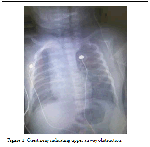
O2 saturation was kept on 97%. Arterial blood gases were pH 7.173, PaO2 67.7, PaCO2 67.6, lactate 0.70 mmol/L. The baby was norm thermic and kept warm with normal blood sugar level all through the resuscitation period. Failed two nasogastric tube insertion attempts. Heart rate was around 134 b/min, respiratory rate was 56 breaths/minute, arterial blood pressure was 45/33 mmHg, and O2 saturation was 97% on 30% of FiO2. Chest x-ray was normal which led the pediatrician not to proceed to ET intubation without multidisciplinary input from the anesthetist and tertiary hospital neonatal specialist with high prediction of upper airway obstruction (Figure 1).

Figure 1: Chest x-ray indicating upper airway obstruction.
The second constraint was that Ajaccio public hospital is situated in the Mediterranean island of Corsica which lacks advanced neonatal unit, Ajaccio public hospital is affiliated to Marseille university hospital in France and difficult neonatal and pediatric pathologies should be referred to the tertiary hospital. The distance between Ajaccio and Marseille is 400 kilometers. Transfer is done by helicopter. Besides, the incident took place during a weekend where there was no enough medical support to summon help.
The baby was born around 10 O’clock in the morning; the Marseille’s neonatal team is arrived to retrieve the baby around 5 O’clock in the afternoon. The baby was maintained on CPAP ventilation in neonatal Intensive Care Unit (ICU). The anesthetist and Ear-Nose-Throat (ENT) specialist surgeon on-call were informed and to be stand-by for ET intubation once the Marseille’s neonatal team arrives on-site. The anesthetist with the anesthetic nurse oncall prepared the difficult intubation drill trolley to deal with the difficult ET intubation; the pediatric video laryngoscope (axess vision bronchoflex agil Glidescope, Ø 3.9, blade size 1 and 2) and fiberoptic bronchoscope (bronchoflex agile and vortex) with different sizes of stylets and bougies were prepared. Once the Marseille’s neonatal team arrived in place, was joined immediately by the anesthetic, ENT and pediatric on call teams.
Marseille’s neonatal team prepared the difficult intubation drill trolley; which included pediatric airway AirTrach Vygon and bougies. They prepared the anesthetic induction agents (intravenous ketamine, midazolam and suxamethonium), calcium, magnesium, sodium bicarbonate, adrenaline syringes and noradrenaline infusion syringe. Midazolam and sufentanil infusion syringes were being prepared as well for intravenous sedation during transfer.
First ET intubation attempt is failed with the identification of upper airway obstruction by voluminous supraglottic cystic mass, however it was easy to ventilate the baby with facial mask and to maintain his airway and O2 saturation, baby was never being hypoxic.
Second ET intubation attempt was successful by the aid of a glidescope and stylet. The baby was intubated by 3 mm Internal Diameter (ID) cuffed ET tube, it was a bit difficult to push the ET tube down the trachea past the laryngeal inlet, however it was successful to push it down the trachea with gentle manipulation, ET tube was secured in-place and baby was successfully transferred by helicopter to Marseille’s neonatal unit around 8 O’clock in the afternoon (Videos 2 and 3).
Video 2: ET intubation attempt was successful by the aid of a glidescope.
Video 3: ET intubation attempt was successful by the aid of stylet
The baby was operated upon next day by the pediatric ENT surgical team whom underwent laser marsupialization of the right supraglottic paralaryngeal cyst and they treated as well a second small subglottic cyst by laser which was communicating with the larger right paralaryngeal cyst via a duct which explains the difficulties to advance the ET tube down the trachea during endotracheal intubation. The anesthetist decided not to extubate the baby following the end of the procedure due to laryngeal edema (Videos 4 and 5).
Video 4: Laryngeal view following ET intubation showing the right paralaryngeal tumor.
Video 5: The end of surgical procedure with evident laryngeal edema
Baby was transferred to neonatal ICU under sedation for postoperative ventilation and management. Baby was successfully extubated on 3rd day postoperative with no complications. Patient survived and pathological analysis showed benign supraglottic paralaryngeal and subglottic vascularized Malpighian cysts.
Results and Discussion
Congenital head and neck tumors can lead to stridor and upper airway obstruction in newborns and might pose consequently massive challenge to both neonatologists and anesthetists to secure their upper airways [1-3]. Differential diagnoses include teratomas, hemangiomas, lymphatic malformations, and neuroglial heterotopias [4-6]. Subglottic stenosis can be congenital or acquired. It is defined as a diameter of less than 4 mm of the cricoid region in a full-term infant and less than 3 mm in a premature infant [4]. Periglottic masses and swellings can effectively cause difficult intubation which may lead to serious adverse outcome including death which requires upper airway management expertise and appropriate skills [1,3,5]. Working in isolated Remote Island with minimal neonatal resuscitation resources adds to the technical difficulties.
A “Difficult Airway Situation” arises whenever face mask ventilation, direct laryngoscopy, ET intubation, or use of supraglottic device fails to secure ventilation. As bradycardia and cardiac arrest in the neonate are usually of respiratory origin, neonatal airway management remains a critical factor [2,3].
Continuous medical education and improving skills and management of neonatal and pediatric disorders should be emphasized for pediatricians, gynecologists and anesthetists working in remote and understaffed hospitals. Availability as well of up-to-date resources, equipment and facilities for neonatal and pediatric management in remote and isolated hospital settings are of utmost importance to save lives [3].
Early resection of head and neck tumors in the infantile period is advocated. Early resection and reconstruction can avoid prolonged intubation or tracheostomy and thus allow early recovery of normal airway function [6].
Recently, these tumors can be detected early in the antenatal period by advanced fetal ultrasound or MRI which necessitates emphasizing in the development of those diagnostic skills and expertise during antenatal care by concerned physicians especially in public and remote hospitals [7].
Well-planned delivery by Ex-utero Intrapartum Treatment (EXIT) could be performed [8]. This is an extension of standard caesarean section with the baby partially delivered while the umbilical cord is kept intact to maintain maternofetal circulation to allow time for intubation and to secure the airway [8,9].
Conclusion
This case report concludes the importance of developing advanced antenatal ultrasonography during antenatal period to detect neonatal congenital malformations in-utero before birth which will result in safe delivery of the neonate by experienced medical team to deal with the challenging pathology and will consequently improve neonatal morbidity and mortality.
The increased use of ultrasonography in the management and evaluation of pregnancy has provided a unique opportunity to observe the anatomy of the developing fetus from 12 weeks gestation until term. These include upper airway tumors, ascites, gastroschisis, omphalocele, sacro-coccygeal teratoma, cystic hygroma, hydrocele, duodenal atresia, jejunal atresia, conjoined twins, uretero-pelvic junction obstruction, urethral valves, urethral agenesis, multicystic kidney and hydronephrosis secondary to reflux.
Prenatal diagnosis by ultrasonographic examination has significantly improved perinatal management. Elective cesarean section has benefited conjoined twins and infants with lesions causing dystocia, such as sacro-coccygeal teratoma, omphalocele. Advance notification of surgeons and neonatologists has reduced the delays of postnatal evaluation and treatment that contribute significantly to complications and death. In addition, transfer of the pregnant mother carrying an infant with a significant surgical anomaly to a center with facilities for neonatal surgery and specialized postoperative care can be properly planned for in advance. In the near future, intrauterine fetal surgery or palliative intervention may provide increased salvage of patients with obstructive uropathy and diaphragmatic hernia, both of which carry high mortality rates secondary to in-utero damage.
References
- Heinrich S, Birkholz T, Ihmsen H, Irouschek A, Ackermann A, Schmidt J. Incidence and predictors of difficult laryngoscopy in 11.219 pediatric anesthesia procedures. Paediatr Anaesth. 2012; 22(8):729-736.
[Crossref] [Google Scholar] [PubMed]
- Robert T. Managing the difficult airway in the neonate-A BAPM Framework for Practice. 2020.
- Berisha G, Boldingh AM, Blakstad EW, Rønnestad AE, Solevåg AL. Management of the unexpected difficult airway in neonatal resuscitation. Front Pediatr. 2021:1103.
[Crossref] [Google Scholar] [PubMed]
- Ida JB, Guarisco JL, Rodriguez KH, Amedee RG. Obstructive lesions of the pediatric subglottis. Ochsner J. 2008; 8(3):119-128.
[Google Scholar] [PubMed]
- Boudewyns A, Claes J, Van de Heyning P. Clinical practice-An approach to stridor in infants and children. Eur J Pediatr. 2010; 169(2):135-141.
[Crossref] [Google Scholar] [PubMed]
- Wong BY, Ng RW, Yuen AP, Chan PH, Ho WK, Wei WI. Early resection and reconstruction of head and neck masses in infants with upper airway obstruction. Int J Pediatr Otorhinolaryngol. 2010; 74(3):287-291.
[Crossref] [Google Scholar] [PubMed]
- Nawapun K, Phithakwatchara N, Jaingam S, Viboonchart S, Mongkolchat N, Wataganara T. Advanced ultrasound for prenatal interventions. Ultrasonography. 2018; 37(3):200.
[Crossref] [Google Scholar] [PubMed]
- García-Díaz L, Chimenea A, de Agustín JC, Pavón A, Antiñolo G. Ex-Utero Intrapartum Treatment (EXIT): indications and outcome in fetal cervical and oropharyngeal masses. BMC Pregnancy Childbirth. 2020; 20(1):1-6.
[Crossref] [Google Scholar] [PubMed]
- Barrette LX, Morales CZ, Oliver ER, Gebb JS, Feygin T, Lioy J, et al. Risk factor analysis and outcomes of airway management in antenatally diagnosed cervical masses. Int J Pediatr Otorhinolaryngol. 2021; 149:110851.
[Crossref] [Google Scholar] [PubMed]
Citation: Bayoumy RE, Coinde E, Nimal M (2023) Difficult Endotracheal Intubation in a Neonate: A Clinical Challenge. J Neonatal Biol. 12:386.
Copyright: © 2023 Bayoumy RE, et al. This is an open access article distributed under the terms of the Creative Commons Attribution License, which permits unrestricted use, distribution, and reproduction in any medium, provided the original author and source are credited.

