Indexed In
- Open J Gate
- Genamics JournalSeek
- ResearchBible
- RefSeek
- Directory of Research Journal Indexing (DRJI)
- Hamdard University
- EBSCO A-Z
- OCLC- WorldCat
- Scholarsteer
- Publons
- MIAR
- Euro Pub
- Google Scholar
Useful Links
Share This Page
Journal Flyer
Open Access Journals
- Agri and Aquaculture
- Biochemistry
- Bioinformatics & Systems Biology
- Business & Management
- Chemistry
- Clinical Sciences
- Engineering
- Food & Nutrition
- General Science
- Genetics & Molecular Biology
- Immunology & Microbiology
- Medical Sciences
- Neuroscience & Psychology
- Nursing & Health Care
- Pharmaceutical Sciences
Research Article - (2021) Volume 10, Issue 7
Anti-Diabetic Effect of Methanolic Extract of Aristolochia ringensLeaf
Rasidat O Tijani*, Lawal SO, Oyekan JO, Koleoso OK, Onasanya SS and Fasasi AAReceived: 17-Jun-2021 Published: 09-Jul-2021, DOI: 10.35248/2161-1009.21.10.397
Abstract
The aim and objective of this study is to investigate the hypoglycemic, hepatoprotective, hypolipidemic and antioxidant potentials of the methanolic extract of Aristolochia ringens leaf (MLAR) in STZ-induced diabetic wistar rats. The airdried and powdered leaf of the plant (Aristolochia ringens) was extracted by cold maceration and concentrated. In vitro analysis was carried out by screening the extract for phytochemical constituents and antioxidant activities using different models. In vivo analysis was also carried out by randomly dividing forty eight (48) male wistar rats (140-170 g) into eight groups of six (6) rats each. Group 1 served as control; group 2 received STZ (60 mg/kg wt i.p); groups 3 to 8 served as the treatment group. The animals were sacrificed without anesthesia after 30 days and their blood were collected for biochemical investigation. The in vitro results showed that MLAR possesses good antioxidant potential when compared with the standard and it is also known to contain a number of phytochemicals. Also, the administration of STZ caused significant (p<0.05) elevation of the serum level of glucose, ALT AST, GGT, ALP, total cholesterol, LDL, TRIGS, MDA, NO and MPO activity by 100%, 47%, 38%, 32%, 51%, 29%, 34%, 41%, 51%, 29%, 54% and 59% respectively and significant reduction in HDL, Catalase, GPx, GSH, SOD, GST and plasma insulin by 34%, 34%, 61%, 46%, 66%, 55% and 52% respectively, but supplementation with MLAR brought all these parameters close to normal. In conclusion, Aristolochia ringens may be used as a prophylactic and an alternative medicine in the treatment of diabetes mellitus as it was established in this study that it possesses antioxidant, antiinflammatory, hypolipidemic and hypoglycemic effects which may be due to the presence of phytochemicals in it.
Keywords
Aristolochic acid; Lipid peroxidation; Streptozotocin; Diabetes
Introduction
Diabetes Mellitus (DM) is a metabolic disorder of multiple etiologies characterized by chronic hyperglycemia with disturbances of carbohydrate, fat and protein metabolism resulting from defects in insulin secretion and/or insulin action (W. H. O., 2019). There are basically two types of diabetes mellitus: type 1 and type 2 diabetes mellitus. Type 1 diabetes mellitus, an autoimmune disease, is characterized by the loss of pancreatic β-cells resulting in absolute insulin deficiency. It accounts for about 5%-10% of all newly diagnosed diabetes mellitus Sundaram et al. [1]. On the other hand, type 2 diabetes mellitus is characterized by insulin resistance and β-cell dysfunction. It remains the most common form of diabetes mellitus and constitutes about 90-95 % of all diabetes cases Parral et al. [2]. The common signs and symptoms are excessive thirst and urination, weight loss or gain, fatigue and influenza–like symptoms. Early diabetes symptoms can be very mild and often even unnoticeable. Diabetes mellitus is one of the common metabolic disorders with micro and macro vascular complications that results in significant morbidity and mortality. It is considered as one of the five leading causes of death in the world Bhupesh et al. [3]. It was estimated in 2017 that type 2 diabetes accounts for 171 million of the total population and is likely to increase by 48% to 360 million by 2030 (I. D. F., 2017).
Presently, there are six classes of available oral antidiabetic drugs in use for the management of diabetes which includes biguanides (metformin), sulfonylureas (glimepiride), meglitinides (repaglinide), thiazolidinediones (pioglitazone), dipeptidylpeptidase IV inhibitors (sitagliptin), and α-glucosidase inhibitors (acarbose) (Nathan, 2007). Apart from high cost of these drugs irrespective of their classes, they are not without one or two side effects, for example, the major side effect of sufonylureas is hypoglycemia while weight gain, headache, dizziness, hypersensitivity reactions and nausea are minor side effects Chaudhury et al. [4].
Over the last few decades, there has been an increasing interest in the use of herbal medicine because medicinal plants have proved useful in the treatment of many diseases among which are inflammatory diseases and diabetes and they have little or no side effects. They provide considerable economic benefit to many rural and poor people who may not be able to afford the costly synthetic drugs. Medicinal plants are also sources for the development of new anti-diabetic drugs. The use of medicinal plants for the treatment of prevailing diseases such as diabetes and cancer has become on the increase Sandikapura et al. and one of such is Aristolociha ringens. Aristolochia ringens is a bushy climber native of tropical America, introduced to most West African countries such as Sierra Leone, Ghana and Nigeria as a garden ornamental [5]. The plant is commonly called “Dutchman's pipe” and “Snake work” but local names in Nigeria include “Ako-igun” (Yoruba, Southwest Nigeria) and “Dumandutsee” (Hausa, Northern Nigeria) Burkill. Aristolochic acid (AA) (3, 4-methylenedioxy-8-methoxy-10- nitrophenanthrene-1-carboxylic acid have been identified to be its active constituent Sonibare et al. [6]. Preparations of the leaves, roots, and whole plant have been reported to be used traditionally in Nigeria for the treatment of diverse ailments including guinea worm, skin diseases, typhoid sores, as an antidote to snake poison and an anti-hermitic remedy De Groot et al. [7]. In South America, the plant is used for the treatment of snakebites, fever, ulcers, and colic Van et al. [8]; while the root of the plant is used in Senegal as an antidote for snakebites Neuwinger [9]. The root extracts of Aristolochia ringens was reported to have anti-asthmatic activities Sonibare and Gbile and also used for the treatment of hemorrhoids Soladoye et al. [10]. The anti-inflammatory and anti-trypanosomal activities of the plant were also reported by Aigbe et al. [11] and Olabanji et al. [12]. Hence, this study is designed to investigate the medicinal value, explore and exploit the anti-diabetic and anti- hyperlipidemic effects of methanolic extract of Aristolochia rigens leaf in Streptozotocin-induced diabetic Rats.
Materials and Methods
Chemicals
All the experiments were performed using 752N UV-visible spectrophotometer. All the chemicals used including the solvents were of analytical grade (Analar). DPPH (2, 2-diphenyl- 1-picrylhydrazyl), ABTS (2, 2-azinobis (3-ethylbenzothiazoline-6 sulfonic acid), Folin ciocalteu, BHT (Butylated Hydroxyl Toluene), Griess reagent Catechin and NBT (Nitroblue Tetrazolium) were purchased from Sigma Aldrich Chemical Company. Trichloroacetic Acid (TCA), ferric chloride, ascorbic acid, sodium nitroprusside, EDTA, ammonium sulphate, Thiobarbituric Acid (TBA), acetic acid, Dimethyl Sulphoxide (DMSO), potassium ferricyanide were of analytical grade.
Collection of plant material and preparation of plant extracts
The plant materials, the leaves of Aristolochia rigens were collected around the vicinity of Cocoa Research institute of Nigeria (CRIN) and Forest Research Institute of Nigeria (FRIN), Ibadan. Fresh plant materials were collected and air dried. The air-dried and powdered leaves of the Aristolochia rigens were extracted by cold maceration. 250 g of the dried pulverized leaves was soaked in 700 ml of 80% methanol for 48 hours at room temperature. The extracts obtained were then concentrated and finally dried to a constant weight. Dried extract was kept at 20°C until it is needed for other studies.
Phytochemical screening
The crude methanol extract of Aristolochia rigens leaf were subjected to phytochemical analysis to screen for the presence of secondary metabolites such as alkaloids, saponins, anthraquinones, tannins, flavonoids, terpenoids, etc. This screening was carried out using standard procedure Harborne et al. [13]
In vitro screening of the leaf extract for antioxidant activity
Free radical scavenging activity (DPPH•): The free radical scavenging activity of methanolic extract of Aristolochia rigens leaf was measured using 2, 2-diphenyl-1-picrylhydrazyl (DPPH•) method of Manzocoo et al. [14]. DPPH solution was prepared by dissolving 0.01 g of DPPH powder in 100 mL of methanol. 1 mL of the plant extract at graded concentrations (100-1500/mL) and (100-1000 /mL) for the standard drug (ascorbic acid) was added into well labeled and arranged test tubes. This was followed by the addition of 3 mL of DPPH solution and the resulting mixture was incubated for 30 mins in the dark at room temperature after which the absorbance was read at 570 nm using methanol as the blank.
Inhibition of Lipid Per-Oxidation (LPO): LPO was carried out according to the method of Abimbola et al. Fresh liver sample of cow was purchased from an abattoir in Omida market Abeokuta, Ogun state. It was weighed and homogenized to get a clear homogenate in a cold phosphate buffer (pH 7.4) using a pestle and mortar. 1 mL of the liver homogenate was added to the different concentrations of the plant extract (100 - 1500 μg/mL) prepared inside well labeled test tubes. Lipid peroxidation was then initiated by adding 100 μL of a reaction mixture. The reaction mixture contained 1.5 mL of 0.67% Thiobarbituric Acid (TBA) in 50% acetic acid. The resulting mixture was incubated in a water bath at 80°C for 30 mins. The intensity of the pink coloured complex formed was measured in a spectrophotometer at 535 nm.
ABTS (2, 2–azinobis (3–ethylbenzothiazoline–6–sulfonic acid) scavenging activity: The ABTS assay was conducted according to the method described by Avallone et al. [15]. Mixture of 3 g/L of ABTS and 132 g/mol NH4SO4 in ratio 1:1 was left to stand at room temperature for 12 hours in the dark before use. To different concentrations (100–1500 μg/mL) of the extract, 0.6 mL of the mixture of ABTS and NH4SO4 was added, followed by the addition of 0.4 mL ethanol. Finally, the absorbance was read at 745 nm.
Reducing power scavenging activity: Reducing power activity was measured by the method described by Jimoh et al. 2 mL of the plant extract was mixed with 2.5 mL of phosphate buffer (0.2 M, pH 6.6) and 2.5 mL of 1% potassium ferricyanide (10 mg/mL). The mixture was incubated at 50ºC for 20 minutes, then rapidly cooled, mixed with 2.5 mL of 10% Trichloroacetic Acid (TCA) and centrifuged at 6500 rpm for 10 minutes. 2.5 mL of supernatant was diluted with distilled water (2.5 mL) and then ferric chloride (0.5 mL, 0.1%) was added and allowed to stand for 10 minutes. The absorbance was read spectrophotometrically at 700 nm.
Nitric oxide scavenging activity: Nitric Oxide (NO) radical scavenging activity was measured by the method described by Duru and Nbata, 2010. Sodium Nitroprusside (1.5 mM) in phosphate buffered saline was mixed with different concentrations of the plant extract (100-1500 μg/mL) dissolved in methanol and incubated at 25°C for 5 hours at room temperature. After 5 hours incubation, 1.5 mL of the incubated solution were removed and diluted with 0.5 mL of Griess reagent. The absorbance was measured at 546 nm.
Superoxide scavenging activity (SO): Superoxide scavenging activity was determined according to the method described by Manzocoo et al. 0.5 mL of the plant extract was taken at different concentrations (100-1500 μg/mL), to the extract 1 mL of Dimethyl Sulphoxide (DMSO) was added then 0.2 mL nitroblue tetrazolium (NBT) was also added. The solutions were prepared in 0.1M sodium phosphate buffer (pH 7.4). The absorbance was read at 540 nm and then 560 nm.
In vivo screening of the leaf extract
Animals: Eighty (80) male wistar rats (four to five week-old), weighing between 150 and 180 g, were purchased from the Animal House of the Physiology Department, University of Ibadan, Ibadan, Nigeria. The animals were kept in well-ventilated cages in the Departmental Animal house at room temperature (28-30ºC) and under controlled light cycles (12-hr light: dark). They were maintained on normal laboratory chow (Ladokun Feeds, Ibadan, Nigeria) and water. All experiments were conducted without anesthesia and the protocol conforms to the guidelines of the National Institutes of Health (NIH publication no. 85-23, 1985) for laboratory animal care and use.
Ethical approval: The ethical approval was obtained from Moshood Abiola Polytechnic Local Committee on the use and care of laboratory animals.
Acute toxicity and dose selection: Acute oral toxicity was carried out according to the procedure of Mohammed et al. [16]. Acute toxicity testing of methanol extracts of the leaf of Aristolochia rigens (MLAR) was performed after fasting the animals for 15 h prior providing only water for them. Thereafter, the animals were administered with MLAR using five doses: 250, 500, 1000, 2000, and 2500 mg/kg B. W. (three rats for each dose) and control group that was given corn oil. The animals were studied continuously during the first hour, and then every hour for the following time intervals (6, 12, and 24 h), and then every 24 h for 3 weeks, to detect any physical signs of toxicity such as writhing, gasping, salivation, diarrhea, cyanosis, pupil size, any nervous manifestations, or mortality.
Induction of Diabetes: Cohort of male wistar rats were fasted overnight for at least 8 hours. Hyperglycaemia was induced in each fasted rats by administering STZ (60 mg/Kg body weight; intraperitoneally) in normal saline. The control cohort was administered normal saline intraperitoneally. At 3 days post- induction of hyperglycaemia, blood glucose was assayed by the glucose oxidase method, using a glucometer. Only those rats with established hyperglycaemia (blood glucose>200 mg/dL) were included for subsequent treatment Selvaraju et al.
Experimental design: Group I contained non-diabetic rats that were given corn oil (control). The Streptozotocin (STZ)-diabetic rats were randomly divided into four groups of seven animals each which formed the Groups II- V. Group II were made up of untreated diabetic rats that were given corn oil, Group III contained the STZ-diabetic animals that received 200 mg/kg of methanolic leaf extract of Aristolociha ringens (MLAR), Group IV contained the STZ-diabetic animals that received Metformin (MET) at a dose of 30 mg/kg body weight (Therapeutic dose) while Group V contained the STZ-diabetic animals that received 50 mg/ kg Quercetin (QUER). MLAR, QUER and MET were dissolved in corn oil and distilled water and given daily to the rats in groups VI to VIII, respectively, by oral gavage for four consecutive weeks.
Preparation of samples: After the last dose of the drugs, rats were fasted overnight and sacrificed by cervical dislocation. Blood was collected from the heart of the animals into plain centrifuge tubes and allowed to stand for 120 minutes before centrifugation to obtain serum. The serum was used for the analysis of glucose, insulin and other biochemical parameters of the animals. The liver from the animals were quickly removed and washed in ice- cold 1.15% Kcl solution, dried and weighed. The tissues were homogenized in 4 volumes of 5 mM phosphate buffer, pH 7.4 and centrifuged at 10,000 rpm for 900 seconds to obtain Post- Mitochondrial Supernatant Fraction (PMF) of the liver, which was used for the assay of antioxidant indices.
Assay methods
Protein serum and hepatic protein levels were determined according to the method of Lowry et al. using bovine serum albumin as standard [17].
Serum glucose was determined using finetest glucometer. Aspertate Transaminase (AST), Alanine Transaminase (ALT), Alkaline Phosphatase (ALP) and Gamma Glutamyl Trasferase (GGT) were estimated by methods described by Adaramoye using RANDOX kits.
High Density Lipoprotein (HDL), Total Cholesterol (TC) and Triglycerides (TG) were estimated by using commercially available kits (RANDOX) according to the method of Narkhede et al. [18].
Plasma glucose insulin was measured using commercially available enzyme-linked immunosorbent assay (ELISA) kits (Elabscience Biotechnology Inc., Texas, United States) according to the manufacturer’s instructions.
Superoxide Dismutase (SOD) was measured by the Nitroblue Tetrazolium reduction method of Abdel-Rahman et al. Catalase (CAT) and Glutathione-S-Transferase (GST) activity was determined by the method of Kasali et al.; the method for GST was based on the rate of conjugate formation between glutathione (GSH) and 1-chloro-2,4-dinitrobenzene, while CAT activity was assayed by measuring the rate of decomposition of hydrogen peroxide at 240 nm as described by Alaofi et al. [19,20].
Hepatic Glutathione Peroxidase (GPx), Reduced Glutathione (GSH) and Malonaldehyde level were quantified according to the method of Adebayo et al. (2018). Reduced GSH level was analyzed by measuring the rate of formation of chromophore formed in a reaction between DTNB (5,5-dithiobis (2-nitrobenzoic acid) and free sulfhydryl groups (such as reduced glutathione) at 412 nm according to the method of Srinivasan et al. [21]. The extent of Lipid Peroxidation (LPO) was estimated by the method of Matsumoto et al. (2003), the method involved the reaction between Malondialdehyde (MDA, product of LPO) and TBA to form a pink precipitate, which was read with spectrophotometer at 535 nm [22].
Statistical analysis
All values were expressed as the mean ± SD of seven animals per group. Data were analyzed using one-way ANOVA followed by the post hoc Duncan multiple range test for analysis of biochemical data using SPSS (10.0; SPSS Inc., Chicago, IL, USA). Values were considered statistically significant at P <0.5 (Tables 1-4)
| Phytochemicals | MLAR |
|---|---|
| Alkaloids | + |
| Saponins | + |
| Anthraquinones | + |
| Couramins | - |
| Terpens | + |
| Flavonoids | + |
| Tannis | - |
| Phlobatanis | - |
| Cardiac Glycosides | + |
+ =Present, - Absent
Table 1: Preliminary phytochemical screening of the crude methanolic extract of Aristolochia ringens leaf (MLAR).
| Conc. (mg/mL) | % Inhibition of DPPH | % Inhibition of ABTS | ||
|---|---|---|---|---|
| MLAR | Catechin | MLAR | Catechin | |
| Control | 0 | 0 | 0 | 0 |
| 100 | 24.23 ± 0.43* | 28.43 ± 1.34* | 12.57 ± 2.34* | 28.56 ± 0.54 |
| 200 | 35.65 ± 1.13* | 39.45 ± 2.52* | 25.43 ± 1.82* | 40.55 ± 0.82 |
| 500 | 51.04 ± 0.54* | 46.57 ±1.64* | 35.82 ± 3.17* | 70.97 ± 1.43 |
| 750 | 70.45 ± 0.49* | 63.87 ± 4.76* | 41.28 ± 5.38* | 78.23 ± 3.57 |
| 1000 | 75.24 ± 1.86* | 75.82 ± 7.29* | 47.83 ± 4.90* | 86.33 ± 7.34 |
| 1500 | 79.26 ± 1.5* | 89.55 ± 5.63* | 58.74 ± 3.63* | 93.67 ± 2.65 |
| IC50++ | 23.54* | 22.43* | 34.63* | 30.32* |
DPPH - 2,2-diphenyl-1-picrylhydrazyl radical, ABTS- 2,2-Azino-bis(3-ethylbenzothiazoline-6-sulfonic acid radical, MLAR-Methanolic extract of Aristolochia ringens leaf, IC50- Inhibitory concentration at 50 μg/ml
Table 2: Antioxidant activity of the Methanolic extract of Aristolochia ringens leaf (MLAR) measured as % inhibition of DPPH and ABTS relative to Catechin.
| Conc. (mg/mL) | % Inhibition of MDA | % Inhibition of NO | ||
|---|---|---|---|---|
| MLAR | Catechin | MLAR | Catechin | |
| Control | 0 | 0 | 0 | 0 |
| 100 | 33.55 ± 0.75* | 21.50 ± 1.04* | 53.82 ± 2.45 | -45.54 ± 1.67 |
| 200 | 53.43 ± 0.61* | 33.48 ± 0.98* | 56.29 ± 4.53 | -31.64 ± 1.49 |
| 500 | 65.35 ± 1.34* | 46.33 ± 3.61* | 19.69 ± 2.89 | -15.24 ± 2.53 |
| 750 | 78.45 ± 0.93* | 69.14 ± 2.65* | -87.92 ± 5.31 | 11.46 ± 1.85 |
| 1000 | 82.97 ±2.43* | 77.36 ± 3.65* | -44.06 ± 4.76 | 32.54 ± 4.23 |
| 1500 | 89.43 ± 1.78* | 92.22 ± 2.41* | -56.99 ± 8.43 | 57.22 ± 3.17 |
| IC50++ | 28.14* | 24.12* | 46.63* | 38.94* |
MDA- Malonaldehyde index of lipid peroxidation), NO- Nitric Oxide, MLAR-Methanolic extract of Aristolochia ringens leaf, IC50- Inhibitory concentration at 50 μg/mL
Table 3: Antioxidant activity of the methanolic extract of aristolochia ringens leaf (MLAR) measured as % inhibition of malon aldehyde and Nitric oxide relative to Catechin.
| Groups/No of days | Blood glucose level (mg/dL) | |||
|---|---|---|---|---|
| Day 1 | Day 10 | Day 20 | Day 30 | |
| Control | 110.03 ± 2.30 | 98.43 ± 5.40 | 102.21 ± 3.20 | 105.52 ± 4.50 |
| STZ | 305.43 ± 6.40* | 340.43 ± 8.20* | 382.54 ± 9.2 | 473.43 ± 5.70 |
| STZ+MLAR | 340.43 ± 8.20** | 305.54 ± 4.30** | 282.65 ± 6.10 | 213.54 ± 9.30 |
| STZ+MET | 334.43 ± 7.70** | 295.23 ± 8.20** | 250.76 ± 6.30 | 185.46 ± 7.90 |
| STZ+QUER | 352.40 ± 6.90** | 318.32 ± 9.00** | 278.34 ± 3.90 | 189.56 ± 6.4 |
| MLAR | 98.15 ± 9.30 | 95.65 ± 7.40 | 91.26 ± 3.90 | 79.59 ± 8.30 |
| MET | 115.43 ± 2.10 | 91.86 ± 6.90 | 75.23 ± 9.20 | 43.75 ± 8.80 |
| QUER | 102.43 ± 7.60 | 92.43 ± 5.20 | 82.22 ± 5.43 | 75.57 ± 7.2 |
MLAR: MLAR- Methanolic leaf extract of Aristolochia ringens; STZ - Streptozotocin; MET- Metformin; QUER- Quercetin * - Significantly different (P<0.05) from the control; ** - Significantly different (P<0.05) from the STZ
Table 4: Effect of methanolic leaf extract of Aristolochia ringens on the serum level of Glucose of STZ-induced Diabetes in male wistar rats.
Results
There were significant progressive increases (P<0.05) in the serum level of glucose (Table 4) following the administration of STZ right from day 1 up to day 30 taken at interval of 10 days. At day 30, the glucose level was elevated precisely 4 times that of the control but supplementation with methanolic leaf of Aristolachia ringens (MLAR), Metformin (MET) and Quercetin (QUER) lead to Progressive decrease inthe serum level of glucose specifically by 50%, 78% and 76% respectively (Table 4, Figure 1).
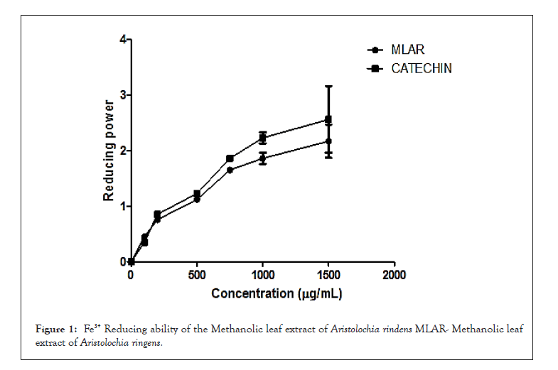
Figure 1: Fe3+ Reducing ability of the Methanolic leaf extract of Aristolochia rindens MLAR- Methanolic leaf extract of Aristolochia ringens.
There were significant increase (P<0.05) in the serum activity of ALT and AST following the administration of STZ precisely by 47% and 38% relative to the control while supplementation with MLAR, MET and QUER specifically lower the serum ALT and AST activities by 45%, 36% and 56% for AST and 32%, 46% and 43% for ALT respectively (Figure 2).
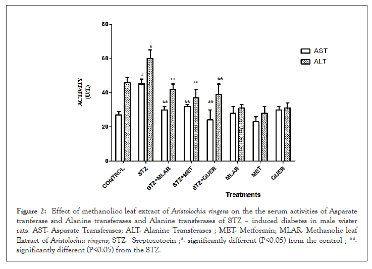
Figure 2: Effect of methanolioc leaf extract of Aristolochia ringens on the the serum activities of Asparate tranferase snd Alanine transferases and Alanine transferases of STZ – induced diabetes in male wisterrats. AST- Asparate Transferases; ALT- Alanine Transferases ; MET- Metformin; MLAR- Methanolic leaf Extract of Aristolochia ringens; STZ- Sreptozotocin ;*- significantly different (P<0.05) from the control ; **- significantly different (P<0.05) from the STZ.
There were significant increase (P<0.05) in the serum activity of ALP and GGT following the administration of STZ precisely by 51% and 32% relative to the control while supplementation with MLAR, MET and QUER precisely reduced the serum ALP and GGT activities by 41%, 32% and 51% for ALP and 35%, 18% and 33% for GGT respectively (Figure 3).
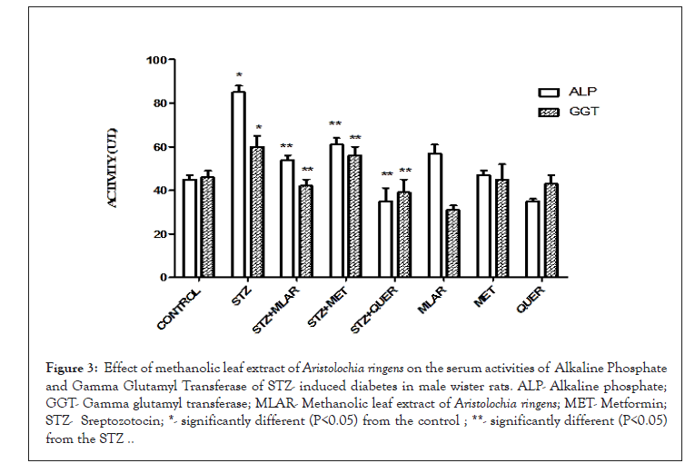
Figure 3: Effect of methanolic leaf extract of Aristolochia ringens on the serum activities of Alkaline Phosphate and Gamma Glutamyl Transferase of STZ- induced diabetes in male wister rats. ALP- Alkaline phosphate; GGT- Gamma glutamyl transferase; MLAR- Methanolic leaf extract of Aristolochia ringens; MET- Metformin; STZ- Sreptozotocin; *- significantly different (P<0.05) from the control ; **- significantly different (P<0.05) from the STZ ..
Intoxication of the animals with STZ caused significant (P<0.05) elevation of the serum level of cholesterol and lowering of LDL precisely by 29% and 34% relative to the control but supplementation with MLAR, MET and QUER precisely lower the serum level of cholesterol by 26%, 46% and 43% and elevated the serum LDL level by 36%, 28% and 48% respectively (Figure 4).
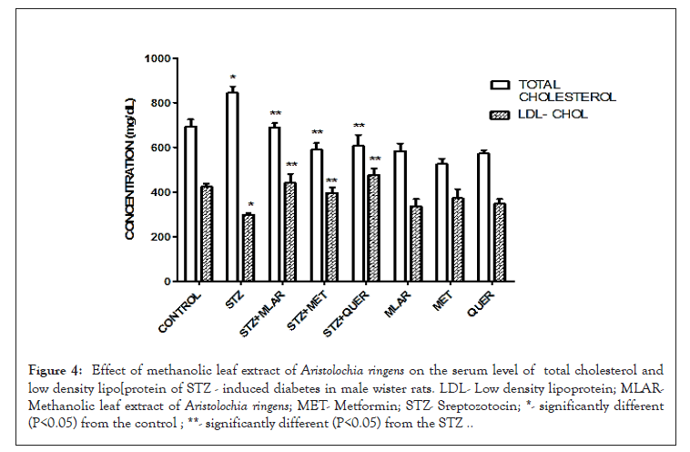
Figure 4: Effect of methanolic leaf extract of Aristolochia ringens on the serum level of total cholesterol and low density lipo[protein of STZ - induced diabetes in male wister rats. LDL- Low density lipoprotein; MLARMethanolic leaf extract of Aristolochia ringens; MET- Metformin; STZ- Sreptozotocin; *- significantly different (P<0.05) from the control ; **- significantly different (P<0.05) from the STZ ..
Administration of STZ to the animals caused significant (P<0.05) increase in the serum level of TRIGS and lower the serum level of HDL precisely by 41% and 34% relative to the control while mediation with MLAR, MET and QUER precisely significantly lowered the serum level of TRIGS by 32%, 35% and 42% and elevated the HDL level by 34%, 53% and 47% respectively (Figure 5).
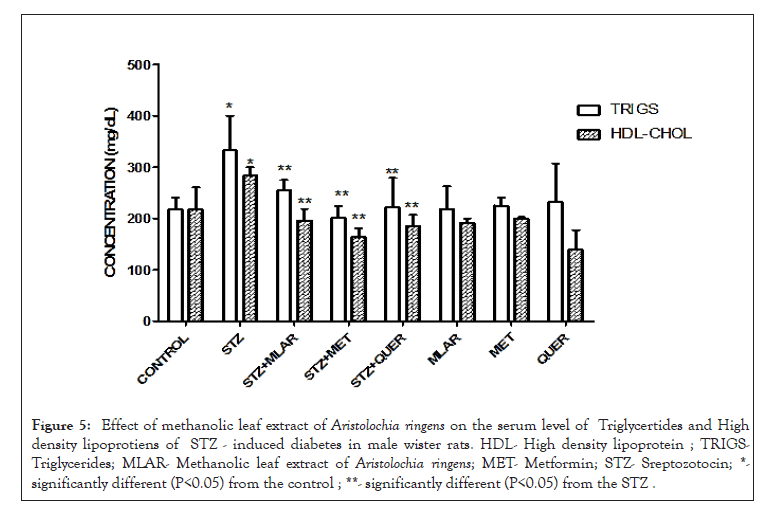
Figure 5: Effect of methanolic leaf extract of Aristolochia ringens on the serum level of Triglycertides and High density lipoprotiens of STZ - induced diabetes in male wister rats. HDL- High density lipoprotein ; TRIGSTriglycerides; MLAR- Methanolic leaf extract of Aristolochia ringens; MET- Metformin; STZ- Sreptozotocin; *- significantly different (P<0.05) from the control ; **- significantly different (P<0.05) from the STZ .
Administration of STZ to the animals caused significant (P<0.05) increase in the hepatic level of Nitric oxide and Myeloperoxidase precisely by 54% and 59% relative to the control while mediation with MLAR, MET and QUER significantly lowered the hepatic level of Nitric oxide by 42%, 53% and 46% and Myeloperoxidase level elevated by 29%, 43% and 30% respectively (Figure 6).
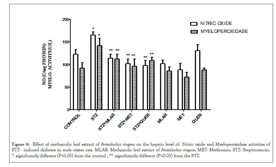
Figure 6: Effect of methanolic leaf extract of Aristolochia ringens on the hepatic level of Nitric oxide and Myeloperoxidase activities of STZ - induced diabetes in male wister rats. MLAR- Methanolic leaf extract of Aristolochia ringens; MET- Metformin; STZ- Sreptozotocin; *- significantly different (P<0.05) from the control ; **- significantly different (P<0.05) from the STZ.
Intoxication of the animals with STZ caused significant (P<0.05) elevation of the hepatic level of MDA and lowering of Catalase precisely by 29% and 34% relative to the control but supplementation with MLAR, MET and QUER precisely lower the hepatic level of MDA by 35%, 22% and 30% and elevated the hepatic level of catalase by 62%, 41% and 56% respectively (Figure 7).
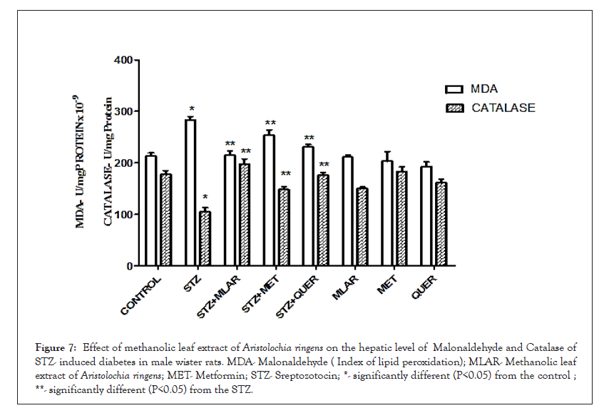
Figure 7: Effect of methanolic leaf extract of Aristolochia ringens on the hepatic level of Malonaldehyde and Catalase of STZ- induced diabetes in male wister rats. MDA- Malonaldehyde ( Index of lipid peroxidation); MLAR- Methanolic leaf extract of Aristolochia ringens; MET- Metformin; STZ- Sreptozotocin; *- significantly different (P<0.05) from the control ; **- significantly different (P<0.05) from the STZ.
Administration of STZ to the animals caused significant (P<0.05) decrease in the hepatic level of SOD and GST precisely by 66% and 55% relative to the control while mediation with MLAR, MET and QUER significantly lowered the hepatic level of SOD by 45%, 31% and 47% and Myeloperoxidase level elevated by 53%, 49% and 42% respectively (Figure 8).
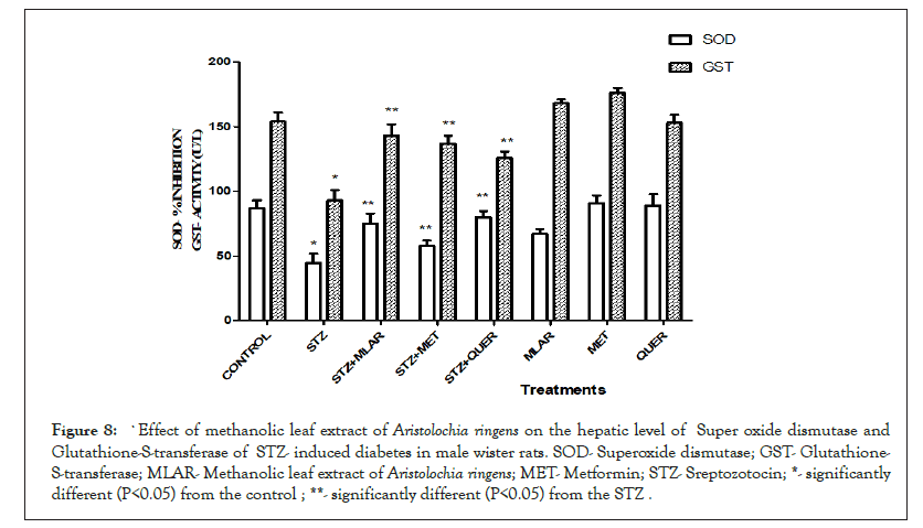
Figure 8: `Effect of methanolic leaf extract of Aristolochia ringens on the hepatic level of Super oxide dismutase and Glutathione-S-transferase of STZ- induced diabetes in male wister rats. SOD- Superoxide dismutase; GST- Glutathione- S-transferase; MLAR- Methanolic leaf extract of Aristolochia ringens; MET- Metformin; STZ- Sreptozotocin; *- significantly different (P<0.05) from the control ; **- significantly different (P<0.05) from the STZ .
Intoxication of the animals with STZ led to significant (P<0.05) decrease in the hepatic level of GPx and GSH precisely by 61% and 46% relative to the control while treatment with MLAR, MET and QUER significantly elevated the hepatic level of GPx by 47%, 63% and 41% and GSH level elevated by 38%, 53% and 49% respectively (Figure 9).
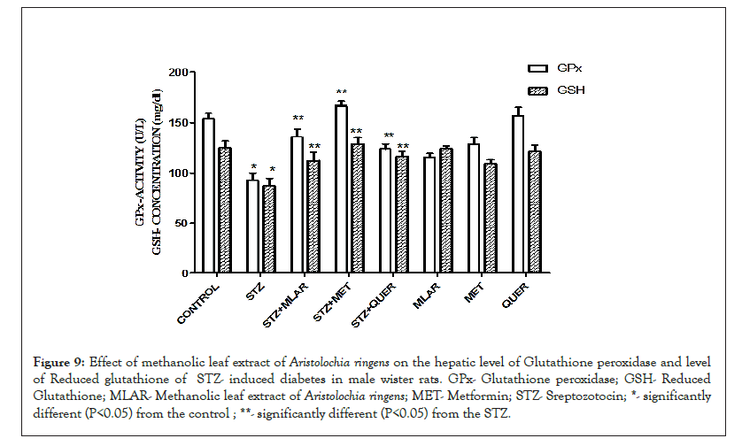
Figure 9: Effect of methanolic leaf extract of Aristolochia ringens on the hepatic level of Glutathione peroxidase and level of Reduced glutathione of STZ- induced diabetes in male wister rats. GPx- Glutathione peroxidase; GSH- Reduced Glutathione; MLAR- Methanolic leaf extract of Aristolochia ringens; MET- Metformin; STZ- Sreptozotocin; *- significantly different (P<0.05) from the control ; **- significantly different (P<0.05) from the STZ.
Intoxication of the animals with STZ led to significant (P<0.05) decrease in the concentration of serum insulin precisely by 52% relative to the control while treatment with MLAR, MET and QUER significantly elevated the serum level of insulin by 51%, 83% and 43% respectively (Figure 10).
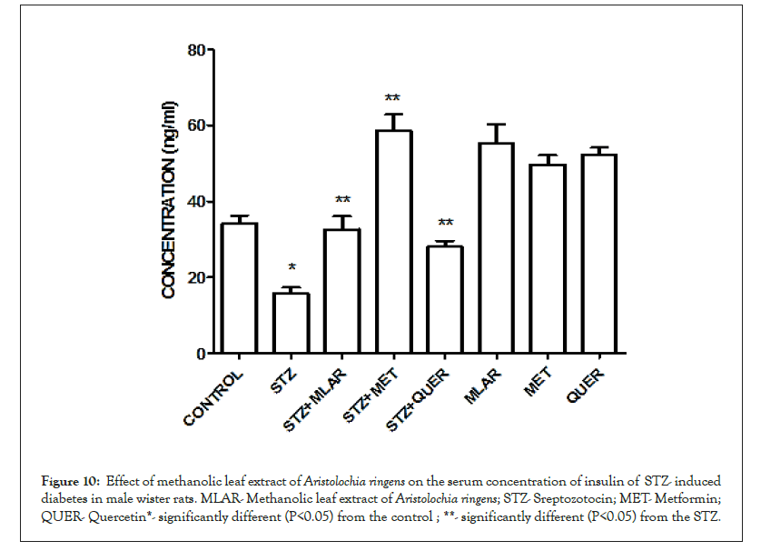
Figure 10: Effect of methanolic leaf extract of Aristolochia ringens on the serum concentration of insulin of STZ- induced diabetes in male wister rats. MLAR- Methanolic leaf extract of Aristolochia ringens; STZ- Sreptozotocin; MET- Metformin; QUER- Quercetin*- significantly different (P<0.05) from the control ; **- significantly different (P<0.05) from the STZ.
Discussion
The interest in natural products for use as medicine has acted as the catalyst for exploring methodologies involved in obtaining the required plant materials for pharmacological screening and drug development Adedara et al. [23]. Medicinal plants are used for discovering and screening of the phytochemical constituents which are very helpful for the manufacturing of new drugs Gottlieb et al. [24]. The result obtained from the phytochemical screening of the Methanolic Leaf Extract of Aristolochia ringens (MLAR) indicate the presence of alkaloids, saponins, anthraquinone, flavonoids, terpene and cardiac glycosides while phlobatannins, couramins and tannis were confirmed absent (Table 1).
Methanolic Leaf Extract of Aristolochia ringens (MLAR) at graded concentrations showed strong antioxidant activity when compared with the standard. It was observed that free radicals were scavenged by the extract in a concentration dependent manner. MLAR scavenged DPPH and ABTS radicals in a dose dependent manner with activity well comparable with the standard catechin as shown in Table 2. The reduction capability of DPPH and ABTS radicals absorbed at 517 nm and 745 nm respectively Lata et al. by MLAR is a measure of its antioxidant activity which implies that the action may be by either inhibiting or scavenging the DPPH and ABTS radicals since both inhibition and scavenging properties of antioxidants towards this radical have been reported in earlier studies Shaukat et al. [25,26].
Initiation of the lipid peroxidation by ferrous sulphate takes place either through the ferryl- perferryl complex or through Hydroxyl (OH) radical by Fenton’s reaction Singh et al. [27]. Thus the ability of MLAR to lower the MDA level in liver phosphatidylcholine in a dose dependent manner as depicted in Table 3 is an indication that it’s a potent antioxidant relative to catechin. The relationship between reducing power and antioxidant as it indicate the potential of an extract to donate electron to radicals and also reduce the oxidized intermediates of lipid peroxidation processes, thus such extract can act as primary and secondary antioxidants Ahmad et al. As observed in this study, the yellow colour of the test solution changed to various shades of green and blue based on the reducing power of MLAR as the concentration increase. Presence of reducers in MLAR caused the conversion of the Fe3+/ferricyanide complex used in this method as such MLAR displayed excellent reducing activity in a dose dependent manner relative to the reference catechin (Figure 1).
One of the useful experimental model used to study hypoglycemic activity is Streptozotocin (STZ)-induced hyperglycaemia Adaramoye et al. STZ is known to be a toxic analogue of glucose that selectively damage pancreatic insulin-producing beta cells when administered to rodent and many other animal species, thus leading to an insulin-dependent diabetes mellitus known as STZ diabetes in these animals. As observed in this study, administration of STZ to the animals caused significant elevation of serum Fasting Blood Glucose (FBS) monitored within 30 days and serum insulin which were statistically significant. This findings conformed with various studies as reported by the studies of Srinivasan, et al. [21,28-30]. However, supplementation with MLAR brought the serum glucose level close to the normal at the 30th day well comparable with the standard.
Liver impairment can be assessed measuring the marker enzymes such as AST (Aspartate Amino Transferase), ALT (Alanine Amino Transferase), ALP (Alkaline Phosphatase) and GGT (Gamma Gluamyl Transferase) which are originally present in high concentration in the cytoplasm and when there is hepatic injury these enzymes leak into blood stream Silver et al. [31]. Elevated levels of these enzymes are indicative of cellular leakage and loss of functional integrity of cell membrane in the liver Evans et al. [32]. As observed in this study, administration of STZ caused liver impairment evidenced by the elevated level of AST, ALT, ALP and GGT, while treatment with the extract significantly reduced their levels. This attuned to previous study of Atef, Senthil, Kavipriya et al. . STZ induced diabetes was further evidenced by significant decreases in the activities of SOD, CAT, GST, GPx, GSH levels and elevated level of Malonaldehyde in STZ treated rats. Similar results were recorded in other published studies such as Atef, Altar et al. [33-35].
On the other hand, Nitric Oxide (NO) is regarded as a proinflammatory regulator that produced inflammation as a result of high production in diseased states and it is synthesized and discharged into the endothelial cells by the help of NOS that changed arginine into citrulline there by producing NO Sharma et al. [36]. Heme-containing peroxidase (Myeloperoxidase [MPO]) is synthesized majorly from polymorphonuclear neutrophils and channeled into the extracellular fluid after oxidative stress where it exhibit inflammatory responses Amjad et al. [37]. As evidenced in this study, high level of NO and activity of MPO was observed following the administration of STZ thus revealed evidence of inflammation as a result of toxicity of STZ while treatment with MLAR significantly reduced their levels. MLAR and Quercetin had displayed good anti-oxidant and anti-inflammatory characteristics because they significantly brought the levels of MDA, NO, MPO, SOD, CAT, GST, GPx, GSH close to normal.
Hyperlipidemia in humans is frequently related to an increased risk for atherosclerosis. STZ have considerable effects on plasma lipoprotein and apolipoprotein concentrations which is evident by the elevated levels of serum Cholesterol (CHOL), Triglycerides (TRIGS) and lowered level of serum High Density Lipoprotein (HDL) Ademoye et al. [38]. Insulin triggers lipoprotein lipase resulting in normal triacylglycerol and increased HDL levels in a state of wellness Mohammed et al. Reduction in the serum level of insulin in STZ-diabetes led to the failure of lipoprotein lipase to observe its normal physiological functions, thus the development of hyperlipidemia and hypercholesterolemia. This current study depicted that intoxication of the animals with STZ significantly increased the serum level of total cholesterol, LDL and TRIGS and lower HDL level while treatment with the extract significantly reduced the serum levels of total cholesterol, LDL, TRIGS and increases HDL level. Similar response were found and reported in previous studies such as Mommed, Atef, Ademoye, Ibrahim, Ayodele, Ogunmefun et al. [16,28,38-40] MLAR acted as an hypolipidemic agent by inducing the response of plasma lipoprotein lipase to insulin action, therefore lowering the TGs, LDL and Total cholesterol levels and increasing HDL levels.
Conclusion
The current study provided a novel strategy and a scientific mechanism of action for the use of Aristolochia ringens leaf as a prophylactic and an alternative medicine in the treatment of diabetes mellitus because this study established that it possess antioxidant, anti-inflammatory, hypoglycemic and hypolipidemic effects. These activities could be due to the presence of phytochemical in it.
Acknowledgement
This research work was fully financed by the research grant from TETFund Abuja, Nigeria under the Management of Moshood Abiola Polytechnic, Abeokuta, Ogun State Nigeria.
REFERENCES
- Sundaram C S, Sivakumar J, Subbiah S K, Zin T, Rao M, Rao U S. Antidiabetic potential and high synergistic antibacterial activity of silver nanoparticles synthesised with Musa Paradisiaca tepal extract. Med J Malaysia. 2021;76(1):80-86.
- Parra AL, Soto-del Valle RM, Ferrer JP, Hang PT, Thi N, Phuong AB, et al. Antidiabetic, hypolipidemic, antioxidant and anti-inflammatory effects of Momordica charantia L. foliage extract. J Pharm Pharmacogn Res. 2021;9(4):537-548.
- Semwal BC, Shah K, Chauhan NS, Badhe R, Divakar K. Anti-diabetic activity of stem bark of Berberis aristata DC in alloxan induced diabetic rats. Int. J. Pharmacol. 2008;6: 1-6.
- Chaudhury A, Duvoor C, Reddy Dendi VS, Kraleti S, Chada A, Ravilla R. et al. Clinical review of antidiabetic drugs: implications for type 2 diabetes mellitus management. Front Endocrinol. 2017;8:6-12.
- Sandikapura MJ, Nyamathulla S, Noordin MI. Comparative antioxidant and antidiabetic effects of Syzygium polyanthum leaf and Momordica charantia fruit extracts. PJPS. 2018.
- Sonibare MA, Gbile ZO. Ethnobotanical survey of anti-asthmatic plants in South Western Nigeria. Afr J Tradit Complement Altern Med. 2008;5(4):340-345.
- De Groot H, Wanke S, Neinhuis C. Revision of the genus Aristolochia (Aristolochiaceae) in Africa, Madagascar and adjacent islands. J Linn Soc Bot.. 2006;151(2):219-238.
- Van Wyk B. & Wink M.. Medicinal Plants of the World: An Illustrated Scientific Guide to the Important Medicinal Plants and Their Uses. Portland, OR, USA: Timber Press. 2004.
- Neuwinger HD. African traditional medicine: a dictionary of plant use and applications. With supplement: search system for diseases. Med-Pharm; 2000.
- Soladoye MO, Adetayo MO, Chukwuma EC, Adetunji AN. Ethnobotanical survey of plants used in the treatment of haemorrhoids in South-Western Nigeria. Ann Biol Res. 2010;1(4):1-5.
- Aigbe FR, Adeyemi OO. The aqueous root extract of Aristolochia ringens (Vahl.) prevents chemically induced inflammation. Planta Med. 2011;77(12):PF28.
- Olabanji SO, Omobuwajo OR, Ceccato D, Adebajo AC, Buoso MC, Moschini G. Accelerator-based analytical technique in the study of some anti-diabetic medicinal plants of Nigeria. NUCL INSTRUM METH B. 2008;266(10):2387-2390.
- Harborne, J. B & Baxter, H. Phytochemical dictionary – a handbook of bioactive compounds from plants, Taylor & Francis. (1993); 2:270.
- Manzocco L, Anese M, Nicoli MC. Antioxidant properties of tea extracts as affected by processing. LWT LWT-FOOD SCI TECHNOL. 1998;31(7-8):694-698.
- Avallone R, Zanoli P, Puia G, Kleinschnitz M, Schreier P, Baraldi M. Pharmacological profile of apigenin, a flavonoid isolated from Matricaria chamomilla. Biochem. Pharmacol. 2000;59(11):1387-94.
- El-Sayed MI, Al-Massarani S, El Gamal A, El-Shaibany A, Al-Mahbashi HM. Mechanism of antidiabetic effects of Plicosepalus Acaciae flower in streptozotocin-induced type 2 diabetic rats, as complementary and alternative therapy. BMC COMPLEM ALTERN M. 2020; 20: 290.
- Lowry OH, Rosebrough NJ, Farr AL, Randall RJ. Protein measurement with the Folin phenol reagent. J Biol Chem. 1951;193:265-275.
- Narkhede MB, Ajimire PV, Wagh AE, Mohan M, Shivashanmugam AT. In vitro antidiabetic activity of Caesalpina digyna (R.) methanol root extract. Asian J Plant Sci. 2011;1(2):101-106.
- Alofi MT, Zaki AA, Abdel-Rahman HA, El Tigani EA. Antioxidant and anti-stress biomarkers of some nutraceuticals in alloxan-induced diabetic rats. JMS. 2016;6:68-81.
- Kasali F. M., Wendo F. M., Muysia S. K., Kadima J. N. Comparative hypoglycemic activities of flavonoids and tannins fractions of Stachytar phetaindica (L) Vahl leaves extracts in guinea pigs and rabbits; Int J Pharm Pharm Res. 2016.
- Srinivasan K, Patole PS, Kaul CL, Ramarao P. Reversal of glucose intolerance by pioglitazone in high fat diet-fed rats. Methods Find Exp Clin Pharmacol. 2004;26(5):327-333.
- Matsumoto S, Koshiishi I, Inoguchi T, Nawata H, Utsumi H. Confirmation of superoxide generation via xanthine oxidase in streptozotocin-induced diabetic mice. Free Radic Res.. 2003 Jul 1;37(7):767-772.
- Adedara IA, Daramola YM, Dagunduro JO, Aiyegbusi MA, Farombi EO. Renoprotection of Kolaviron against benzo (A) pyrene-induced renal toxicity in rats. Ren Fail. 2015 Mar 16;37(3):497-504.
- Gottlieb OR, Borin MR. Natural products research in Brazil. Ciênc. cult.(Säo Paulo). 1997:315-320.
- Lata N, Dubey V. Preliminary phytochemical screening of Eichhornia crassipes: the world’s worst aquatic weed. J Pharm Res. 2010;3(6):1240- 1242.
- Shaukat SS, Siddiqui IA, Ali NI. Nematicidal, allelpathic and antifungal potential of Launaea procumbens. Pakistan J Phytopathol. 2003; 2(3): 181-193
- Singh R, Singh S, Kumar S, Arora S. Evaluation of antioxidant potential of ethyl acetate extract/fractions of Acacia auriculiformis A. Cunn. Food Chem Toxicol. 2007;45(7):1216-1223.
- Adaramoye OA, Lawal SO. Effect of kolaviron, a biflavonoid complex from G arcinia kola seeds, on the antioxidant, hormonal and spermatogenic indices of diabetic male rats. Andrologia. 2014;46(8):878-886.
- Berg T, DeLanghe S, Al Alam D, Utley S, Estrada J, Wang KS. β-catenin regulates mesenchymal progenitor cell differentiation during hepatogenesis. J Surg Res. 2010;164(2):276-285.
- Bastaki, S. Review Diabetes Mellitus and Its Treatment. Int J Diabetes Metab. 2005;13:111-134.
- Silva EM, Souza JN, Rogez H, Rees JF, Larondelle Y. Antioxidant activities and polyphenolic contents of fifteen selected plant species from the Amazonian region. Food Chem. 2007;101(3):1012-1028.
- Evans JL, Goldfine ID, Maddux BA, Grodsky GM. Are oxidative stress activated signaling pathways mediators of insulin resistance and β-cell dysfunction?. Diabetes. 2003;52(1):1-8.
- Al-Attar AM, Alsalmi FA. Effect of Olea europaea leaves extract on streptozotocin induced diabetes in male albino rats. Saudi J Biol Sci. 2019 Jan 1;26(1):118-128.
- Dharmalingam SR, Madhappan R, Ramamurthy S, Chidambaram K, Srikanth MV, Shanmugham S. et al. Investigation on antidiarrhoeal activity of Aristolochia indica Linn. root extracts in mice. AFR J TRADIT COMPLEM. 2014;11(2):292-294.
- Kavipriya S, Tamilselvan N, Thirumalai T, Arumugam G. Anti-diabetic effect of methanolic leaf extract of Pongamia pinnata on streptozotocin induced diabetic rats. J Coast Life Med. 2013;1(2):113-117.
- Sharma SR, Dwivedi SK, Swarup D. Hypoglycaemic, antihyperglycaemic and hypolipidemic activities of Caesalpinia bonducella seeds in rats. J Ethnopharmacol. 1997;58(1):39-44.
- Khan AA, Alsahli MA, Rahmani AH. Myeloperoxidase as an active disease biomarker: recent biochemical and pathological perspectives. Med Sci. 2018;6(2):33.
- Adewoye EO. Effect of methanol extract of musa sapientum leaves on protein glycation and erythrocyte antioxidant status in alloxan-induced diabetic Wistar rats. Afr J Med Med Sci. 2015;44(3):261-268.
- Ibrahim JA, Ayodele AE. Taxonomic significance of leaf epidermal characters of the family Loranthaceae in Nigeria. World Appl Sci J. 2013;24(9):1172-1179.
- Ogunmefun OT, Fasola TR, Saba AB, Oridupa OA. The toxicity evaluation of Phragmanthera incana (Klotzsch) growing on two plant hosts and its effect on Wistar rats’ haematology and serum biochemistry. AJPS. 2013;6:92-98.
Citation: Tijani RO, Lawal SO, Oyekan JO, Koleoso OK, Onasanya SS, Fasasi AA (2021) Anti-Diabetic Effect of Methanolic Extract of Aristolochia ringens Leaf. Biochem Anal Biochem. 10:397.
Copyright: © Tijani RO, et al. This is an open-access article distributed under the terms of the Creative Commons Attribution License, which permits unrestricted use, distribution, and reproduction in any medium, provided the original author and source are credited.
