

Indexed In
- Open J Gate
- Genamics JournalSeek
- Academic Keys
- JournalTOCs
- The Global Impact Factor (GIF)
- China National Knowledge Infrastructure (CNKI)
- Ulrich's Periodicals Directory
- RefSeek
- Hamdard University
- EBSCO A-Z
- OCLC- WorldCat
- Publons
- Geneva Foundation for Medical Education and Research
- Euro Pub
- Google Scholar
Useful Links
Share This Page
Journal Flyer
Open Access Journals
- Agri and Aquaculture
- Biochemistry
- Bioinformatics & Systems Biology
- Business & Management
- Chemistry
- Clinical Sciences
- Engineering
- Food & Nutrition
- General Science
- Genetics & Molecular Biology
- Immunology & Microbiology
- Medical Sciences
- Neuroscience & Psychology
- Nursing & Health Care
- Pharmaceutical Sciences
Research Article - (2026) Volume 17, Issue 3
The Fc Domains in the Monoclonal Antibody and Fusion Protein Significantly Boost the Biocomposite Suspension's Power to Combat Harmful Microorganisms, Enhance Drug Delivery, and Improve Protein Stability
Roongnapa Suedee*, Watchara Pholthien, Nanticha Kaewsud, Krit Prakannoppakun, Khanittha Santipiboon, Pimpisut Getsuvan, Kanok-on Jaisawan, Jittiya Rodruksa and Pistawus KhomintrReceived: 17-Aug-2026, Manuscript No. PAA-26-32433; Editor assigned: 19-Aug-2026, Pre QC No. PAA-26-32433 (PQ); Reviewed: 02-Sep-2026, QC No. PAA-26-32433; Revised: 09-Sep-2026, Manuscript No. PAA-26-32433 (R); Published: 16-Sep-2026, DOI: 10.35248/2153-2435.26.17.866
Abstract
Monitoring protein interactions provides vital insights into how structural changes affect the interactions between soluble Fc fragments and various plasma proteins. The use of biocomposite suspensions has been shown to enhance antimicrobial activity, improve drug delivery systems, and maintain protein stability by ensuring a uniform preservation of protein integrity within these complexes. In-depth analyses of amino acid abundance reveal critical information about the incorporation of protein additives and osmolytes into the final proteinenriched suspension, particularly when co-administering biotherapeutics with Cannabidiol (CBD). These studies reveal how water influences surface modifications and the role of bioorganic molecules in altering membrane properties. Exploring protein interactions reveals critical insights into how structural changes affect relationships between soluble Fc fragments and plasma proteins. The integration of biocomposite suspensions enhances antimicrobial activity, drug delivery, and protein stability, thereby preserving protein integrity. Studies on amino acid abundance shed light on the incorporation of protein additives and osmolytes, particularly during the co-administration of biotherapeutics such as CBD. The application of Molecularly Imprinted Polymers (MIPs) is vital for tracing complex protein interactions, in which three-dimensional structures significantly influence antibody reactivity and binding sites. Ongoing research explores how factors like Polycaprolactone (PCLT)/Cannabidiol (CBD) ratios and additives influence release behavior while mitigating microbial risks. Findings from high-resolution Liquid Chromatography-Quadrupole Time-of-Flight Mass Spectrometry (LC-QTOF-MS) across different formulations are helping to unravel the complex interactions of proteins and polymers, ultimately aiming to improve access to innovative therapies for individuals facing serious health challenges. Flexible amino acids concentrate reactive species during MIP immunocapture, while the challenges posed by salt constraints under osmotic stress provide a vivid picture of protein refolding and structural changes, driven by local water adsorption on the surface of target proteins. Notably, cysteine levels remain stable through recycling, aiding post-translational modifications. The purification process plays a crucial role in maintaining insulin's native structure, preventing irregular oligomer formation. Advanced techniques, including atomic force microscopy, Raman mapping, and fluorescence analysis, provided tracking of antibody internalization studies that transported to HSA FcRn via plasma MIP and how the dispersed phase affects enzyme-buffer interactions that support microbial inhibition, confirming the presence of key proteins like FcεRI on immune cell membranes. Quanta Scanning Electron Microscopy (QuantaSEM) imaging highlights structural dynamics within dense PCL-T oily gels, affecting Complementarity-Determining Region (CDR) binding dynamics. Interactions between histidine protonation and FcRn binding can significantly impact antibody efficacy, explored through charge-state analysis and dual-mode LCQTOF-MS. Analysis of a high-protein suspension yields a liquid-phase fluorescence image of post-immunocapture processes. Our proteomic profiling rigorously examines the effects of Post-Translational Modifications (PTMs), while fluorescence microscopy clearly demonstrates structural relaxation and amino acid movements. By leveraging NMR signals, scientists can measure local concentrations and structural changes in PMIP environments, particularly in varying CBD-to-MIP ratios. MIPs also enhance antibody recognition and inhibit microbes, while flexible amino acids concentrate reactive species during immunocapture. Ultimately, this research contributes to stabilizing suspensions and understanding the impact of osmotic stress on drug efficacy. Gelled coacervates enhance cell interactions and optimize antibody transport in biofilms, highlighting the role of water in maintaining structural integrity and facilitating binding interactions essential for biopharmaceutical development.
Keywords
Biopharmaceutical characterization; Chiral recognition; Charge-dependent solvation; Ligand-based NMR; Hydrogel suspension
Abbreviations
Fc: Fragment crystallizable; MIPs: Molecularly Imprinted Polymers; NIP: Non-Imprinted Polymer; CBD: Cannabidiol; LC-QTOF-MS: Liquid Chromatography-Quadrupole Time-of-Flight Mass Spectrometry; HSA: Human Serum Albumin; FcRn: Neonatal Fc Receptor (also called the neonatal fragment crystallizable receptor); PTMs: Post-Translational Modifications; NMR: Nuclear Magnetic Resonance; PMIP: Plasma Molecularly Imprinted Polymer; CDR: Complementarity-Determining Region; FcεRI: High-Affinity Immunoglobulin E Receptor (the Fc epsilon receptor I); mRNA: Messenger Ribonucleic Acid; ATP : Adenosine Triphosphate; MRSI: Microscopy and Super-Resolution Multiparametric Imaging; PBS: Phosphate-Buffered Saline; AFM: Atomic Force Microscopy; PCL-T: Polycaprolactone-T; mV: milliVolt; PTM: Post-Translational Modification
Introduction
Variations in antibody processes have gained attention due to their impact on biopharmaceutical manufacturing and storage [1,2]. These variations pose contamination risks and require techniques such as co-admixing or gel suspension, making it difficult to maintain consistent proteomic profiles. Biocomposite suspensions enhance antimicrobial activity, improve drug delivery, and stabilize proteins by uniformly preserving protein integrity within complexes. The use of immune cells for personalized therapy requires consistent batch production to maintain functional activity, with MIP droplets protecting enzyme-sensitive antibody regions [3]. The lipophilic HSA-MIP facilitates the release of hydrophobic drugs, ensuring stability and proper refolding. Regulating binder density during nanocoating helps manage temperature, supporting effective enrichment and formulation of multibiotherapeutics. These innovations enhance enzyme stabilized nanocarriers, improving drug delivery and cellular immunotherapies. Anti-IgE antibodies can reduce asthma and allergies by modulating IgE activity. They allow IgE to bind to high-affinity FcεRI, a mast cell multicomplex with an alpha chain that binds ligands and a beta chain that activates immune responses, even in the presence of elevated allergenspecific IgE levels. The interaction of FcεRI with IgG occurs within its subunit and extracellular structure. Current research examines how antibodies are transported to HSA FcRn via plasma MIP and how the dispersed phase affects enzyme-buffer interactions that support microbial inhibition. Effective allergy treatments require stringent contamination prevention with appropriate materials, promoting their use in pharmaceuticals and immunotherapy. Additionally, increased proinflammatory cytokines and altered immunity in migraine have drawn attention, as migraine sufferers often have comorbid atopic disorders and elevated plasma IgE without typical type I hypersensitivity, that antibody-advanced therapy for severe migraine is another option [4]. Advanced drug delivery platforms that could revolutionize healthcare, particularly in cellular immunotherapies for cancer, autoimmune diseases, and neurodegenerative disorders. These targeted treatments involve using immune cells for personalized therapy. Supportive care is crucial for improving outcomes and managing adverse effects, such as cytokine release syndrome and neurotoxicity. Antibody therapies face challenges related to efficiency, safety, and manufacturing, and recent studies aim to address these issues. Advances in metabolic engineering and imaging have accelerated progress in precision immunotherapies; however, high costs remain a barrier to access, highlighting the need for strategies to make treatments more affordable. Manufacturers are increasingly focusing on patient-centered outcomes, with value-based agreements that link treatment success to costs, reflecting a shift toward sustainable healthcare. This approach enhances therapy adherence and underscores the significance of accurately measuring clinical markers through reliable blood or plasma tests. Research is focused on improving patient access and quality of life for individuals with complex conditions. Advances in metabolic engineering are driving the development of biotherapeutics using techniques such as plasma desorption, additive interactions, Fc receptor binding, adsorption, and protein self-assembly, which help maintain cellular equilibrium and ensure protein functionality in immunotherapies [5]. Reevaluating manufacturing processes, such as lyophilization, and managing lactate levels can optimize protein production and treatment consistency. Adequate lighting in fluorescence imaging facilitates detailed sample analysis. Developing cost-effective strategies is crucial because targeted therapies are often expensive. The molecular structure significantly influences charge-based solvation in plasma-MIPs. Innovations involving co-biotherapeutic forms, droplet suspensions, and MIPs are advancing targeted drug delivery, particularly in tumor immunotherapy. Synthetic MIPs stabilize proteins, including CBD diastereomers bound to HSA, thereby impacting immune responses. Modern template-free MIPs offer enhanced selectivity and recognition compared to traditional methods. Molecular dynamics play a role in designing probes for enantiomerization and catalysis. This also provides screening insights into the ability of plasma MIPs to be customized to address biological needs, a pharmaceutical concept involving hydrogels, MIPs, and nanostructures, with the advantage of enabling atomic scale and conformational changes. Establishing charge-dependent behaviors that are beneficial for cellulose bionanofiltration. Opposing charge effects influence protein segregation and particle dispersion, which are crucial for drug absorption and powder flow. Factors such as PCL-T/CBD ratios, additives, and enzyme inhibitors impact release behavior, while pH adjustments and excipients help mitigate microbial risks. Furthermore, MIP and native materials demonstrate variations in plasma redistribution at protein-rich cellulose membranes, encountering the patch effect of ion salts post-purification. Soluble Fc and other macromolecules may interact with plasma proteins, which are subsequently recognized as foreign by the body, leading to their elimination by RES cells and microbes during production, or in combination with biopharmaceuticals.
Epitope screening offers a fascinating insight into the world of dimeric membrane proteins, tracking their interactions, monitoring drug diffusion, and observing the structural relaxation that occurs during the intricate process of protein refolding. It's a dynamic exploration of how these proteins function and respond, shedding light on critical biotherapeutic processes. Techniques such as AFM, Raman mapping, and fluorescence confirm the integrity of the FcεRI chain. Changes in histidine protonation can affect FcRn binding and reduce antibody efficacy, studied with charge state analysis and LC-QTOF-MS. The droplet proteinrich particles vary in nanoscale size, influenced by PCL-T density, which may also affect histidine protonation. Membrane-bound organelle labeling and localization are studied through various analyses. Recycled samples prevent clumping, stabilize insulin, and reveal the protonation behavior of CBD. Hydrogel elasticity and solubility are affected by oxidation at enzyme-inhibitor sites; ATP binding aids phosphorylation and ligand attachment, supporting mAb stability. LC-QTOF-MS helps identify peptide biomarkers in the final protein-enriched suspension from proteins and the Extracellular Matrix (ECM), reflecting unbound CBD. Since CBD doesn’t dissolve easily, it remains well distributed without diffusing too much. Various components, such as FcRn receptor-targeting dimers, FcεRI chains, hinge regions, and exposed proteins, are involved; notably, the FcεRI chain has two IgG-binding sites that interact with FcIgE, cleave glycosidic bonds, and lower pH to maintain stability and shape. This process supports nutrient absorption and helps maintain enzyme stability within hydrogels. Fluorescence imaging reveals how ligands are arranged on surfaces and the different states they adopt. When we purify protein clusters, they demonstrate Fc functions, and the FcεRI α chain moves within the protein-rich droplet after purification. Under osmotic stress, salts induce structural changes in proteins, causing them to absorb water at their surfaces, thereby affecting co-administered proteins and drugs. This quick, charge-based method allows us to evaluate how peptide structures change in water hydrogels with high CBD absorption under biorelevant media, which raises concerns about the effects of filled-pore-charge electrolytes on conformational changes and protein interactions [6]. Disordered regions of proteins tend to resist phase separation, influencing interactions at interfaces and with amino acid motifs. Detecting elements at phase boundaries supports the development of the administered co-biotherapeutic drug products, that is, highly personalized drug treatments, paving the way for targeting cellular immunotherapies.
This study investigates how charge-based control of solvation in enzymatic biosynthesis and energy flow can alter protein structures and improve selectivity, notably for the neonatal Fc receptor. It demonstrates that hydrogels created via solvent-assisted suspension printing and imprinting enable functional separation, enhance flexibility and flow, and boost performance in cellulose biofilms. Enhanced coatings from cellular matrix lysates and optimized PCL-T content also contribute to these improvements. Current research mainly examines protein interactions, stability, and antibody functions, with the Hydrodynamic Radius (RH) as a key metric. Tests with Human Recombinant Insulin (HRI) and lysozyme show that complexed PCL-T gels withstand freezing and maintain thermal stability, improving delivery in the Gastrointestinal Tract (GI), reducing microbial risks, and ensuring sterility during flow production. Target binding site of the antibodies by nanopore membrane can provide the development of (bio) therapeutics of individual analyse within the filled pore and therapeutic potential of antibody drug application [7]. Controlling biotherapeutics, additives, and contamination during post-exposure steps like product examination and transfer is essential, especially amid changing productivity and biotherapeutic developability. New approaches aim to improve liquid-liquid phase testing by integrating the MIP system, protein assembly, formulation additives, membrane interfaces, and nanopores on High-Protein Suspensions (HPS). HMIP is planned to continuously monitor productivity changes. This complex process involves mixtures rich in active ingredients and therapeutics to enhance biological efficacy. Short peptides of 8-10 amino acids can be designed to block epitopes or direct immune responses, depending on whether soluble or insoluble macromolecules in co-administered biotherapeutics are targeted. These strategies, as explored in recent studies, aim to advance cellular immunotherapy, though reliable efficacy testing remains essential for clinical success and potentially life-changing therapies.
Cellulose lysate matrices used for antibody nanocapsules leverage their high affinity to optimize mixing and amino acid purification. Nanofibrils with interparticulate interactions can prevent dye penetration, as shown in past studies [8]. Organic nanofiltration membranes enable precise separation of target molecules [9]. Dynamic molecules may change shape or cluster due to degradation or contamination; stabilizers such as osmolytes and sugars improve antibody fragment stability through volume exclusion, thereby preventing unwanted interactions and aggregation in hydrogels. Studying these interactions and employing membrane filtration enhances analyte performance and helps remove impurities and bacteria.
Analyzing methacrylate-PCL-T hydrogel matrices with MIPs reveals that charge-based solvation notably influences the conformational dynamics and binding specificity of hydrophobic amino acids, as noted in prior our studies [10]. This insight is crucial for improving protein stability and particle formation, particularly for key elements like the neonatal Fc receptor when loaded with agents such as Human Recombinant Insulin (HRI) and CBD. Such benefits are increasingly vital given the global surge in allergic conditions like asthma, rhinitis, and eczema. Factors such as pollution, allergen exposure, dietary changes, early infections, and atopic sensitivities elevate allergy risks. Forecasting aims to guide present decisions rather than predict the future, especially in public healthcare. Advances in managing asthma and allergies rely on improved industrial practices and developability processes. Early infections in life are likely to stimulate immune responses. Additionally, these sophisticated hydrogels, produced via solvent assisted suspension printing and selective membranes [10], significantly improve the performance of concentrated protein-loaded cellulose biofilms by modulating their elasticity and flow through hydrophobic interactions with biotherapeutic proteins. The pH-responsive Fc binding enhances MIP biocompatibility by reducing hydrophobic amino acid aggregation and increasing drug delivery effectiveness.
Multispecific anti-IgE fusion proteins, such as scFv, use flexible glycine-rich linkers to preserve structure and improve therapeutic performance. Identifying Complement Component 1Q (C1Q) interactions with the complement component and the IgG crystallization fragment (Fc) is crucial for assessing the effector function of biotherapeutic Monoclonal Antibodies (mAbs) [11]. The binding affinity of FcεRI for IgE is essential to the effector function and persistence of the antibodies; FcεRI is a monomer composed of two light chains and two epsilon (ε) heavy chains, forming part of the cell receptor; thus, identifying the protein structure is essential during the process and validation. The epsilon Fv region binds allergens, while the Fc region interacts with the receptor. FcεRI is embedded in immune cell membranes with an α chain, a β chain, and disulfide-linked γ chains. FcεRI chains have a D1-D2 hinge, while the FcεRIα chain contains two extracellular IgG-binding sites that bind to FcIgE. The gamma chains transmit signals through Immunoreceptor Tyrosine-based Activation Motifs (ITAMs); allergen cross-linking initiates cascades leading to degranulation and histamine release. Bannert, et al. identified FcεRIα on cells in the esophagus, stomach, and duodenum and found that modulation of FcεRIα leads to GI inflammation [12].
Incorporating CDR sequences enhances the bioavailability of poorly soluble drugs, allowing higher doses with a lower infusion volume. FcRn affinity modulates effector functions by binding the Fc region [13]; structure-function relationships of human IgG subclasses show that renewable polysaccharides improve bioprocess stability and reduce contamination. Peptide modifications regulate immunogenicity, ensuring predictable or masked responses. Ultrashort blocks facilitate neoantigen discovery. Ensuring purity and validating immunogenicity are vital for hydrophobic peptide immunotherapies. Proper design with specific molecules improves stability, reduces self-interaction, and minimizes contamination, supporting drug development, therapies, recycling, and ecoefficiency. Factors such as PCL-T/CBD ratios, additives, and enzyme inhibitors affect release behavior; pH adjustments and excipients mitigate microbial risks. Techniques like solvent nanofiltration preserve therapeutic molecules while allowing smaller byproducts and single molecule to pass. Under high-ionic conditions, organisms may modify the levels of acidic amino acids to maintain protein stability, thereby impacting CBD and co-loaded biotherapeutic release. Managing phase transitions reduces protein variability relative to static controls, thereby enhancing efficacy and safety.
Platform technologies, including messenger RNA (mRNA), recombinant proteins, and nanoparticles, provide unique immunological advantages that enhance therapeutic applications. Targeted immunotherapies designed to enhance adhesion and stability are employed in peptide and protein vaccines. The complement system, which involves interactions between singlechain Fc and IgG, plays a critical role in immune defense and can also trigger inflammation and immune responses [14]. The activation of this system is dose-dependent. Furthermore, variations in the physicochemical properties of biotherapeutic macromolecules, along with co-loaded drugs, can significantly influence their distribution and the formation of polymolecular aggregates, which are investigated in vitro under diverse excipient conditions.
Advancements in isotope research have deepened our understanding of antibodies by identifying isomers of active pharmaceutical compounds. Techniques aimed at strengthening materials, such as coalescence and crosslinking, enhance the durability and stability of biotherapeutic suspensions and the performance of PCL-T hydrogels. These methods support suspension gels and particulate suspensions, with sedimentation behavior influenced by factors such as coating thickness and desolvation, both of which can alter the conformational stability of protein therapeutics and the chemical composition, leading to (bio) interactions with additives in response to (de)solvent effects.
The composition and quantity of the hydrogel, including any additional ingredients, contribute to improved resilience, thickness, and volume, thereby facilitating sustained protein production and responsiveness to environmental conditions (such as pH, salts, and ions) and temperature, thereby aiding in maintaining longterm stability. Additionally, research into the disruption of protein complexes during the transfer of macromolecules into the gas phase of epitopes within the gel-embedded phase-considering different void volumes-enhances measurement accuracy, particularly for compact molecules in the gas phase of epitopes in the gel. The high dynamics of proteins may lead to local binding and immunogenic responses [15].
Managing immune-related therapy and its complications relies on the precise control of neural signals and immune pathways. A critical factor in enhancing the efficacy of protein biotherapeutics, such as recombinant insulin and FcRn-binding antibodies, is the interaction between enzyme catalytic sites and high-energy cofactors like ATP. Recent advancements in MIPs have significantly improved targeted drug delivery. Innovations in liquid-phase oligonucleotide synthesis, mRNA technology, and membrane systems are revolutionizing biopharmaceutical manufacturing by enhancing solubility and efficiency [16]. Plasma-MIP droplets help maintain protein stability during delivery, while new MIP designs with complex three-dimensional binding pockets enhance selectivity in biorelevant environments. By polymerizing around protein templates stabilized by polysaccharides and water, these designs improve selectivity and facilitate quicker detection of molecular probes. MIP technologies depend on molecular recognition and exhibit variable behaviors in hydrogel matrices, which can induce conformational changes that affect stability. The integration of MIP matrices with nanocoatings enhances the accuracy of molecular interaction predictions and preserves protein integrity. Additionally, analytical techniques such as LC-MS and spectroscopy provide valuable insights into enzyme and antibody behavior, helping ensure protein stability through water dynamics. In the world of experimental optimization, we’re diving deep into the nuances of cell matrix lysate coatings and the intricate quantities of PCL-T. To unlock the secrets of protein stability, focusing on the essential role of molecular interactions-especially in those protein hydrophobic regions. The fascinating pH-dependent binding of Fc not only enhances biocompatibility at the HSA. binding site of MIPs but also minimizes aggregation, paving the way for superior drug delivery systems. However, as we utilize microand nanosuspensions in printing, we must navigate the challenges posed by pH changes, which can lead to precipitation and reduce overall efficiency. A plethora of factors-such as density, methods, solvents, ionic strength, temperature, and additives-interact to influence risks such as dissolution, adsorption, and aggregation. These dynamics can significantly impact lipid absorption, lipid properties, enzyme modulation, and even how biological pathways respond to external stimuli or microbes [17]. Enter in situ probes, our rapid-response tools for monitoring biopharmaceutical stress markers and drugs during processes like bionanofiltration. These tools are game-changers for formulation and monitoring, enhancing clinical relevance while reducing artifacts in our findings. As we explore recycling techniques, we must remain cautious; they can inadvertently alter active ingredients-affecting MIP separation and overall efficacy. This exploration leads us to cutting-edge techniques such as virtual screening and structure-activity analysis, which are essential for pinpointing drug targets and developing innovative inhibitors. In the realm of neuronanomedicine, cell culture and organ-on-chip models are revolutionizing drug delivery across the blood-brain barrier and enabling in vivo testing-crucial steps that accelerate drug discovery and the evaluation of biotherapeutics. Advanced microscopy introduces a brand new lens through which to visualize and manipulate dynamic pathways. By leveraging these powerful tools, we can significantly elevate the efficacy and stability of engineered systems, enhance the predictability of biological models, and ultimately instill greater confidence in Research and Development (R&D), thereby attracting industry investment. Technologies such as Stimulated Raman Scattering (SRS) microscopy, super-resolution MRSI, and multiparametric imaging are at the forefront of real-time observation of metabolic activity, helping us mitigate contamination risks. Research shows that the properties of porous solids-strength and permeabilityare heavily influenced by pore structure. By leveraging Fc's pHdependent binding to enhance biocompatibility, we reduce the aggregation of hydrophobic amino acids and improve drug delivery systems. Ensuring stability in Fc and antibodies is key to meeting biopharmaceutical standards. Techniques like solvent nanofiltration adeptly isolate proteins and co-payloads, thereby advancing drug formulation and the production of biologic products containing Active Pharmaceutical Ingredients (APIs) within biocomposite pharmaceutical formulations for Suspension Drug Products (SPDs). These suspensions often cleverly incorporate CBD diastereomers at antibody Fc sites, amplifying stability and minimizing protein adsorption all while improving delivery. Our innovative approach is about more than just efficiency; we are optimizing performance by cleverly attaching FcRn receptor-targeting dimers, hydrolyzing glycosidic bonds, and lowering pH to achieve protein stabilization and the vital surface modifications needed for enhanced nutrient uptake and enzyme stability in hydrogels. The interactions between therapeutic proteins and Protein Molecularly Imprinted Nanoparticles (PMIPs) are fundamental, ensuring both safety and efficacy by preventing protein aggregation. By incorporating MIP-based suspension hydrogels tailored to specific molecules and making strategic adjustments, we not only boost stability but also reduce contamination-benefiting pharmaceutical manufacturing, cell therapies, ecological recycling, and sustainability initiatives. We find that charged proteins exhibit distinct behaviors in antibodybased solutions when compared to hydrogels, all due to charge-based solvation effects. Immunobridging plays a crucial role in confirming that new biotherapeutics align with existing ones in terms of safety and efficacy, necessitating a nuanced approach to meet regulatory standards and build trust within the industry. Overcoming these immunobridging challenges is vital for establishing universally accepted standards and ensuring global access to new therapies. We also find that charged proteins are remarkably sensitive to ionic strength, which affects their drug delivery and overall stability. Customizable 3D binding sites in plasma MIP templates enable tailoring of solutions to specific biocatalytic states and energy requirements. Hydrogel matrices imbued with MIPs and nanostructures reveal unique charge-driven solvation behaviors, distinguishing them from standard suspensions, and provide the transport flux of the molecules and ions. When we combine these cutting-edge methods with cellulose bionanofiltration, we achieve a breakthrough in efficient processing. By employing a cellulose lysate matrix for antibody and ligand interactions, we tap into its high affinity to optimize hydrogel mixing and amino acid separation during purification. This technique is instrumental in identifying biotherapeutics and antibodies, especially at crucial sites such as the Fc and FcR interfaces and HSA interactions, propelling us toward groundbreaking advancements in therapeutic development.
Experimental optimization is uncovering the intricate relationships between cell matrix lysate coatings and PCL-T quantities to enhance protein stability, particularly in hydrophobic regions. Fc’s pH-dependent binding boosts biocompatibility at the HSA-binding site of MIPs, reducing aggregation and improving drug delivery. However, micro and nanosuspensions in printing face challenges due to pH fluctuations, which can reduce efficiency. A range of factors-density, solvents, temperature, and additives-affect risks such as dissolution and aggregation, impacting lipid absorption and enzyme modulation. In situ probes shed light on critical processes by monitoring biopharmaceutical stress markers during bionanofiltration, thereby improving formulation and monitoring. As we explore recycling techniques, we must be cautious, as they can alter active ingredients and affect MIP separation. Cuttingedge methods like virtual screening and structure-activity analysis help identify drug targets. In neuronanomedicine, cell culture and organ-on-chip models advance drug delivery across the bloodbrain barrier, expediting drug discovery. Advanced microscopy enables visualization of cellular pathways, increasing the efficacy of engineered systems and boosting R&D confidence. Technologies such as Stimulated Raman Scattering (SRS) and multiparametric imaging enable real-time metabolic observations, while the properties of porous solids are governed by pore structure. Stability of Fc and antibodies is essential for biopharmaceutical compliance, with techniques such as solvent nanofiltration used to isolate proteins and enhance drug formulation. Our approach incorporates CBD diastereomers at antibody Fc sites, optimizing stability and delivery. By utilizing FcRn receptor-targeting dimers and adjusting pH, we stabilize proteins and enhance nutrient uptake in hydrogels. Interactions between therapeutic proteins and PMIPs are essential for preventing aggregation and ensuring efficacy. Immunobridging is key in establishing safety and efficacy in new biotherapeutics, fostering trust and regulatory compliance. Charged proteins are sensitive to ionic strength, which influences stability and drug delivery. Customizable 3D binding sites in plasma MIP templates cater to specific biocatalytic needs, while PMIP-loaded hydrogel matrices exhibit unique solvation behaviors. Customizable hydrogel coatings modulate matrix interactions and respond to charged amino acids and buffer ions-key factors in phase transitions. Understanding these interactions aids in designing formulations with enhanced stability, controlled risk of microbial contamination, and thermal resistance, supporting continuousflow processing in production and testing. Customizable hydrogel coatings that skillfully modulate matrix interactions, responding dynamically to charged amino acids and buffer ions-essential players in the phase transition and changes of concentrations in biomacromolcules and transported ions by high-resolution analysis and precision method. By unlocking the secrets of these interactions, we can design formulations that boast improved stability, control microbial contamination risks, and exhibit thermal resistance, all vital for continuous-flow processing in production and testing. Water emerges as a key stabilizer of biocomposite materials, influencing reactivity through intricate protein interactions and osmotic pores. However, we must remain vigilant, as mechanical forces and plasma proteins can threaten stability. By examining the physicochemical properties of biotherapeutics, we can uncover how charge affects protein enrichment in micro- and nanovesicles and modulates CBD release profiles. Our commitment to researching amino acid dynamics in PCL-based hydrogels enhances our ability to detect specific residues and organize short-chain peptides. We’re making strides in the rapid detection of small-molecule drugs using in situ probes with plasma-MIP droplets, addressing manufacturing hurdles efficiently. Collecting sparse concentrationtime data on drugs, understanding prognostic factors, and closely monitoring various proteins are essential. Timing is everythingcarefully planning sampling at strategic intervals can reveal the intriguing relationships between CBD release from plasma MIPs and recycling filtrate. Moreover, we’re exploring how changes in bacterial inhibition and amino acid fold domains can affect ligand interactions and stabilization, emphasizing the importance of precise measurement timing. Our innovative approach uses a MIP probe to detect and assess the stability of macromolecules such as recombinant insulin and monoclonal antibodies. This technique aids in assessing binding performance; we compare amino acid levels between recycled feed and lysate and report percentage differences that provide valuable insights. We’ve observed mobile, homogeneous protein clusters within subgroups that contribute to impressive Fc effector function and increased membrane diffusion permeability. Water, with its unique ability to disrupt hydrogen bonds, alongside surface amino acids-both hydrophobic and polar-facilitates modifications and binding interactions, enhancing targeting capabilities. Freezing can alter protein epitope arrangements, while effective cold-chain maintenance is critical to preserving biotherapeutics and ensuring equitable access through immunobridging-the process of confirming that new biotherapeutics match the safety and efficacy of existing treatments. Despite advances in metabolic engineering and imaging that enhance cellular immunotherapies, high treatment costs remain a barrier to widespread adoption. There is a growing emphasis on patient-centered outcomes and value-based agreements linking treatment success to costs. Reliable clinical markers and accurate testing methods are crucial for evaluating therapeutic effectiveness. By comparing peak heights in Fourier maps and Attenuated Total Reflectance-Infrared (ATR-IR) analysis, we highlight differences in binder densities across formulations. Our engineered 3D binding sites within plasma MIP templates are tailored to specific biocatalytic needs in hydrogels, with charge effects influencing their performance. Molecular dynamics studies, combined with advanced techniques in solid-state TRM-IR, enable us to predict interactions, while monitoring post-translational modifications or protein tracking helps us swiftly evaluate mobility, catalysis, and enzyme activity, and detect rapid degradation or contamination via light and fluorescence. Ultimately, the interactions between surface proteins, water, ATP on HSA, and temperature all come into play, affecting target epitope binding within colloids via lipid membranes, enriching Fc receptor binding, and ensuring the high-order structure of encapsulated biotherapeutics respond to the atomic layer of the atomic layer epitope-embedding nanogel pores demonstrating sensing. Our ongoing discoveries continue to enhance our understanding of component interactions within concentrated suspension gels, paving the way for breakthroughs in the field.
High-resolution LC-QTOF-MS analysis across various MIP/PCL-TCBD ratios has produced significant insights into the interactions between antibodies and ligands, the mechanics of CBD release, and the effectiveness of ATP-driven nanomotors. Utilizing ESI techniques to compare dual-mode LC-QTOF-MS profiles, as highlighted in a recent study, showcases our advancements in this field. Furthermore, precise droplet pinhole control enables targeted delivery of human serum albumin, offering promising avenues for treating diseases. Our proteomic profiling rigorously examines the effects of PTMs, while fluorescence microscopy clearly demonstrates structural relaxation and amino acid movements critical for photoswitching. The data derived from LC-QTOF-MS and fluorescence measurements across various MIP/PCLT-CBD ratios sharply delineate the dynamics of antibody interactions, CBD release, and nanomotor activity-key components for ensuring drug stability and effective delivery. The relationship between MIPs and Fc ligands profoundly influences antibody structure; positively charged molecules are essential for stabilizing suspensions by preventing unwanted adhesion to negatively charged particles, thereby maintaining the desired configuration.
Additionally, NMR spectroscopy proved instrumental in assessing the extended bioactive conformational changes in proteins associated with uniformly isotopically enriched FcRn encapsulation. The discussion unequivocally highlights the formation of patches resulting from the adsorption and desorption of HSA and oily PCL-T, which are vital for maintaining cold-chain environments and driving progress in biopharmaceutical protein development.
Materials and Methods
Lysozyme from hen egg white (muramidase) was obtained from Fluka Chemie (Buchs, Switzerland). Perfluoromethylcyclohexane (PMC), pentafluoropropionic anhydride (PPA), methacryloyl chloride, Ethylene Glycol Dimethacrylate (EDMA), human serum albumin (lot# SLB 68395), wheat germ agglutinin, recombinant crystalline human insulin, and the particular anti-IgE antibody in human serum; fusion protein (peroxidase) found in the IgG fraction of antiserum, as well as a robust protease inhibitor cocktail designed for use in mammalian cell and tissue extracts in DMSO solution. Polycaprolactone-T (PCL-T) and dopamine hydrochloride were sourced from Sigma-Aldrich (Milwaukee, WI, USA). Potassium peroxidisulfate, sodium phosphate dibasic anhydrous, and sodium chloride were also obtained from Fluka Chemie (Buchs, Switzerland). N, N'-azobisbutyronitrile was supplied from: Acros organics (Geel, Belgium). Furthermore, potassium chloride was sourced from Univar (NSW2147, Australia), and sodium monophosphate monohydrate was obtained from Sigma-Aldrich (St. Louis, MO, USA).
The preparation of the suspension containing the charged protein complex
The Franz diffusion cell has two chambers: The donor chamber with analytes and the reservoir chamber containing PBS filtrate. Cholate-derivatized methacrylate was synthesized by reacting 4.5 mmol of cholic acid with 24 mM methacryloyl chloride in the prepolymer mixture, following established procedures. This mixture included 15 mM ethylene glycol dimethacrylate as a crosslinker and 10 mM potassium peroxidisulfate as a radical initiator, dissolved in 200 mL of Milli-Q water with 70 mg (0.37 mM) of β-D-glucopyranoside. The reaction was stirred at 125 rpm and 85°C for 3 hours, then filtered under reduced pressure. The particles were washed with a 1:1 ethanol-Milli-Q water solution, followed by three water washes. Non-Imprinted Polymers (NIPs) were prepared similarly but without the template. Rebinding and diffusion studies used an HSA template in PBS (pH 7.4) at room temperature for 24 hours, with the filtrate analyzed via fluorescence spectroscopy. High-concentration protein suspensions were characterized for their chemical and physical properties through in-process controls, including immunohistochemistry and continuous process monitoring. This involved membrane diffusion of the hydrogelbased retentate feed solution, which consistently showed that solvent-assisted printing eliminated the peak observed in MSQTOF analysis.
The dynamic uptake studies of antibody-based suspension hydrogel
We used Franz diffusion cells with 3.20 cm² membranes to examine cellulose biofilm fragments from the plant site. Samples tested at a 1:3 CBD-to-MIP/PCL-T gel ratio with 200 mg of CBD. Each experiment had a donor chamber with formulation containing 500 mg of HSA MIP or NIP, CBD, ATP (20 mg), HSA (40 mg), sodium alginate (350 mg), D-Maltose (150 mg), and a receiver chamber with 15 mL PBS at pH 7.4, stirred at 130 rpm. PBS was prepared with disodium phosphate, sodium monophosphate, potassium chloride, and sodium chloride, diluted to 1 L. Filtrates from four Franz cells were collected over two weeks to improve CBD binding HSA enhanced therapeutic stability and interaction with FcRn. Binding behaviors differed between FcRn-HSA in plasma and MIPbound HSA, influenced by manufacturing processes and premixes, and were further evaluated by in vitro release testing. Each Franz cell had two chambers: one for analytes and one for PBS. CBD was quantified by fluorescence using a Horiba instrument calibrated with standards. Membranes were analyzed post-experiment to confirm CBD and the FcRn and recombinant protein in the complex presence. This facilitated detailed study of monoclonal antibody CDRs and recombinant insulin in coating matrices, improving formulation. Similar fluorescence in the filtrates and the vehicle led to the selection of the PCL-T gel for better drug delivery. Routine sampling involved filtering supernatant and measuring fluorescence in a 96-well plate. Multiple filtrations tracked molecular interactions and biotherapeutic behavior in media with cellulose biofilms by combined techniques using high-resolution SRS, Fluorescence microscopy, and spectroscopy. Changes in fluorescence indicated ligand binding over time. Fluorescence from MIP and NIP interfaces was compared, normalized by solvent. Mutual diffusion coefficients were plotted against CBD concentration, modeled as Dm=Ds(1+kDC), indicating epitope clustering. Each system was tested 8-9 times.
The characterization of antibody-based nanocapsule vesicles after dissolution and under sink conditions
Stimulated Raman Scattering (SRS) Microscopy and Super-Resolution Multiparametric Imaging (MRSI): Liquid-phase Stimulated Raman Scattering (SRS) obtained from highconcentration protein suspensions after post-purification was examined using air-liquid interface experiments on drop-coated glass slides. Raman imaging with a Raman Microscope Spectrometer (Raman Force, Nanophoton, Japan) was used to probe specific interactions between the antibody and antigen, as well as in a suspension containing charged protein complexes. Raman imaging was conducted after incorporating, via octanol, the retentate from the suspension containing a charged protein complex into the feed layer following cellulose-based bionanofiltration. Solution-Raman mapping of the XY plane and net displacement during solventassisted printing with HRI, mAb, and CBD, along with a heatmap for suspensions of the tested samples. In addition, microstructural examination of antibody-based suspension for non-encapsulation has been studied. This was achieved using a Raman Microscope Spectrometer with an excitation wavelength of 785 nm and an excitation power of 70.0 mW for mapping measurements (vertical scan) for 50 sec at a readout rate of 2 MHz, using a TUPlan Fluor 20x/NA 0.45 lens (Nikon, Japan). This instrumentation bolsters our confidence in visualizing the chemical properties of the tested materials using the dye, thereby supporting the reliability of the observed assembly processes in the absence and presence of PCL-T. Dynamic force microscopy was performed using a Nanosurf C3000 (FlexAFM, Nanosurf C3000, Germany) to investigate protein pair interactions in solution. The samples obtained from drop-coated glass and electrodes were examined in dynamic force mode at a vibration frequency of 197.23 kHz and a tip voltage of zero V. The determination of mean force for two proteins from the separation function to a centroid-to-centroid distance (rc-to-c) yielded the Potential Mean Force (PMF) as suggested by Poliukhina, et al [28]. Although Δ Z values varied, consistent measurements of distance, roughness, and amplitude along the Z-axis were observed across batches. The PMF of the protein nanocolloids from various suspension experiments was calculated using the following equation:
W (rc-to-c, C)=-kBTln [g((rc-to-c, C)]
Where kB is the Boltzmann constant and T is the temperature.
ATR-FT-IR and Quanta-SEM: This study examines the water evaporation and adsorption properties of a ready-to-use suspension hydrogel to explore the specific interaction between the IgG proteoform and the Fc gamma receptor (FcR). Time-resolved FT-IR spectra of all samples were obtained using a PerkinElmer Fourier Transform Infrared Spectrometer (Model Spectrum One, MA, USA) with a diamond reference, covering the 4000-600 cm-1 range. Before measurement, the protein lysate in the feed layer was dried under vacuum at 0°C during setup. Samples containing CBD/protein-loaded MIPs for tailored drug loadings, as well as reference internal proteins (lysozymes and CPI incubated with insulin), were drop-coated onto gold-sputtered glass slides and secured with carbon tape for imaging with the FEI Apreo C microscope (Thermo Fisher Scientific, MA, USA). All procedures were performed at 0°C to prevent degradation and maintain the native conformation of the biotherapeutic mAb mixture proteoform. The SEM, equipped with field emission and Energy-Dispersive X-ray Spectroscopy (EDX), analyzed the recycled composite samples after adsorption and subsequent sink dissolution using MIP, CBD, lysozyme, and a protease inhibitor designed to inhibit viral virulence factors and disrupt critical steps in the frozen state. This enabled accurate 3D imaging of the samples to determine their centroid positions. The X-ray analytical microscope (XGT-5200WR, HORIBA Scientific, Tokyo, Japan), coupled with an Energy-Dispersive X-ray Fluorescence (EDXRF) spectrometer for high-resolution micro-XRF analysis, was used to perform rapid elemental imaging, mapping, and point analysis of the recovered suspension gel’s solid phase. Elemental mapping with EDX was performed under low-vacuum conditions with carefully regulated beam current to prevent charging. Imaging took place in the FEI Apreo C chamber at 0°C and 1000 Pa. The X-ray EDX microscopy in mapping mode identified the locations of atomic binding energies of the protein matrix in the test sample under in situ conditions. Residues of the recycled lysate protein, both before and after vacuum drying at 0°C, were mapped using an SEM with a Nicole electron column (Thermo Fisher Scientific, MA, USA).
Color spectrophotometry: Color spectrophotometry was employed to measure the percentage of light reflected across various wavelengths, assessing how the solvent-assisted suspension printing process-combining insulin, antibodies, and CBD at physiological pH-impacts solid formation. The stress-relaxation behavior of the final protein concentrate in a hydrogel suspension was examined to provide insights into the particles reflected at the hydrogel interface. Using the Reflectance Specular Included Mode (RSIN), which captures both diffuse and mirror-like reflections, the heterogeneous sample was analyzed for reflected light at different wavelengths with a HunterLab Ultrascan Pro (USA) under Illuminant D65/10°, with measurements approximately every 33 seconds. All tests were conducted in triplicate. The spectral data were recorded and transformed into chromaticity coordinates (L for brightness, a* for the red-green axis, and b* for the yellow-blue axis) using the software via a HunterLab Ultrascan Pro (USA).
Quadrupole Time-of-Flight Mass (QTOF-MS) of the suspension containing a charged protein complex
A thorough, multifaceted chemical analysis was conducted at several stages of the experimental process, focusing on the examination of ready-to-use suspension gel derived from various sample types, including samples within the octanol-layered feed phase after permeation and Franz’s diffusion. The analysis involved close monitoring of the clustered drug, employing direct-probe quadrupole time-of-flight mass spectrometry (TOF/Q-TOF-MS) in conjunction with advanced microscopy techniques to enhance visualization and characterization. The investigation used a stateof-the-art TOF/Q-TOF mass spectrometer, specifically the Agilent 1290 Infinity II coupled with the LC-6545A model, which is renowned for the high sensitivity and resolution required for complex sample analysis. This system was integrated with an Ultra-High-Performance Liquid Chromatography (UHPLC) setup (Model G71168) featuring a Dual Agilent Jet Stream (AJS) Electrospray Ionization source, which facilitated ionization of analytes for mass spectrometric measurement. For the mass spectrometry operations, specific parameters were meticulously set: an absorption threshold of 200 was established to ensure sufficient signal detection. In contrast, the MS/MS absorption threshold was set to 5 to optimize sensitivity for fragment analysis. All operational thresholds were calibrated to a stringent 0.010% level to enhance measurement reliability. To maintain optimal ionization conditions, the gas temperature was precisely set to 325°C, the flow rate to 13 liters per minute, and the nebulizer pressure to 35 psig. The sheath gas, essential for efficient ion transport, was heated to 275°C with a flow rate of 12 liters per minute. Source settings were defined to maximize ionization efficiency, with a nozzle voltage of 2000 volts and a fragmentor voltage of 175 volts. For accurate mass measurements, internal reference masses were used: m/z 121.05087300 and 922.00979800 for positive Electrospray Ionization (ESI+), and m/z 112.9856 and 1033.9881 for negative Electrospray Ionization (ESI-), which were essential to ensure alignment with peptide fragments derived from detailed amino acid analysis. The highresolution mass spectrometric data generated during the analysis were processed using the MassHunter Workstation Software, a powerful tool for qualitative and quantitative data interpretation. A constant flow rate of 0.2 mL/min was maintained, with the system pressure fixed at 1300.00 bar and the mobile phase set to 100% water. An injection volume of 2.00 microliters was applied for each analytical run. For tandem Mass Spectrometry Tandem Mass Spectrometry (MS/MS) analysis, Navigator V8 software was used to identify peptide fragments and detect CBD in the readyto-use antibody-incorporated suspension, thereby enhancing the specificity and sensitivity of the detection process. Moreover, LCQTOFMS was used to investigate Fc domain-ligand interactions in MIPs derived from recycled lysate protein residues. The study also considered uncomplexed cholic-derived MIPs and serum albumin, focusing on their interactions with potential ligands, local binding sites, and protein stability. The overarching research objective was to conduct a comprehensive protein analysis of the in situ lysate retentate within the octanol-layered feed phase. As part of this, samples were centrifuged at 10,000 revolutions per minute for 5 minutes, separating cellular components and enhancing the purity of the analytical samples for subsequent investigation. This detailed approach provided valuable insights into the biochemical interactions and structural characteristics of the target compounds being studied.
Microstructural examination of antibody-based suspension and adsorption after water evaporation
At the solid drug products of the antibody-based nanocapsule vesicles of the Franz diffusion study, amino acid concentrations in the recycling sample residues (retentate) and the lysate reservoir samples for bionanofiltration through porous cellulose membranes were measured after sink dissolution. Antibody protein activity was assessed via amino acid analysis using an amino acid analyzer (L-89000, Hitachi, Japan). The adsorption capacity was presented as % adsorption purity, calculated as the difference in amino acid content between the recycled feed phase and the lysate, expressed as a percentage of the remaining amino acid.
Solution NMR and ligand-based NMR
We conducted comprehensive 1H and 13C NMR experiments and detailed the equipment and parameters used to demonstrate the technical rigor of our findings. For each set of tests, after bionanofiltration of suspensions containing CBD/proteinloaded MIPs for customized drug loadings, with and without DA-permeated treatment, as well as reference internal proteins, we meticulously extracted a small aliquot of the sample and combined it with deuterated water (D2O), thereby enabling an indepth analysis of interactions among the various components at the material surface. This detailed approach aims to reassure the audience about the robustness of our methodology, emphasizing the importance of understanding how temperature affects stability and efficacy during storage and transport. Our equipment of choice was a 500.1520 MHz Nuclear Magnetic Resonance (NMR) spectroscopy system, specifically the AVANCE NEO model equipped with a cryo-probe, and data acquisition was performed with TOPSPIN software (Bruker, Germany). For our experiments, we configured specific parameters: The Spectral Width (SWH) was precisely set to 30120 Hz, while the frequency offset (SFO 1) was tuned to 125.775 MHz. We also optimized our pulse length (PLW 1) to 155.0000 microseconds to enhance signal clarity. For data processing, we used a spectral width (SI) of 32768 and a Frequency (SF) of 125.7624 MHz, ensuring accurate capture and representation of the NMR signals. Each sample weighed 3.1 mg and was carefully dissolved in 600 μL of D2O before scanning. This preparation was crucial for evaluating the adsorbed components on the membrane surface, enabling a thorough understanding of how temperature variations might affect the stability and interactions of the biotherapeutic substances with their environment.
A series of temperature-dependent 1H-NMR, 13C-NMR, and DEPT-90 experiments was meticulously conducted on a solid multiplebiopharmaceutical formulation at 0°C. In addition, NMR helped detect band broadening caused by distinct relaxation properties within the uniformly isotopically enriched FcRn encapsulation in the presence of a dynamic molecular landscape, with microbe and protein interactions in cold D2O preserving the proteomic profile.
Liquid-phase fluorescence microscopy analysis and hydrodynamic size
Fluorescence Lifetime Imaging (FLT) confirmed the presence of Molecularly Imprinted Polymers (MIPs) and Non-Imprinted Polymers (NIPs) embedded in and co-loaded into our membranes, using laser fluorescence and optical imaging. Bright-field and fluorescence microscopy revealed that our samples, including dissolution tests and immuno-nanofiltration, typically contained between 10 and 100 particles. Using the Leica system, we obtained high-resolution Z-stacks under transmitted light and fluorescence excitation at 375 nm. Morphological and microstructural analyses were conducted using a Leica M205 fluorescence laser microscope with 10 x, 40 x, and 100 x objectives across red, blue, and green fluorescence channels. Autofluorescence at 477 nm, associated with uniformly isotopically enriched FcRn encapsulation, was also noted. We employed threshold quantification to measure the total fluorescence of MIPs in specific regions, ensuring precise measurements from dissolution samples. This advanced setup allowed detailed visualization of the chemical properties of the tested materials, including the presence of dye (S-pyrene butyryl propranolol, 0.1% w/w) and PCL- T, thus confirming the reliability of the observed assembly processes. The Leica M 205 Fluorescence Correlation Analyzer (FCA) facilitated evaluation of particle morphology, enabling particle counting and sizing at 20x (or 40x, 100x) magnification. Our analytical window was set at 10 for fluorescence laser measurements, with optimized thresholds for MIP quantification. Fluorescence Lifetime Imaging (FLT) data highlighted differences in particle concentration and hydrodynamic size, which were analyzed in Excel. The study examined folding and biomolecular concentrations, revealing significant differences among subsample fractions. Sphere volume was calculated and plotted against radial nanoscale for all samples, including aqueous and in situ lysates. Reporting information was illustrated using a 3D surface on a 2D phase-contour graph, analyzing single dissolution and absorption events and the diffusion coefficient of the solid vesicle, thereby supporting the development of biopharmaceuticals and sustainability. Each experiment was conducted in triplicate, with hydrodynamic gyration radius results presented as mean ± Standard Deviation (SD).
Bioactivity testing
We assessed bioactivity by measuring the growth of E. coli, S. aureus, coliforms, Pseudomonas aeruginosa, Bacillus spp., and Salmonella spp. at various stages of product testing of MIP or NIP on membrane, as described in BP 2023. The microbial testing process includes analyzing drainage from DPs using membrane filtration at OSIT, Prince of Songkla University. A positive result for coliforms and E. coli is indicated by a color change from yellow to green-blue in standard Fluorocult® LMX broth (LMX broth) and confirmed by Matrix-Assisted Laser Desorption/Ionization (MALDI) Biotyper after 24 hours of incubation at 41.5°C. Quantitative assessment of S. aureus was conducted by colony spot counting on Becton-Dickinson media. E. coli was enumerated using a fluorometric method followed by standard plate count agar. A color change from yellow to green-blue in LMX broth indicates the presence of coliforms or E. coli after 24 hours of incubation at 41.5°C. After performing a turbidity test in RVS broth, microbes were quantified using Xylose Lysine Deoxycholate (XLD) culture medium, with soft agar incubated at 41.5°C for 24 hours. Colony-Forming Units (CFU) counts for Salmonella spp. were validated using MALDI (Bruker Daltonik GmbH, Germany). We also ensured that the sample meets the endotoxin limit of 5 IU/kg (0.4 IU/mg). The appearance of Pseudomonas aeruginosa and Bacillus spp. is indicated by a color change and confirmed with MALDI. Pseudomonas aeruginosa and Bacillus spp. were examined in the pooled sample of concentrating as confirmed by MALDI. Microbial evaluation for Bacillus cereus was conducted, as outlined by BP 2025. The suspension gel samples were reconstituted with 1 mL sterile deionized water for nine independent bioproduction runs on a cellulose membrane using nanofiltration lysates. B. cereus was plated on Mannitol Egg Yolk Polymyxin (MYP) agar to identify total Bacillus colonies below 105 CFU/g by surface spreading (0.3-0.4 mL), incubated at 30 ± 2°C for 18-24 hours. B. cereus was identified with Matrix-Assisted Laser Desorption/Ionization–Time-of-Flight Mass Spectrometry (MALDI-TOF-MS). All experiments were repeated three times.
Statistical analysis
Release rates were reported as mean ± Standard Error (SE), where SE denotes the Standard Error at the 95% confidence level, calculated in Excel using the t-test formula and ad hoc as presented. The number of replicates is specified. Three key parameters were evaluated: the standard deviation of the regression, the standard deviation of the slope, and the correlation coefficient. We used Microsoft Excel for statistical analysis. We included plots of experimental values versus residuals. This assessment evaluated how well the data from the processed samples matched, with verification through analysis of variance. The significance level was set at p-values less than 0.05, considered indicative of a statistically significant difference. For the calculated adsorption capacity, 17 amino acid recovery calculations and error propagation calculations were carried out.
Results and Discussion
Solid-state analysis of powder antibody suspension
This study investigates the role of osmolytes and ions in the extraction and separation of CBD and related compounds, as well as co-loaded biopharmaceuticals such as proteins and antibodies. The primary objective was to enhance excipient density and coating thickness to improve protein attachment during initial mixing. Innovative membranes made from MIPs play a crucial role in effective separation and purification. Protein stabilization is achieved through pH control and amino acid regulation, which are vital for maintaining protein integrity. Under conditions of high ionic strength, organisms adjust the abundance of acidic amino acids to stabilize proteins. This approach involves FcRn receptortargeting dimers linked to hydrogels that cleave glycosidic bonds and lower pH to stabilize proteins, thereby facilitating surface modifications. We examined a conformation-stable antibody in the presence of the HRI suspension in which charged amino acids naturally accumulate at room temperature, thereby slowing their movement in PCL-T and MIP systems, particularly at lower temperatures. The nanogel-aerogel insulation patch/depatch effect may influence protein adsorption or orientation on the glass substrate, and this can be differentiated from the gel adhesion film, as outlined in Table 1.
| Ingredient/process | MIP/PCL-T CBD (1:1) |
MIP/PCL-T CBD (1:1) |
MIP/PCL-T CBD (1:3) |
NIP/PCL-T CBD (1:1) |
|---|---|---|---|---|
| Customized gel-embedded layer |
- | - | + | - |
| The adding DA incubation | - | + | - | - |
| Multiple bionanofiltration | + | + | + | + |
| The antibody-based suspension |
+ | + | + | + |
Note: MIP: Molecularly Imprinted Polymer; NIP: Non-Imprinted Polymer; PCL: Polycaprolactone; CBD: Cannabidiol
Table 1: The composition components and process of the antibody-based suspension hydrogel of the biotherapeutic proteins upon the recycling filtration.
Figure 1A illustrates differences in dynamic force and AFM images across three configurations. Smaller proteins, affected by formulation conditions, were tested at a 3:1 PCL-T to CBD ratio (LM-SPD), along with NIP and imprinted polymers at a 1:1 ratio (NM/SPD and HM/SPD). NM/SPD and LM/SPD layers are micrometer-thick with a patchy, barrier-like, globular appearance, while HM/SPD exhibits uniformly sparse globular eruptions that show some serum layer desorption, despite both being nanometer-thick, in contrast to the coarse globules in LM/SPD. Dynamic force data from 3D structures formed by drop-coating during shear-stress purification (see Table 2) were used for comparison. Results show NIP surfaces are rougher, with lines containing added substances, as reflected by a larger interatomic centroid radius around proteins. However, 10-fold-larger line roughness indicates shine and roughness caused by the smectic mesophase, the same as in LM/SPD, as evidenced by the Raman signal image. Dynamic force amplitudes for NIP were significantly two orders of magnitude higher than for MIP across different gel systems, although NM/SPD displayed similar area roughness of two orders of magnitude to LM/SPD than HM/SPD. This contrasts with fluffy protein foam particles observed at a 1:1 PCL-T to CBD ratio, HM/SPD. The imprinted polymer at a 1:3 ratio showed similar features but different structures and dynamic forces, with transient surface eruptions from protein interactions and additives, evidenced by amplitudes between (-45) and (-47) mV downward, and 47 to 72 mV upward, roughly six times the line roughness of LM/SPD versus HM/SPD. A larger magnitude of area roughness for NM, which showed prominent dynamic force at 507 mV to (-539 mV). These differences likely result from procedural variations, additives, and biorelevant media, contributing to the sterile environment of the HPSs. Buffer salts appear to enhance the lipophilicity of recombinant insulin and enzymes through electrostatic interactions, thereby aiding transport under shear stress and maintaining nanoscale monomer levels at antibody-binding sites on the HSA-MIP template [9].
| Gel system | Δ Z interatomic distance (nm) | Z axis (nm) | Area roughness (nm) | Line roughness (nm)*** | Amplitude-upward (mV) | Amplitude-downward (mV) |
|---|---|---|---|---|---|---|
| HM/SPD* | 4.4072 | 53.5 | 21.719 | 36.5 ± 2.5 | 71.9 | -45.2 |
| NM/SPD** | 300.22 | 1210 | 1233.3 | 2720 ± 0.0 | 507 | -539 |
| LM/SPD* | 35.42 | 150 | 1110 | 233 ± 0.0 | 47 | -47.8 |
Note: *Scan area of 3 × 3 μm; **Scan area of 10 × 10 μm; ***mean ± Standard Deviation (SD), n=2.
Table 2: Appearance characteristics of the post-purified protein-enriched suspension containing the co-administered biotherapeutics and CBD, analyzed by AFM and dynamic force analysis after manifold bionanofiltration.
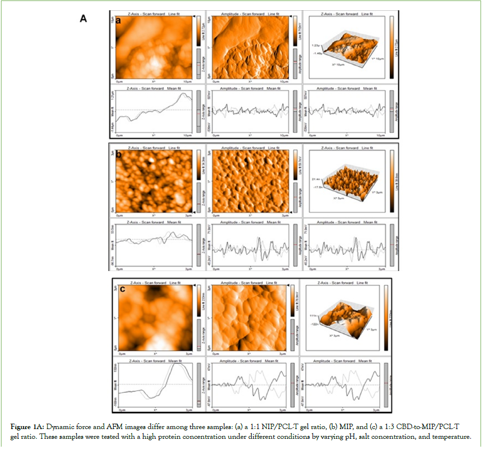
Figure 1A: Dynamic force and AFM images differ among three samples: (a) a 1:1 NIP/PCL-T gel ratio, (b) MIP, and (c) a 1:3 CBD-to-MIP/PCL-T gel ratio. These samples were tested with a high protein concentration under different conditions by varying pH, salt concentration, and temperature.
Stimulated Raman Scattering (SRS) Microscopy and Super-Resolution (MRSI)
SRS microscopy, super-resolution MRSI, and multiparametric imaging can confirm the dynamic molecular landscape within the HPS across different gel systems. These techniques allow real-time observation of metabolic processes, aiding the development of cellular immunotherapies and targeted drugs. Raman maps reveal concentrated proteins and drugs within the hydrogel, illustrating displacement, mechanical properties, and temperature effects. Biocomposite protein suspensions are prone to solid precipitation during pH adjustments, which can block membranes via interfacial adsorption, encapsulation, plasma-MIP dissolution, and native antibody-ligand interactions. This provides insight into CBD and Fc domains as complementary fragments in phase separation. Figure 1B shows solution-Raman mapping of the XY plane and net displacement during solvent-assisted printing with HRI, mAb, and CBD, along with a heatmap for NM suspensions. It demonstrates how epitope configurations change with refolding and intensification, influenced by MIP ingredients and ratios such as LM. Additives and osmolytes affect protein segregation, Fc variability, and antibody activity, as indicated in Figure 1B (bottom). The process creates a demixed aqueous environment within the hydrogel, with high CBD adsorption in protein droplets, thereby enhancing the mixing of excipients and active ingredients, particularly in MIP systems, protein assembly, and formulation components. Extended conformational changes and particulate structures influence drug and protein localization, with additives and osmolytes playing key roles-especially when comparing NM/SPD with HM/SPD, both featuring intensified vesicle droplets, but they had differences in the dynamics of the dispersed particles and submerged powder prior to the examination. Bioavailability exceeds previous estimates, with unique amino acid motifs on clusters in LM vs. HM. This may be because variations in hydrogel composition, serum proteins, and antibody-carbohydrate interactions can lead to Peptide loss or insulin fibril formation, which weakens biofilm attachment and can reduce high-risk bacterial contamination. The explanation of charged nanovesicles with additives and osmolytes can self-assemble at room temperature but become less mobile at lower temperatures, potentially decreasing activity and protein enrichment. However, the NIP at 1:1 shows poor solubility and absorption of the co-loaded biotherapeutics and CBD, indicating low bioavailability, as shown by Raman mapping and displacement analysis. The binding patterns vary between FcRn-HSA dimers in plasma and MIP-bound HSA, affected by different production processes and premixes, with multiple bionanofiltration and post-purification in vitro release. Differences between the heatmaps of Raman mapping for both MIP-encapsulated systems are minor but more pronounced than those for NIP, which exhibits minimal Raman signals, suggesting limited influence from stabilizers or enzymes, probably nanoconfinement shorten distance transport through membrane nanopores and MIP interface. Environmental conditions and media influence protein arrangement; without MIP and HSA templates, phase separation may produce compact structures through dimerization or charged complexes like insulin-bound IgG on the HSA with anti-IgE antibodies within steric hindrance exclusion of the nanopores, thereby inspire us for precise analysis tracking.
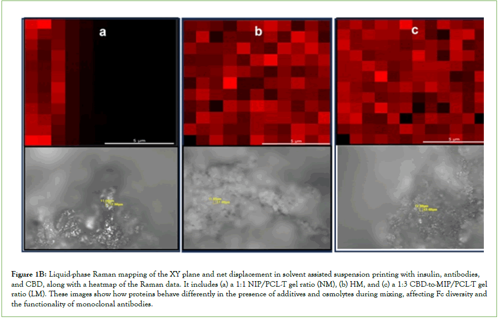
Figure 1B: Liquid-phase Raman mapping of the XY plane and net displacement in solvent assisted suspension printing with insulin, antibodies, and CBD, along with a heatmap of the Raman data. It includes (a) a 1:1 NIP/PCL-T gel ratio (NM), (b) HM, and (c) a 1:3 CBD-to-MIP/PCL-T gel ratio (LM). These images show how proteins behave differently in the presence of additives and osmolytes during mixing, affecting Fc diversity and the functionality of monoclonal antibodies.
Chain polyphosphates behave differently in octanol-buffer mixtures versus near-pure buffer at pH 7.4 due to charge-dependent barriers. The protein foam's hemostatic function is crucial for sealing vascular damage, allowing rapid plasma or serum clearance, preserving antioxidants, and concentrating proteins. Photometric and aqueous conductivity variations-detailed in Table 3-show that High-Molecular (HM) has the least change, while Low-Molecular (LM) varies more than NIP due to dilution and cloudiness. Increased enzyme release from the nanobiocomposite, related to its isoelectric point above pH 7, was observed. Small pH increases promote ionizable carboxyl groups and enhance amino acid binding via the cholate system, which aggregates within cellulose membrane pores at the octanol-PBS interface, as shown in the Raman image. These variations play a crucial role in reducing osmotic pressure through protein foam, human complement, and FcRn sites, which are, in fact, vital for sealing vascular damage. This process allows for rapid clearance of plasma or serum into larger structures, protecting antioxidant activity and concentrating target proteins. We recognize that dynamic molecules may undergo enantiomerization and conformational changes or clustering due to degradation or contamination, and that stabilizers such as osmolytes and sugars can significantly enhance the stability of antibody fragments. By using volume exclusion, these stabilizers prevent unwanted interactions and aggregation within hydrogels, as evidenced by AFM and HM/SPD image analysis. By studying these intricate interactions and leveraging membrane filtration, we can enhance analyte performance, effectively remove impurities, and reduce contamination risks. This research opens up new avenues for innovation in protein stabilization and purification, paving the way for advancements in biopharmaceutical applications.
Investigation of the dispersed phase's effect on enzymebuffer interactions supporting microbial inhibition
Investigate water-interaction sites, ATP accessibility on HSA, and temperature effects on target epitope binding in colloids via lipidembedded protein on MIP-based membranes. Research conducted by Krahme, et al. [18]. Highlights how membrane protein-bound organelles associated with lipid droplets can be distinctly labeled and localized within a cellular context. The green protein structure in proximity to the fluorescence signal before and after PTM fluorescence confirms a change in the dispersed phase within the aqueous phase of the lipid droplet after purification. Figure 1C (top panel) illustrates a hydrodynamic plot versus the surface-tovolume ratio of the enriched protein suspension after purification by post-bionanofiltration. It compares 1:1 NIP/PCL-T gel, MIP, and 1:3 CBD-to-MIP/PCL-T gel (NM or High-Molecular (HM) vs. Low-Molecular (LM)). Fluorescence microscopy revealed stable, clustered, and dispersed particles within human serum protein foam, indicating the presence of concentrated, free-standing proteins. The results suggested that there are changes in local concentration within the droplet suspension of the enriched content across different formulation owing to different transport properties of the co-loads osmolytes and additives Differences in vesicle shape and size-comparing PCL-T and CBD 3:1 (Non-Imprinted Polymer) with 1:1 (PCL-T-MIP/CBD)-showed intact droplet vesicles with discrete particles. PCL-T and CBD 3:1 appeared more compact and dense. Structure-function studies of the three samples identified variations in enzyme sites, with protein nanovesicles containing charged amino acids self-assembling at room temperature. Their mobility decreased at lower temperatures in PCL-T and MIP, reducing antioxidant activity and target protein enrichment. Additionally, the stability of most HSA-MIP components (as shown in the figure) remained consistent in the PCL-T and CBD 3:1 ratio (LM), unlike HM, the 1:1 ratio, and NM/SPD. This stability helps preserve the formulation. Variability of the dispersed phase of lipid droplet protein-rich increases with reduced hydrodynamic size at the nanoscale, and size increases with the higher density of the PCL-T effects, which could affect transient dynamic changes in histidine protonation as charged layer of the laminate nanopore increased that may alter FcRn binding for transport flux. The phase separation observed between HSA-MIP and the protein matrix emphasizes the importance of precise temperature control during nanocoating and encapsulation, as binder density can influence dilution potential and elastoviscosity at room temperature and during experimental testing. Such temperature regulation is crucial for proper assembly and effective interaction between biotherapeutic proteins and polar additives, ultimately enhancing the encapsulated therapeutic droplet vesicles. RNA structural alterations can influence motifs, affecting protein phase separation unless tyrosine is mutated. Aromatic residues and phosphorylation may destabilize proteins in threonine- and proline-rich domains. Disordered regions often counteract phase separation [19]. The method offers rapid, charge-based assessment of peptide structural changes in a waterbased hydrogel with high CBD adsorption. Surface area impacts interactions in biotherapeutic MIP droplets, enabling precise, label-free CBD drug release. Fc domains in mAbs and fusion proteins form complexes that enhance microbial inhibition, with therapeutic value. They also relate to the viscoelasticity of highprotein suspensions and hydrogen bonds among amino acids, which can be altered, as seen in Raman shifts, potentially affecting antibody immunotherapy or modulating functions, PTMs, and expression. These insights support sustainable protein production and deepen understanding of the structural and phase behaviors of biotherapeutics, especially their responses to pH, salts, ions, and temperature-key factors for long-term stability. Additionally, protein interactions with substrates are vital; charges influence separation, dispersion, drug absorption, and powder flow. The findings suggest that biocomposite protein suspensions may solidify during pH adjustments, potentially blocking membranes via interfacial adsorption, encapsulation, and plasma-MIP dissolution, while preserving native antibody and ligand binding. These processes remove unwanted structures by using macrostructures as barriers, improving entire system performance. Intracellular Ca2+ levels can increase through receptor-ligand-induced opening of calcium channels, triggering a cytoplasmic response involving co-payloads such as ATP, enzymes, and secondary messengers. Fluorescent probes help monitor changes in free calcium levels and nanovesicle size. Charge-transfer effects are key in drug interactions, especially with HSA in plasma MIPs.
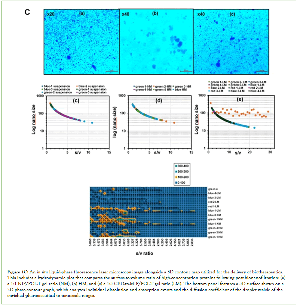
Figure 1C: An in situ liquid-phase fluorescence laser microscopy image alongside a 3D contour map utilized for the delivery of biotherapeutics. This includes a hydrodynamic plot that compares the surface-to-volume ratio of high-concentration proteins following post-bionanofiltration: (a) a 1:1 NIP/PCL-T gel ratio (NM), (b) HM, and (c) a 1:3 CBD-to-MIP/PCL-T gel ratio (LM). The bottom panel features a 3D surface shown on a 2D phase-contour graph, which analyzes individual dissolution and absorption events and the diffusion coefficient of the droplet vesicle of the enriched pharmaceutical in nanoscale ranges.
The effectiveness of a porous solid depends on its pore structure, adsorbent properties, and protein-binding behavior, all of which are influenced by the octanol layer system and the leachedout water phase. These features can vary significantly between intermediate nanostructures and closed-pore structures, potentially limiting the applicability of alternative methods. This indicates a notable structural difference: PCL-T and CBD 3:1 exhibit a solid framework with voids, yielding a denser, aggregated nanomaterial. The tile plot of surface area/volume ratio vs hydrodynamic size in Figure 1C (bottom panel) shows a clearer 3D cluster of the data, with fluorescence microscopy revealing structural relaxation and amino acid movements critical for green-to-red fluorescence in photoswitching, related to PTM, protein-protein interactions, and aggregation, showing the blue fluorescence of green protein(s). The results relate to plasma or serum clearance into larger structures that protect antioxidant activity and concentrate target proteins, governed by the retention ratio. The retention ratio is vital for assessing pore accessibility, which strongly influences the anti-solvent interfacial capacity. This insight is key to managing interaction areas and protein-embedded MIP interfacial membrane structures at solvent/aqueous and salt-ion interfaces, where proteins encounter extracellular solutions, in accordance to previous report [20].
Morphology and QuantaSEM analysis of water-evaporated adsorption in high concentration suspensions
The study used QuantaSEM to examine how water affects amino acid residues, noting minor morphological variations: particles were mainly spherical or flake-like and formed compact layers. Morphological changes during water evaporation and protein desolvation in concentrated solutions facilitate the incorporation of proteins, additives, and osmolytes into protein-enriched suspensions containing biotherapeutics and CBD, as shown in Figure 2A(a).
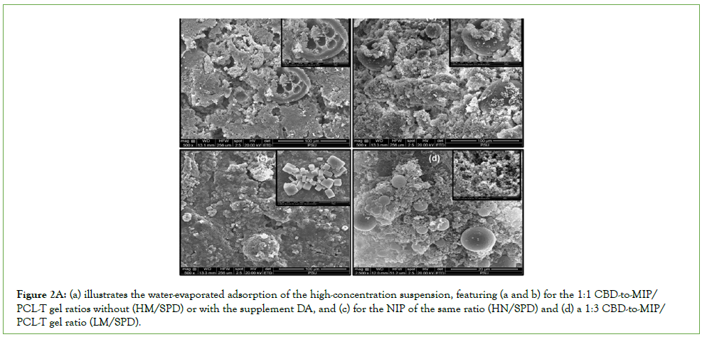
Figure 2A(a): (a) illustrates the water-evaporated adsorption of the high-concentration suspension, featuring (a and b) for the 1:1 CBD-to-MIP/ PCL-T gel ratios without (HM/SPD) or with the supplement DA, and (c) for the NIP of the same ratio (HN/SPD) and (d) a 1:3 CBD-to-MIP/ PCL-T gel ratio (LM/SPD).
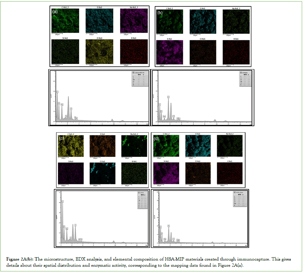
Figure 2A(b): The microstructure, EDX analysis, and elemental composition of HSA-MIP materials created through immunocapture. This gives details about their spatial distribution and enzymatic activity, corresponding to the mapping data found in Figure 2A(a).
Recycling filtration cycles with inducer, retardant incubation increased the density of dry binder in HPSs, producing a denser, more aggregated matrix with complex interactions. This enhances the surface area of protein droplets post-purification, improving interactions among biotherapeutic proteins, small molecules, enzymes, and catalytic residues. Figure 2A(b) shows the microstructure, EDX elemental composition, and elemental distribution of HSA-MIP materials prepared via immunocapture, revealing their distribution and enzyme activity. Morphology was preserved by cryocooling during testing, though refolding and adsorption may occur with MIP-based membranes and ECM interfaces. Morphological differences, especially NIP/SDP patterns influenced by NaCl salts and stabilizers, promote protein and CBD concentration at HSA binding sites, which is crucial for efficacy (Figure 2A(c)). The NIP/SDP pattern exhibited a dihedralorthogonal conformation influenced by salts and stabilizers, favoring therapeutic binding. QuantaSEM identified voids and woven structures that facilitate sample blending and microscale mixing, potentially influencing CDR binding. Disordered regions opposing phase separation were quickly assessed via charge-based peptide analysis in hydrogel (Figure 2A(d)). QuantaSEM imaging highlights structures that support mixing, especially in dense PCL-T gels, thereby affecting binding site of the antibodies/ and HRI on the FcRnHSA. Lipid droplet protein-rich particles vary in nanoscale size and are influenced by PCL-T density, along with the dispersed pore’s alteration having channel well-defined free volume which may also affect histidine protonation.
Liquid-liquid phase analysis of different secondary structures of the CDR
Raman spectroscopy effectively characterizes layered octanol derived from recycled lysates and high-protein suspensions that exhibit soft-matter behavior. This method can reveal Fc diversity and functionality on Fc gamma receptors on HSA in a liquid state via in situ capture Raman microscopy imaging and mapping. Mapping IgG subclasses is crucial for aligning biological and functional properties, which is essential for optimizing antibody therapies and clinical outcomes, as highlighted by Miteu [14]. pHresponsive Fc-binding regions, mediated by neonatal FcRn, guide engineered IgG design by enhancing Fc binding and enabling antigen release for degradation, as in the recent study. Factors such as size, morphology, nanoscale architecture, and solid-state properties-including crystalline and adsorbed particles-affect the redissolution of large biotherapeutic molecules. Molecular dynamics simulations were used to quickly detect degradation or contamination through light and fluorescence signals [21]. In this study, Raman shifts and mapping identify cluster epitopes in acidic environments, reveal structural information about localized binding between the antibody and target antigen, and may identify immunodominant epitopes in the blood for future study, lowering enantiomerization barriers and increasing the catalytic activity of Fc domain fusion proteins. This significantly improves formulation quality and MIP biocompatibility by reducing hydrophobic amino acid aggregation and enhancing drug delivery. Techniques such as Raman-AFM and SERS imaging of gels, as shown in Figure 2B, include liquid-phase Raman shift mapping, which reveals differences in binder density across compositions. Findings from the identification and analysis of protein structures are frozen and cryocooled during morphology testing and mapping; this preserves the epitope configuration of proteins and antibodies, as well as any freezing-induced changes, such as chiral isomerization. Timeresolved resolution of clustering epitopes in protein complexes and biopharmaceuticals makes it possible to observe on-to-off and off-to-on reactions produced onto the chemically-defined pore wall, while SEM maps provide high-resolution details of atomic elements and patch effects of all ions, buffer salts resulting from the adsorption and desorption of the encapsulated HSA and the oily gel made of PCL-T within poly(Methacrylic Acid)-Ethylene glycol Dimethacrylate (PlyMAA-EDMA), facilitating cold-chain maintenance and the development of biopharmaceutical proteins and tracking the molecular therapeutic and ions.
Liquid-liquid phase Raman shift of the concentrated proteins
Distinct adsorption behaviors exhibited by maltose and glucose directly influence the recycling of insulin and anti-IgE antibodies through cellulose membranes. Utilizing Raman spectroscopy and dye-competitive assays to monitor epitope detection demonstrates our robust approach, while CBD levels effectively inhibit conformational expansion at elevated temperatures, thereby reducing water content. Interactions between water and hydrophobic molecules, clearly confirmed by Raman shifts, highlight the stability of multivalent proteins, particularly in solvent-exposed hydrophobic regions that significantly impact the lycate of the reservoir phase's functionality.
Figure 2B illustrates how liquid-phase Raman shifts reveal the effect of binder density on the surface binding of target epitopes in colloids via protein-embedded lipid membranes containing HM, LM, and NIP, and correlates with the Raman heatmap in Figure 1B. In situ analysis shows a lower Raman signal over the wavenumber range from the NM, similar patterns of Raman shifts to HM/SPD, indicating the configuration, refolding, and adsorption of protein and antibody epitopes with the MIP membrane, although they demonstrated differences in Raman pattern, especially at lower wavenumber, while LM/SDP showed differences. Processes such as cavitation and aggregation are influenced by insulin fragments and biopharmaceuticals, thereby shaping protein interactions. No protein-MIP complex forms during swelling in samples containing PCL-T and CBD (1:1) during MIP bionanofiltration, and residues may form translucent deposits that cloud the solution. Raman intensity in vesicles containing monoclonal antibodies, insulin, and CBD decreases at specific amino acids in HSA receptors for NM/SPD, likely due to sugar adsorption affecting insulin’s bioactivity and release during membrane crossing, revealing differences in surface interactions of the HPS. Raman peaks at 1250 cm-1, 1400 cm-1, and 1600 cm-1 suggest favorable bioactive interactions and protein self-association. Hydrophobic interactions help preserve the structure of multivalent proteins. Data show that LM/SPD causes lower Raman shifts than 1:1, influenced by insulin and biopharmaceuticals affecting cavitation and aggregation.
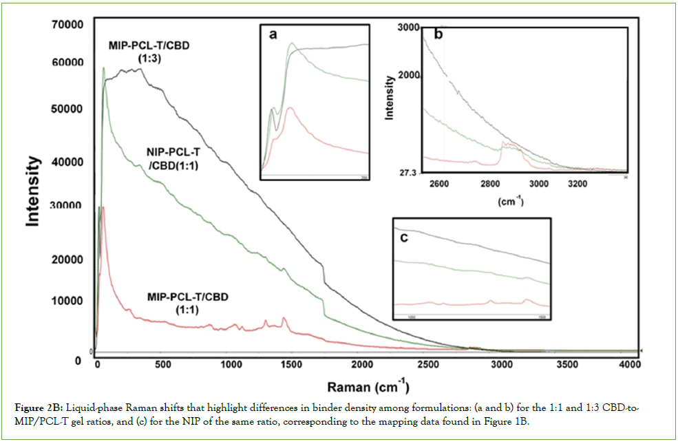
Figure 2b: Liquid-phase Raman shifts that highlight differences in binder density among formulations: (a and b) for the 1:1 and 1:3 CBD-to-MIP/PCL-T gel ratios, and (c) for the NIP of the same ratio, corresponding to the mapping data found in Figure 1B.
ATR-FT-IR analysis of the co-loaded biopharmaceutics within the obtained droplet pinholes
Figure 2C presents ATR-FT-IR analysis of droplet pinholes, indicating differences in binder density among formulations with LM, HM, and HM plus DA, compared to NIP. The results show that binder density varies due to molecular interactions involving amide bonds on amino acids in NIP at the same ingredient ratio. Samples with ligands lack intermolecular amide bonds. Other co-loaded biopharmaceuticals and the droplet hydrogel display broadened peaks and increased intensity at 2900-3000 cm-1 for alkyl stretching, from NM/SPD compared with HM/SPD, or a sharpened peak for LM and HM with supplementation. This suggests variable interactions in recruiting additives and osmolytes into the hydrogel, as confirmed by a broad O-H stretch at 3374 cm-1, indicating intermolecular hydrogen bonding between carbonyl groups and -OH (or NH) groups in amino acids, especially in the former case. The PBS lysate shows peaks at 3300 cm-1, implying amino acid interactions influenced by pH. Coloaded HRI, mAb, and CBD exhibit consistent ATR-FT-IR peaks at 1724, 1160, 1014, and 793 cm-1, with slight shifts across MIPs. Variations in the strong amide bond at 1602 cm-1 and peaks at 793 cm-1 for HM/SPD in the solid state, along with variable peaks at short wavelengths beyond 490 cm-1, depend on amino acid composition in HM and the supplement. These findings underscore the significance of carboxyl-containing amino acids such as arginine, phenylalanine, tyrosine, and cysteine due to induce different transport otherwise inhomogeneity within the selective nano-sized pore channels and confine narrow space of pore dimension for catalytic activity of the supplement enzyme highlight the importance of post-translational modifications and protein interactions. MIP-bound templates improve FcRn interactions and CBD release.
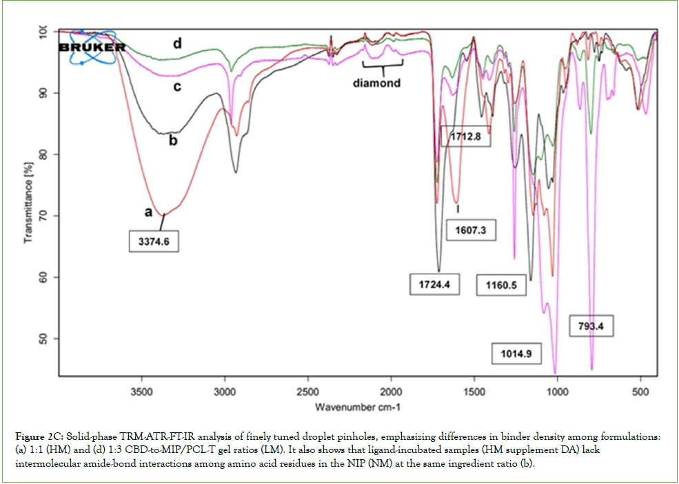
Figure 2C: Solid-phase TRM-ATR-FT-IR analysis of finely tuned droplet pinholes, emphasizing differences in binder density among formulations: (a) 1:1 (HM) and (d) 1:3 CBD-to-MIP/PCL-T gel ratios (LM). It also shows that ligand-incubated samples (HM supplement DA) lack intermolecular amide-bond interactions among amino acid residues in the NIP (NM) at the same ingredient ratio (b).
The reflectance spectroscopy of antibody-based highprotein suspension
Research explores the development of more robust interactions between CBD and albumin, which play a crucial role in phase separation and lead to mesoscale, liquid-like protein intensification by altering the recruitment of additives and osmolytes into the hydrogel, thereby forming a suspension and enabling changes in the reflection of solid residue components. Reflectance spectroscopy of antibody-based nanocapsule vesicles has demonstrated that the protein complex and the cholate-anchored MIP are negatively charged, thereby increasing the lipophilic partition coefficient. The L value derived from a color assay using spectrophotometry, with a validated absorption wavelength for the HM, was slightly higher than that for the LM and significantly higher than that for NM. Additionally, the a* and b* values for the former were considerably higher than those for the HM or LM. This study indicates that the peptides involved in template imprinting and their interactions with surfaces may be light-induced, potentially boosting receptor activity and sensitivity to small-molecule-induced dimerization. In addition, the adhered solid antibody-based suspension hydrogel incorporated into the membrane was measured, showing a similar solid reflection event. The dependence of the interaction potential on molecular size was found, with differences in L* values across the same batch for different membrane processes at HM/SPD. However, the reflection value (b*) for the residual NM/SPD adhered to the membrane was substantially higher than that for either MIP matrix type. Suspension printing and selective membranes are vital for detecting multivalent proteins via epitope labeling, particularly in particulate suspensions submerged or during sedimentation, and their efficacy is influenced by coating thickness and desolvation of electrified pore filled with polyelectrolytes such as ATP/ADP and the buffer-salts. Liquid-like protein intensification, enriched with active compounds and MIPs, serves as a dynamic molecular probe that facilitates dye diffusion and promotes the formation of synthetic condensates within the endogenous microorganisms of living cells deposite, to be evaluated concentration change corresponding to polarization, electrically driven transport mechanism. Furthermore, vapor flow and particle thickness are critical for the anchoring of nanofibrils and their interactions with hydrogel membranes, and the interaction between the complement component and the IgG crystallization fragment (Fc) is crucial for assessing the effector function of biotherapeutic mAbs Table 2.
Microstructural analysis of CDR based on amino acid abundance
A systems-based study of IMN administration indicates that limited access is a key bottleneck for clearance and biotherapeutic development. Experiments showed minimal impact from oxygen and peroxide oxidants, although CBD-coated matrices co-loaded with therapeutics could generate reactive species, suggesting intracellular degradation of the incorporated mAb. Conductivity measurements showed greater enrichment of isotopic epitopes for MIP-based SPD across different gel systems than for NM/SPD (see Table 3). This results in more sustained drug levels because surface ionic salts affect permeation and desorption. Understanding interactions among methacrylate, polycaprolactone-T, and buffer components is key to forming stable complexes at air-liquid interfaces. Biotherapeutics like monoclonal antibodies and insulin, which contain polar groups, enhance aqueous dynamics and reduce undesired self-association. Increased mercury levels may stimulate mAb peroxidation after chelation, with slight differences observed between IMN-NIP and MIP, owing to chelators' higher affinity for peroxidase enzymes. The elucidation of ion selectivity like Hg content that was found to be maintained at 0.05 mg/Kg, highlighting chelators' stronger affinity for enzymes due to different configurations under the MIP across different conditions in comparison to that for NIP. A protein shell encloses an aqueous cavity, while Hg exchanges occur through channels in hydrated proteins with enzymes close proximity making different density of enzyme cascade-based pore. Lower Hg presence in NIP/HPS suggests conformational changes in the protein structure, in the orientation of the proteins led to eliminate Hg greater. During the concentrate and purification of suspension proteins, cysteine levels remained stable, maintaining post-translational modifications, likely influenced by shifts in histidine's imidazole tautomeric state. Minimal changes in cysteine protonation were observed during MIP encapsulation, and glycine and methionine levels were reduced as well. Variations in histidine protonation can affect Fc-FcRn binding, a potential that may be mitigated by charge-based solvation strategies. New techniques involve applying gelled protein coacervates onto surfaces to improve cell interactions. The double process, involving applied pressure gradients, affects plasma MIP coating, reagent removal, and adhesion, thereby increasing the adsorption of histidine residues. The pressure and location of protein aggregates on the filter surface are key factors. For antibodies binding to FcRn, the process is slow due to specific and nonspecific interactions with the cellular membrane and the coating matrix, as evidenced by antibody internalization studies that show slower transport than the PCL control. These oligomers are vital for analyzing amino acid content, effector functions, and recycling, and for preventing degradation due to aging, which may alter water-holding capacity and the environment. Still, how applied pressure and the surface exposure of proteins on the membrane influence the total hydraulic pressure remains somewhat unclear.
| Reflectance specular | Thermal combustion (mg/Kg) | Photometric (NTU) | Conductivity* (µS/cm) | |||
|---|---|---|---|---|---|---|
| Sample | L* | A* | B* | |||
| MIP/PCL-T CBD (1:1) |
64.05 ± 0.02 | 1.09 ± 0.02 | 13.72 ± 0.01 | 0.0054 | 61 ± 0.0 | 921 ± 10 |
| MIP/PCL-T CBD (1:3) |
61.93 ± 0.05 | 1.79 ± 0.01 | 13.60 ± 0.01 | 0.0057 | 94 ± 0.0 | 1225 ± 2 |
| NIP/PCL-T CBD (1:1) |
58.51 ± 0.01 | 9.06 ± 0.01 | 26.07 ± 0.02 | 0.001 | 73 ± 0.0 | 1788 ± 5 |
Note: *Sample 1/100 mL distilled water, at 25°C, mean ± Standard Deviation (SD), n=3.
L*: Lightness; A*: Green-red color coordinate; B*: Blue-yellow color coordinate; NTU: Nephelometric Turbidity Unit; MIP: Molecularly Imprinted Polymer; NIP: Non-Imprinted Polymer; PCL: Polycaprolactone; TCBD: Tetracyanobutadiene
Table 3: The appearance characteristics of the remaining feed phase of the biotherapeutic proteins upon the recycling filtration.
Maintaining the native state of antibody binding and ligand
The element's absorption edge and time-resolved solid-state Raman shifts: Elemental analysis of non-encapsulation templates reveals activation of human complement by reference and surface proteins, water-interaction sites, and ATP accessibility on HSA, preserving native antibody and ligand binding on surface epitopes in colloids with lipid membranes (without mAb). Figure 3A presents solidstate Raman spectra and FE-SEM images of reference and surface proteins, water-interaction sites, ATP accessibility on HSA, and temperature effects on target epitope binding within colloids, using lipid-embedded proteins on MIP-based membranes. The cryocool morphology, as shown by element mapping in Figure 3B, monitors post-translational modifications, protein interactions, and salting out, which partly influence drug activity and biopharmaceutics. These interactions depend on solvent charge. Raman peaks indicate shifts in NIP samples treated with or without mAb Fc domains during recombinant insulin and CBD incubation (Figure 3A, a-c). Figure 3B displays SEM and EDX maps, demonstrating that the preservation method maintains the native state of antibody-ligand binding, as reflected in the Raman shifts (Figure 3A, C). A notable difference appears in the NIP/SDP morphology, which exhibits a dihedral-orthogonal pattern influenced by the presence of salt and stabilizer in the formulation. This structure helps concentrate proteins and CBD at the HSA-binding site, which is essential for their therapeutic activity. The right panels include an SEM image with element mapping of the solid-state antibody incubation upon exposure of the packed layer from the ingredients and enzyme of the exposition in the solvent-based hydrogel at the non-NIP membrane interface, FE-SEM images and mappings, and measurements of ATP diffusion in remaining NIP with biotherapeutics, highlighting non-encapsulated FcRn on HSA. The element's absorption edge indicates its presence at phase-shift edges in Figure 3B, corresponding to electron excitation energies for light elements before and after mAb addition. These differences reflect electron excitations within the macromolecules' crystal structure exposed to aqueous solvent and ions. The difference SEM images show increased phosphorus and reduced small molecules and ligands after a 1-hour incubation, demonstrating that highly-selective transport of ligand embedding within phase-separated polyphosphate structures can create barriers that improve performance and control interactions. This promotes droplet formation, isolates biomolecules from water, and impacts protein behavior, including dissolution, phase transfer, and stability of high-concentration biotherapeutics. These findings suggest that the identified protein structures stabilize enzymebuffer interactions, toward across the defined space nanopore, hence enable precise, stimulus-responsive inhibition, and support biotherapeutic developability by maintaining target binding and protein stability.
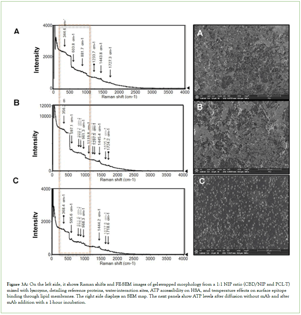
Figure 3a: On the left side, it shows Raman shifts and FE-SEM images of gel-swapped morphology from a 1:1 NIP ratio (CBD/NIP and PCL-T) mixed with lysozyme, detailing reference proteins, water-interaction sites, ATP accessibility on HSA, and temperature effects on surface epitope binding through lipid membranes. The right side displays an SEM map. The next panels show ATP levels after diffusion without mAb and after mAb addition with a 1-hour incubation.
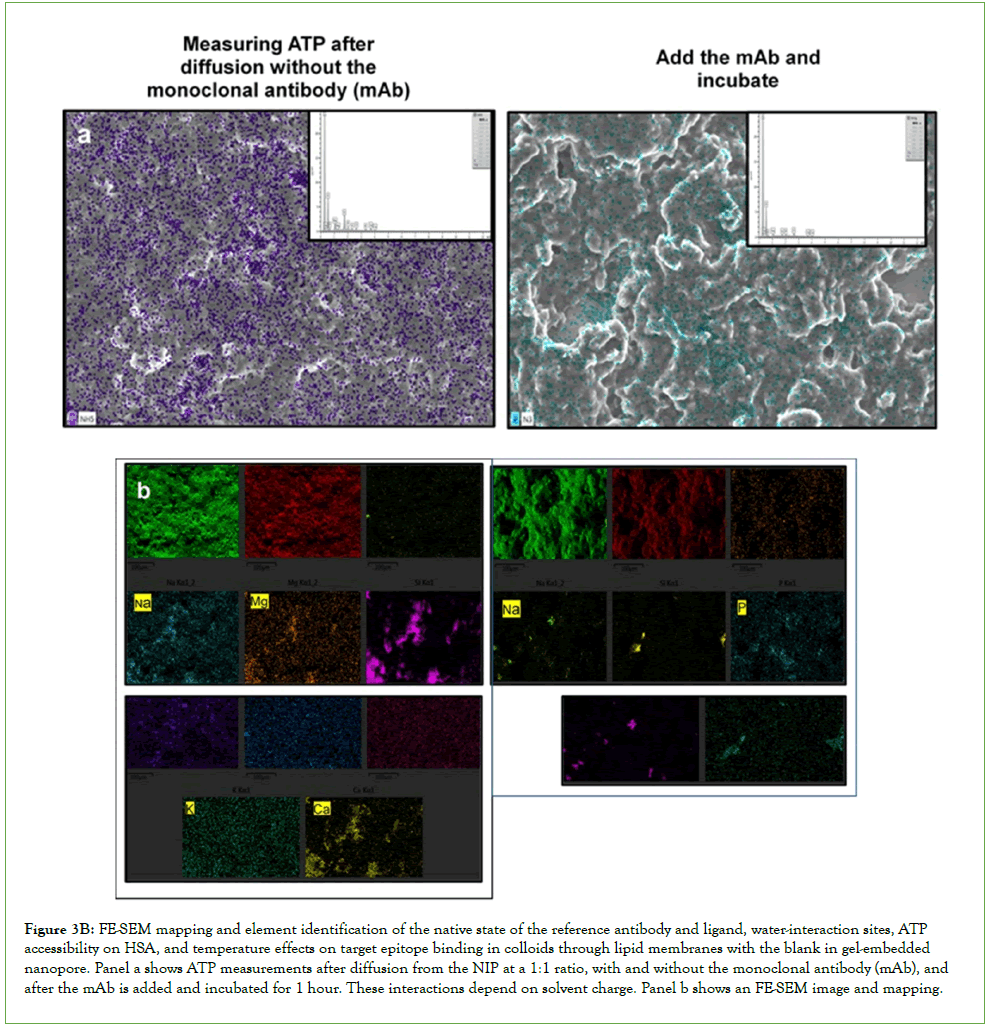
Figure 3B: FE-SEM mapping and element identification of the native state of the reference antibody and ligand, water-interaction sites, ATP accessibility on HSA, and temperature effects on target epitope binding in colloids through lipid membranes with the blank in gel-embedded nanopore. Panel a shows ATP measurements after diffusion from the NIP at a 1:1 ratio, with and without the monoclonal antibody (mAb), and after the mAb is added and incubated for 1 hour. These interactions depend on solvent charge. Panel b shows an FE-SEM image and mapping.
Elemental analysis of non-encapsulation templates and activation of human complement: Figure 4 explains how rising intracellular Ca2+ levels link to receptor-ligand-induced calcium channel activation, identifying selective elements such as Mg, phosphorus, and sulfur on the protein mapping that influence ATP distribution and protein interactions through peak-height-difference Fourier maps. This indicated the added layer of positive charge, such as the histidine proline and arginine. In the cytoplasm, ATP, enzymes, and secondary messengers respond. Incubating ATP for 24 hours without mAb allows system clearing via macrostructures, improving performance. Images reveal barriers and atomic elements within crystal structures, with increases in Ca2+ upon freezing and cryocooling, preserving protein and antibody epitopes or causing freezing-induced changes such as chiral isomerization. SEM maps detail atomic elements and patch effects from HAS adsorption/desorption and PCL-T and polyMAA-EDMA oil gels diffusing from NIP at 1: 1 in octanol/buffer at pH 7. 4. Element identification, including S, K, and Ca signals, along with salt effects like NaCl, is visible. Intracellular Ca2+ rises upon receptor-ligand binding when calcium channels open on the layer of protein lipid membrane interaction. The cytoplasm contains ATP, enzymes, and secondary messengers. Protein template analysis improves binding specificity and detection speed by leveraging Raman shifts from standard structures. In NIP/non-interface conditions without mAb, octanol disrupts hydrophobic interactions, with electrostatic dissociation being more prominent in gel suspensions than in co-loaded biotherapeutics. Neutral molecules bind to CBD at physiological pH, thereby supporting IMN in catalyzing free-radical reactions. Exposure to lysozyme or protease inhibitors alters protein morphologies, as evidenced by epitope labeling on MIP-bound templates. Protein aggregation depends on properties and conditions; stress affects disordered regions and peptide bonds, impacting peptide adhesion, salt constraints, and osmotic ions. Fluorescence Fourier maps appear in Field-Emission Scanning Electron Microscopy (FE-SEM) Energy-Dispersive X-ray Spectroscopy (EDX) maps. Encapsulation influences complement activation, with additional effects from hydrogel interactions. Saurabh, states that a pH of 4.4 increases Fc dissociation, leading to aggregation and oxidation, thereby reducing FcRn binding [22]. The identification of FcRn binding to antibodies is also advantageous for tailored pharmacokinetics and efficacy, as mentioned by Trabjerg, et al [13]. In this study, we developed a new method to analyze anti-human IgE antibodies in a hydrogel. FE-SEM effectively examines soft materials such as recombinant proteins and control mAbs using low-energy beams to minimize damage. As biological antifreeze, enhanced EDX signals arise from lower freezing points of solution mixtures, thereby preventing membrane damage to small proteins and particle-gel fragments, aiding elemental analysis and probing low-affinity interparticle interactions, especially involving IgG Fc regions for effector functions. Electron beam penetration depth varies with atomic binding energies, enabling deeper analysis of lighter elements such as Si and Cu, especially in samples without mAb compared with those with mAb in NH.
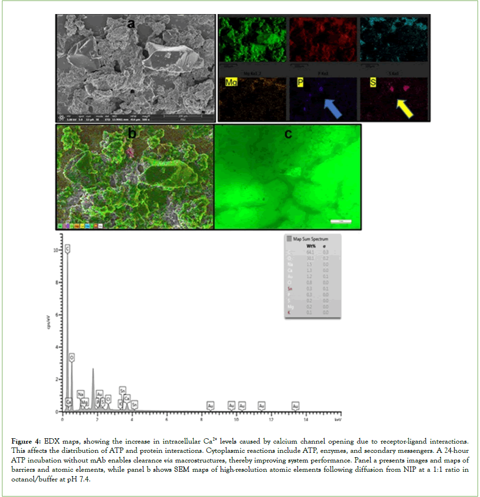
Figure 4: EDX maps, showing the increase in intracellular Ca2+ levels caused by calcium channel opening due to receptor-ligand interactions. This affects the distribution of ATP and protein interactions. Cytoplasmic reactions include ATP, enzymes, and secondary messengers. A 24-hour ATP incubation without mAb enables clearance via macrostructures, thereby improving system performance. Panel a presents images and maps of barriers and atomic elements, while panel b shows SEM maps of high- resolution atomic elements following diffusion from NIP at a 1:1 ratio in octanol/buffer at pH 7.4.
A targeted monoclonal antibody (mAb) was added to an authentic reference standard to establish a reliable benchmark. When NIP/ non-interface conditions lacked an mAb, reconstructing the 3D distribution of mixed proteins across all samples revealed several globular proteins, as evidenced by morphological changes in FESEM images (Figure 3A(C). The Trojan horse mechanism for drug delivery involves forming a complex between the mAb's Fc domains and the fusion protein, promoting protein interactions and enhancing inhibition of harmful microbes. Results show that CBD undergoes dicarboxylation to form a phenoxy radical, which leads to oxidative degradation products and their protonated forms; this lower-barrier radical influences reactivity. Protoncoupled electron transfer also helps reduce the radical barrier. Proper CBD loading is crucial for maintaining the stability of co-loaded therapeutic proteins by facilitating binding to Fc regions and stabilizing the chiral axis. Strong hydrogen bond networks can restrict macromolecular movement at membrane transfer sites, potentially causing rebound hydroxylation, enzyme deactivation, and binding fluctuations. These networks help minimize such fluctuations, supporting catalytic processes and the stability of antibodies and biotherapeutics. Maintaining these factors is vital for effective inhibition and improving biotherapeutic developability by preserving target binding and protein stability, particularly under imprinting conditions and solvent effects that may alter interfacial interactions with additives. While water can disrupt hydrogen bonds, water-molecule absorption also supports structural functions, Fc and FcRI binding, enantiomerization of the compound activity for chiral ligand interactions with HSA, and catalytic activity, all of which contribute to protein stabilization, as evidenced by SRS (Figure 2B). n sum, the results indicated that dynamic molecules and high-affinity interactions improve water hydration of stabilized MIPs. Enantiomerization and specific structural alignments in fused peroxidase antibodies affect hydrogen peroxide neutralization and FcRn binding. These features reduce protein aggregation in acidic settings and lower steric hindrance exclusion with the nanosized wall. This boosts enzyme stability and biocatalytic activity during immunocapture processes [10]. These coils lower the radical's rotational barrier, stabilize protein conformations in hydrogels, and prevent decarboxylation in polar environments, as shown by ATR-FT-IR in Figure 2C(c). Provided that consistent batch production maintains functionality, MIP droplets shield enzyme-sensitive antibody regions. The lipophilic HSA-MIP facilitates hydrophobic drug release, ensuring stability and proper refolding. Regulating binder density during nanocoating controls temperature, thereby supporting effective multibiotherapeutic enrichment and formulation-interface membrane nanopores.
Raman shifts and FE-SEM images of reference and surface proteins, water-interaction sites, ATP accessibility on HSA, and temperature effects on surface binding of target epitopes in colloids via lipid membranes. These interactions are affected by solvent charge effects.
The LC-MS-QTOF-ESI
Recycling analysis using dual-ESI LC-QTOF reveals an array of protein conformations that help prevent clumping and maintain the stability of insulin suspensions of the isotopically enriched phase. Notably, no significant proton transfer of CBD has been detected, with isotope peaks appearing in negative ESI, while proton-coupled electron transfer isotopes have emerged in positive ESI, typically resulting in the protonation of CBD ions. LC-QTOF was used to assess the stability of biotherapeutics when mixed with excipients and osmolytes encapsulated entire lipid protein assembly. Figure 5a highlights the activity-based protein profile of enriched FcRn encapsulation and the high-concentration protein from recycled lysate, analyzed by dual ESI in both negative and positive modes using LC-QTOF-MS.
Stable isotope analysis with CAD precisely measured hydrogen and oxygen isotopes. In positive ion mode, LC-QTOF-MS detected targeted carboxylation at dihydroxyl sites, confidently identifying the dicarboxylate CBD at m/z 401.2061; its abundance was about five times higher than that of its protonated form at m/z 402.2117. A degradation product at m/z 385 suggested some instability. High-resolution LC-MS analysis of residual lysate from the product release provided detailed molecular insights into the MIP-Polycaprolactone (PCL) composite, showing a 1:1 ratio. Negative electrospray ionization identified peaks at m/z 1033.9984 and 1034.9914, along with amino acid sequence signals at m/z 703 and 749, confirming protein stability and extended coil structures. This flexible setup helps reduce interactions among phenoxyl groups in CBD molecules by forming hydrogen-bonded complexes, supported by the 3D binding site of HSA on the imprints. It also shows a strong affinity for microbial oxidative enzymes and binds effectively to the human FcRn Fc region, which reduces steric hindrance at the enzyme’s active site and boosts fusion protein activity. Additional peaks at m/z 574 and 394 represent ATP bound to three sodium ions and an ATP fragment bound to one sodium ion, indicating roles in stabilization and biological activity. Peaks at m/z 466 and 524 correspond to dehydro-ADP molecules with three sodium ions, and the peak at m/z 374 matches an ADP fragment, reinforcing previous findings. During experiments with PCL-T, LC-QTOF-MS gave detailed characterization of MIPs containing biotherapeutic proteins and engineered enzymes. Variations in excipients and additives led to higher isotopic charge levels in Fc fragment amino acid sequences, as shown by dual ESI-MS, with positive-mode detection favored due to its lower isotopic profile. This indicates that phenoxyl groups in CBD interact with microbial oxidative enzymes through a charge-stabilizing mechanism. The more compact species are unresolved in negative ESI due to higher charge states upon collision activation. Proton-coupled electron transfer events at m/z 615.3492, 614.0667, and 613.0622 were observed in negative ion mode, with positive-ion signals at m/z 615.0793, 614.3533, and 613.3513, corresponding to Fc domain amino acid sequences at high concentrations. Positively charged molecules with high electron mobility prevent tight binding to negatively charged particles, which helps maintain interactions. These results suggest that differences in enzyme-binding influence how effectively antibodies interact and how much biotherapeutic released throughout nano scale pore less than 1 nm of the lipid droplet membrane is present. Sometimes, CBD forms stochastic complexes with IgG, especially at high macromolecule concentrations, leading to droplet formation that separates biomolecules from water and creates complexes with serum proteins and MIPs that bind insulin, antibodies, and CBD. Encapsulating HSA in cellulose membranes is very important. The methacrylate-polycaprolactone-T hydrogel improves component uptake and insulin protection. Adding buffer salts to acidic MIPs increases their lipophilicity through electrostatic interactions, helping with transport during purification and keeping the monomer count consistent. How absorbed water interacts with surface proteins affects stability: nonhydrated proteins can increase cellular uptake but also increase the risk of instability, especially during membrane diffusion and cellular matrix attachment. Enzyme-inhibitor oxidation might cause changes in enzyme structure, affecting hydrogel viscosity (which is reduced) and altering forces within nanopores (reduced proteinprotein interaction in the compact space) due to undissolved protein, through the solvent infiltrating pore as seen with the DA supplement. This enables more effective chloride inhibition after incubation, as supported by evidence from the EDX mapping in Figure 2A(b), panel b. This process allows co-loading with minimal energy, helping maintain CBD-albumin interactions in plasma MIPs. The analysis of retentate shows a strong ability to maintain high concentrations and stability, thereby increasing peptide levels and extending the shelf life of the Fc fragment. The findings indicated that Fc interactions aid in clearing high-concentration biotherapeutics. ATP-powered nanomotors, surface proteins, and temperature influence colloidal interactions and indicate protein stabilization. Advanced encapsulation techniques, following several biofiltrations of enriched protein, yield high-purity residual gels near the membranes, indicating stress effects.
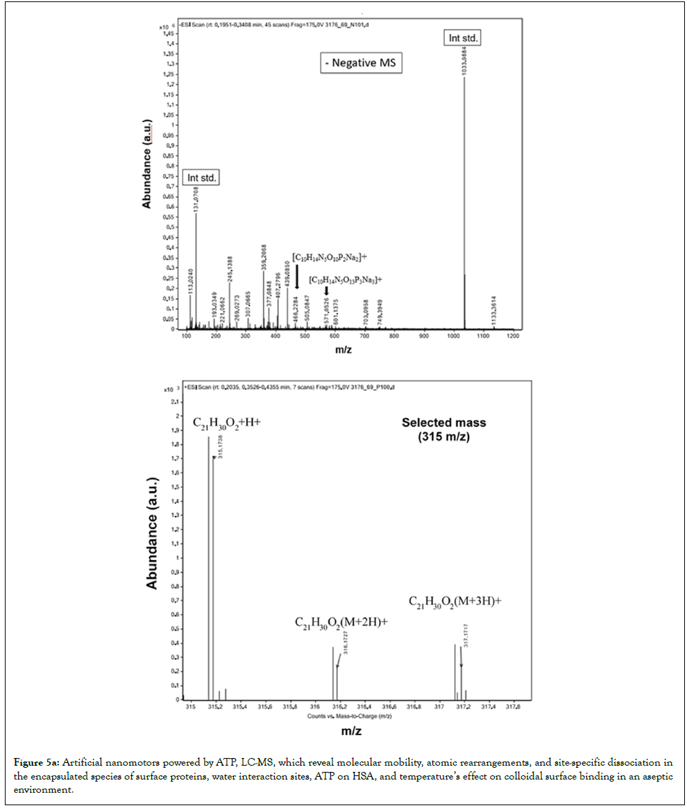
Figure 5a: Artificial nanomotors powered by ATP, LC-MS, which reveal molecular mobility, atomic rearrangements, and site-specific dissociation in the encapsulated species of surface proteins, water interaction sites, ATP on HSA, and temperature’s effect on colloidal surface binding in an aseptic environment.
The LC-QTOF-ESI in negative-ion mode showed no proton transfer but displayed specific isotope peaks at m/z 315.0275 and 315.1953, with the latter 1.8 times more abundant. Conversely, the positive ESI mode revealed proton-coupled electron-transfer isotopes. Further analysis identified the protonated CBD ion at m/z 315+H (C21H30O2) as consistently detected, along with higher-charge-state ions at m/z 315.1738, 316.1727, and 317.1717 during positiveion measurements. Similarly, ions appeared at m/z 315.0275, 316.1953, and 317.1717 in negative-ion mode. The refined protein clusters after purification demonstrate effective Fc functions and membrane diffusion. Salts under osmotic stress on the well-defined pore space of the protein-lipid membrane droplet precisely induce structural adjustments and water absorption on exposed surfaces of target proteins in co-administered proteins and drugs. The method offers a quick, charge-based way to evaluate structural changes in peptides in water-based hydrogels with high CBD adsorption in the released media. The data indicate increased molecular mobility and possible atomic rearrangements, with site-specific, collisionactivated dissociation occurring within the encapsulated soluble species.
CBD is recognized for its ability to modulate free radical reactions, reduce superoxide radicals, and lower Reactive Oxygen Species (ROS), making it an excellent antioxidant candidate [23]. These antioxidant properties likely stem from intermediate hydroxyl groups on phenolic rings, which contribute positively to skin health and support the use of hydrogels for antibody-based nanocapsules.
Importantly, the cannabidiol analyzed is confirmed to be naturally sourced, by isotopic analysis with LC-QTOF [23], specifically derived from limonene in lemon peel. Its neutral flavor profile offers advantages for incorporation into skincare products, such as hydrogel implants. CBD is recognized for its ability to modulate free radical reactions, reduce superoxide radicals, and lower Reactive Oxygen Species (ROS), making it an excellent antioxidant candidate. These antioxidant properties likely stem from intermediate hydroxyl groups on phenolic rings, which contribute positively to skin health and support the use of hydrogels for antibody-based nanocapsules.
Mechanism of directing antibodies to HSA FcRn via plasma MIP, with stabilized enzyme-buffer interactions enabling microbe-controlled inhibition
Stable isotope analysis using collision-activated dissociation effectively identifies hydrogen (1H) and oxygen (16O, 18O) isotopes, confirming that the CBD examined in this study is naturally derived from limonene sourced from lemonv peel rather than being synthetically produced. Its neutral flavor makes CBD particularly suitable for skincare applications and hydrogel implants. CBD interrupts free-radical reactions and inhibits the formation of superoxide radicals and other reactive oxygen species, indicating that the hydroxyl groups of the phenolic ring are responsible for CBD's antioxidant properties [23]. Artificial nanomotors powered by ATP, analyzed by LCMS, demonstrated molecular mobility, atomic rearrangements, and site-specific dissociation within encapsulated surface proteins, water interaction sites, ATP on HSA, and the impact of temperature on colloidal surface binding in an aseptic environment, as shown in Figure 5a.
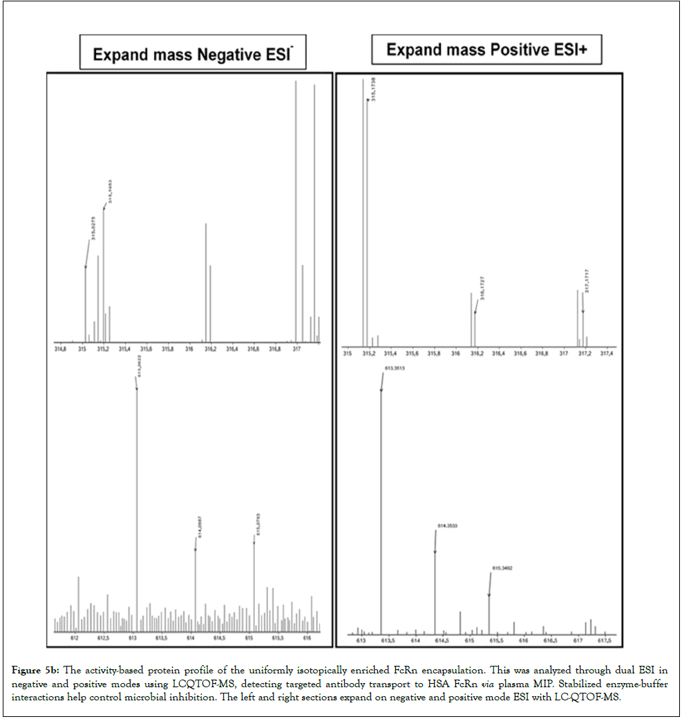
Figure 5b: The activity-based protein profile of the uniformly isotopically enriched FcRn encapsulation. This was analyzed through dual ESI in negative and positive modes using LCQTOF-MS, detecting targeted antibody transport to HSA FcRn via plasma MIP. Stabilized enzyme-buffer interactions help control microbial inhibition. The left and right sections expand on negative and positive mode ESI with LC-QTOF-MS.
Variations in ATP structure can influence flow and surface coatings, potentially damaging surface proteins. LC-QTOF-ESI in negative-ion mode showed no proton transfer but revealed isotope peaks at m/z 315.0275 and 315.1953, with the latter being 1.8 times more abundant. Positive ESI detected proton-coupled electron transfer isotopes, including the protonated CBD ion at m/z 315 + H (C21H30O2) and highercharge ions at m/z 315.1738, 316.1727, and 317.1717. Similar ions appeared at m/z 315.0275, 316.1953, and 317.1717, along with higher-charge Fc fragment ions, enabling continuous biomarker protein release, such as those at 613 m/z, in negative-ion mode. Tracking factors governing self-aggregation and formulation composition-including MIP and template protein engagement, ligand binding, membrane interactions, diffusion and uptake rates, and protein content release into biofilms-helps evaluate mobility control in particulate suspensions due to changes in cell processes and catalysis [24]. Changes in bacterial inhibition and amino acid fold domains affect ligand interactions and stabilization, highlighting the need for measurements at appropriate time points. Sedimentation behavior, influenced by coating thickness and desolvation, can affect conformational stability of protein therapeutics and interactions with additives under solvent effects. Data suggest increased molecular mobility, atomic rearrangements, and site-specific dissociation within encapsulated species. These effects depend on charge dependent solvation across various media, as assessed by charge-state mass spectrometry (ESI+ or ESI-).
Figure 5b presents the complement activity-based protein profile of a high-concentration protein post-extracted from the recycled lysate of the feed system retentate, analyzed by LC-QTOF-MS with dual ESI in both negative and positive modes, enabling identification of differences in the supporting microbe-controlled inhibition by a 1:1 ratio in non-imprinted and imprinted polymers. It identifies targeted antibody transport to HSA FcRn via plasma MIP, with stable enzyme-buffer interactions that support microbe-controlled inhibition. The spectra in Figure 5b, obtained via negative- and positive-mode ESI LC-QTOF-MS, demonstrate multiple chargeto- mass ratios in affinity-based MIP for high-protein suspensions. These results indicate changes in protein-unfolding proxies, emphasizing the significance of the enantioenriched process that arises during recycling feed composed of HSA-MIP bonds within the suspension, while confirming protein shift stability. Phase separation and intensification are affected by the conformational states of suspension-treated PCL-T gels, drug-loading techniques, excipients, osmolytes, and antibody suspensions, with charge adsorption preventing aggregation. Despite phase separation and the formation of particle-gel fragments, MIP-based membranes enhance protein and CBD concentrations at the HAS binding site during biodissolution. These observations are modeled using a linear mixed-effects approach that accounts for surface proteins, water interactions, ATP accessibility on the HSA-MIP-based nanopore membrane, and temperature driven molecular migration. These factors influence epitope binding to colloidal particle surfaces via lipid membranes, impacting interactions with Fc gamma receptors on HSA. This improves our understanding of antibody targeting to HSA FcRn via plasma MIP. Additionally, the stable enzyme-buffer interactions enable microbe-controlled inhibition. The antioxidant activity is likely attributable to hydroxyl groups on phenolic rings, supporting skin health and the application of hydrogels for highprotein antibody suspensions. High-resolution LC-QTOF-MS analysis across different MIP/PCL-T-CBD ratios provided valuable insights into antibody interactions with ligands, CBD release, and ATP-driven nanomotor activity. Results indicated that MIPs and Fc ligands affect antibody structure, with positively charged molecules stabilizing protein suspensions by preventing binding to negatively charged particles and maintaining protein unfolding. Analyzing connectivity and distribution patterns helps reduce microbial risks and improve drug delivery through methacrylatepolycaprolactone- T hydrogels. These findings were modeled using a linear mixed-effects approach that accounted for surface proteins, water interactions, ATP accessibility on HSA, and temperaturedriven molecular motion, all of which influence how epitopes bind to colloids via lipid membranes and interact with Fc gamma receptors on HSA.
Time-resolved infrared spectroscopy (TRM-IR) for the analysis of the hydrophobic sites of the native state of IgG and FcεR
Figure 6 shows the transient dynamics of epitope clustering in protein complexes, with biopharmaceuticals observed as on-to-off reactions during a 24-hour incubation of ATP after diffusion, measured by time-resolved multiple ATR-FTIR (TRM-IR) for repeated measurements. Figure 5b presents theThe dashed blue trace represented the TRM-IR spectra before/after the protonation state change of the histidine residue in the clustering epitope on FcRn receptor-targeting dimers attached to hydrogels capable of breaking glycosidic bonds and lowering pH for protein stabilization and surface modifications. Colloidal stability can be affected by the extended conformational states of suspension-treated PCL-T gels, drug-loading techniques, excipients, osmolytes, and antibody suspensions through charge adsorption processes that inhibit aggregation, as reported in a prior study. Water evaporation during adsorption and protein desolvation aids the integration of additives and osmolytes into biotherapeutic suspensions, while molecular interactions influence Fc antibody functions by modulating surface hydrophobicity. This influences protein stability and the formation of extended coil structures. Furthermore, a flexible configuration reduces intermolecular interactions among phenoxyl groups in CBD molecules by forming hydrogen-bond complexes, as evidenced by the 3D binding site of HSA on the imprints under a dense phase.
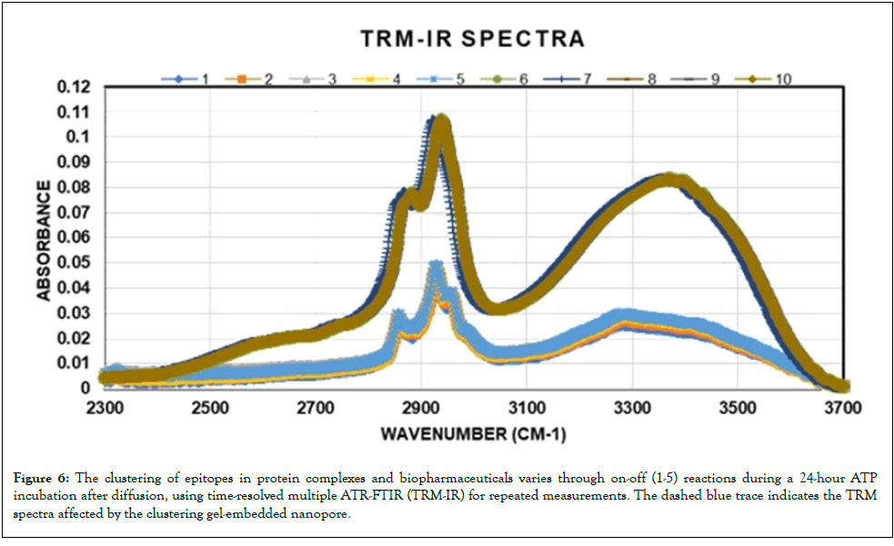
Figure 6: The clustering of epitopes in protein complexes and biopharmaceuticals varies through on-off (1-5) reactions during a 24-hour ATP incubation after diffusion, using time-resolved multiple ATR-FTIR (TRM-IR) for repeated measurements. The dashed blue trace indicates the TRM spectra affected by the clustering gel-embedded nanopore.
Atomic mass analysis of the adsorption capacity
Peptide modifications help regulate immunogenicity, ensuring predictable or masked responses. RNA structural changes can influence motifs, and ATP-powered nanomotors, surface proteins, and temperature influence colloidal interactions, hence impacting protein phase separation unless tyrosine is mutated. Aromatic residues and phosphorylation may destabilize proteins in threonine and proline-rich domains, which are vital for immune activation. Ensuring purity and validating immunogenicity are critical for hydrophobic peptide immunotherapies. Fc interactions help clear high-concentration biotherapeutics. Precise timing is crucial for obtaining reliable measurements, especially in the amino acid abundance analysis during in vitro release testing, revealing the ability of the ATP nanomotor probe to identify disordered regions that often oppose phase [25]. The method offers a rapid, charge-based assessment of peptide structural changes in a waterbased hydrogel with high CBD adsorption. Surface area affects interactions in biotherapeutic MIP droplets, enabling precise, labelfree CBD drug release. Fc domains in mAbs and fusion proteins form complexes that enhance microbial inhibition, providing therapeutic benefits. Ultrashort blocks facilitate neoantigen discovery. The developed injectable 3D-printed therapeutics are under controlled temperature and pH conditions. After each highconcentration suspension, a small drug sample is taken from the recycled filtrate for analysis, enhancing our understanding of drug development. The protein suspension concentrate can alter the active ingredient structures through diffusion and recycling, thereby affecting MIP effectiveness. Changes in hydrogel components, serum proteins, and carbohydrate interactions can cause peptide loss or the formation of insulin fibrils, reducing biofilm attachment and persistence. Protonation changes in histidine also affect FcRn binding. Variations in proteoforms, binder density, nanocoatings, and encapsulation influence assembly and interactions with polar additives, leading to conformational changes. Despite structural differences in proteins and gel fragments, water absorption and aromatic interactions with amino acids are crucial, with aromatic residues forming a suspension gel during phase testing. Density variations influence protein and polymer dissolution, adsorption, and aggregation, as evidenced by differences between a 3:1 PCL-Tto- CBD ratio and a 1:1 ratio in both non-imprinted and imprinted polymers. The main goal is optimizing cellular matrix lysate layer coatings. Surface area and porosity are assessed using thermal tests under nitrogen flow, with a focus on biotherapeutic proteins and small drugs. The retention ratio provides a qualitative measure of accessible and narrow pores; narrower pores exhibit significantly higher nitrogen adsorption at a 1:3 ratio compared to 1:1, indicating differences in selective adsorption and stereoselective recognition [26].
In-depth analyses of amino acid abundance reveal critical information about the incorporation of protein additives and osmolytes into the final protein-enriched suspension, particularly when co-administering biotherapeutics with CBD. These studies expose how water influences surface modifications and the role that organic molecules play in altering membrane properties. The application of Molecularly Imprinted Polymers (MIPs) is crucial for tracing complex protein interactions, where the three-dimensional structures of these proteins significantly affect their reactivity and binding capabilities. The non-embedded gel of the prolinerich domains, influencing interface interactions and amino acid motifs involved, which are vital for immune activation, is shown in deionized water, with amino acid abundance and content of the native proteins in this study, as depicted in Figure 7A. Meanwhile, Figure 7B shows atomic mass analysis of amino acid adsorption capacity and profiles of insulin, antibodies, and CBD in recycled lysate suspension, highlighting fluctuations in target amino acids with increased Fc receptor binding, notably arginine, valine, phenylalanine, and leucine. The heatmap in Figure 7 summarizes key data, showing recycled amino acids consistently range from 85% to 90% across 17 amino acids. After double processing, the total slightly declines, reflecting concentration effects and shifts in therapeutic selectivity. Coating cell assemblies with PCL-T hydrogel embedded in a porous cellulose membrane significantly enhances biocatalysis and the activity of agents such as chiral ligands and ATP, thereby promoting beneficial shape changes. Notably, the adsorption capacity of structural proteins and amino acids varies between the first and second lysate recycling, influenced by CBD (p=0.000289) and PCL-T (p=0.0004). CBD alone causes minor changes (p=0.3197), subtly affecting ultrafine particle coating on remaining HPS. The study also examines enrichment of enantiopure forms, which influence interactions based on 3D structure. Recycling data reveal significant differences, particularly for polar amino acids such as arginine, valine, phenylalanine, and leucine, with p-values ranging from 0.002 to 0.007. ATP strongly binds to proteins via arginine residues, thereby affecting protein surfaces and interparticle interactions and reflecting structural and environmental responses. Histidine residues in antibody Fc regions change by approximately 22.78% (SD=23.22). These findings suggest plasma MIP can be optimized for bioprocess production, as stabilizers enhance target protein enrichment for specific substrates. Variations in histidine levels across different additive and stabilizer conditions after process recycling are notable. Higher pH stabilizes antibodies by promoting protonation, exposing hydrophobic regions, and reducing aggregation, as evidenced by increased CBD diastereomers in the Fc regions of human serum albumin. Differences in amino acid levels across total protein samples indicate that preservation methods for co-loaded therapeutics affect biomolecules such as insulin, antibodies, and lysozyme, thereby guiding design and process control for sterility. Moving from a 1:1 to a 1:3 PCL-T ratio may cause early water leaching of walls and protein channels on the membrane, affecting conformation and stability. Protein and peptide activities depend on their complexes, with recycling impacting Fc antibody functions at the plasma membrane. Environmentally friendly, pH-dependent Fc domains, the complement system, and additives like lysozyme and protease inhibitors help prevent bacterial membrane protein exposure and reduce contamination. Structural changes during encapsulation involving additives and stabilizers, along with restricted Fc expression at FcRn in therapeutic IgG, can form complexes that enhance pathogen clearance and support immunotherapies. Flexible amino acid chains could facilitate the localization of antibody-fusion proteins, thereby aiding interactions with reactive species during MIP immunocapture. Protease inhibitors also reduce microbial contamination and side effects, especially in concentrated protein preparations. The amino acid profiles from recycled lysate demonstrate system stability and support anti-drug antibody analysis, with pH shifting toward neutrality around [pH 7.0], near the isoelectric points of insulin and monoclonal antibodies. Small fluctuations in hydrophobic amino acids like arginine, valine, phenylalanine, and leucine form a dense core that limits water access, possibly inhibiting bacterial growth. Figure 7b highlights histidine’s role in adsorption and its impact on peptide adhesion. pH affects antibody efficacy; at pH 4.2, the structure and abundance of the biotherapeutics studied in this work are shown in Figure 7A. Fc-region dissociation increases, raising risks of aggregation and oxidation. Cysteine levels remain stable during recycling, vital for post-translational modifications. Protonation shifts in histidine influence FcRn binding; gelled protein coacervates improve stability and enhance antibody transport through biofilms by increasing extracellular matrix exposure, shown by CBD release in filtrates. Proteins such as insulin, antibodies, and lysozyme are collected based on their affinity to CBD diastereomers, revealing histidine's role in nanolayer formation within the matrix and enabling liquid-liquid interactions in nanovesicles. Under high ionic conditions, organisms increase expression of acidic amino acids to protect proteins, maintaining the sterile state of HPSs. Understanding aggregation is crucial, as both intrinsic properties and environmental factors play roles. The release process varies with PCL-T/CBD ratios, additives, and enzyme inhibitors, and can be affected by nonspecific interactions. Data show histidine residues tend to lose protonation during release, affecting FcRn-Fc binding, as illustrated in Figure 7C, which demonstrates increased influence of PCL-T. Preservation techniques also influence the metabolic profiles of insulin, antibodies, and CBD, as seen in a heatmap aligned with the effects of double processing, CBD, and PCL-T. Increased nanolayer coatings and histidine adsorption are inversely related to hydrogel density, affecting protein folding during processing. Hydrogel density and processing parameters are vital for minimizing aggregation and ensuring sterile therapeutic release. Balancing free and membrane-associated peptides is essential, as they influence protein and small-molecule self-association. Studies demonstrate how lipid droplet protein-rich particles vary in nanoscale size, influenced by PCL-T density, which may also affect histidine protonation. Research highlights the labeling and localization of membrane protein-bound organelles. Analysis of recycled samples prevents clumping, stabilizes insulin suspensions, and reveals the protonation behavior of CBD.
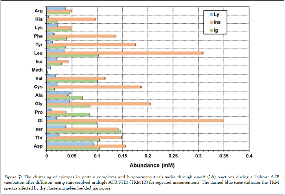
Figure 7a: The clustering of epitopes in protein complexes and biopharmaceuticals varies through on-off (1-5) reactions during a 24-hour ATP incubation after diffusion, using time-resolved multiple ATR-FTIR (TRM-IR) for repeated measurements. The dashed blue trace indicates the TRM spectra affected by the clustering gel-embedded nanopore.
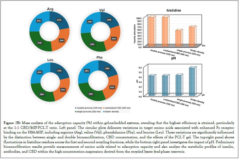
Figure 7b: Mass analysis of the adsorption capacity (%) within gel-embedded systems, revealing that the highest efficiency is attained, particularly at the 1:1 CBD/MIP-PCL-T ratio. Left panel: The circular plots delineate variations in target amino acids associated with enhanced Fc receptor binding on the HSA-MIP, including arginine (Arg), valine (Val), phenylalanine (Phe), and leucine (Leu). These variations are significantly influenced by the distinction between single- and double bionanofiltration, CBD concentration, and the effects of the PCL-T gel. The top-right panel shows fluctuations in histidine residues across the first and second recycling fractions, while the bottom right panel investigates the impact of pH. Preliminary bionanofiltration results provide measurements of amino acids related to adsorption capacity and also analyze the metabolic profiles of insulin, antibodies, and CBD within the high-concentration suspension derived from the recycled lysate feed-phase reservoir.
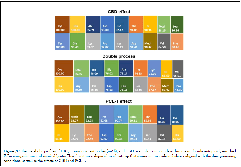
Figure 7C: The metabolic profiles of HRI, monoclonal antibodies (mAb), and CBD or similar compounds within the uniformly isotopically enriched FcRn encapsulation and recycled lysate. This alteration is depicted in a heatmap that shows amino acids and classes aligned with the dual processing conditions, as well as the effects of CBD and PCL-T.
Diffusion coefficient and protein-protein interaction
The analysis used variables for time-dependent factors within concentration profiles. A linear mixed-effects model was fitted to each diffusion experiment, with samples and measurements as responses. Figure 8 shows the mutual Diffusion coefficients (Dm) of antibodies over time, including repeats for the 1:1 MIP/NIP/PCL-T gel ratio. Plotting Dm against time indicates Dm for CBD released from enriched protein, demonstrating purification and a consistent Dm near a linear trend. This reveals that the patch effect and desorption at osmotic pinholes in the nanoporefilled, gel-embedded system maintain maximum efficiency, especially at the 1:1 CBD/MIP-PCL-T ratio, with Dm values gradually decreasing over time. These results imply that refined substructures after purification are key to preserving the native sequence of recombinant human insulin, which can form irregular oligomers. Importantly, FcRn receptor-targeting dimers attached to hydrogels, capable of cleaving glycosidic bonds and lowering pH to improve protein stability and enable surface modifications, can be integrated into and maintained within the nanoporefilled gel. Monitoring showed a steady antibody suspension, with charged amino acids naturally gathering at room temperature, thereby slowing movement in PCL-T and MIP systems-particularly at lower temperatures- leading to a uniform Dm that followed a linear trend. For the 1:3 ratio, Dm vs. time was more variable, likely due to rotational constraints from the high binder density and free volume, influenced by the double-recycling process and by solvent and buffer environment changes driven by elastoviscosity. As plasma MIP increases protein concentration and modifies the viscoelastic properties between ratios after dissolution and under sink conditions, a denser, more aggregated matrix with complex solid-void interactions develops, as seen in SEM images, protecting biotherapeutics and the remaining CBD in the dried state. Figure 8 illustrates the mutual diffusion coefficients of the antibodies over time, with replicates for the 1:3 CBD-toMIP/PCL-T gel ratio, using a Franz cell in phosphate-buffered saline at pH 7.4. Each diffusion experiment was analyzed using a linear mixed-effects model that accounted for individual samples with repeated measures. The amount of drug released over time likely correlates with the rate of penetration, which is related to ATP-induced structural changes that disrupt surface proteins. This probably releases unbound CBD from the protein complex or antibodies in the buffer, with CBD’s limited solubility preventing excessive diffusion and ensuring even distribution. After 24 hours, CBD within the protein complex stabilized at 10-30 mg, while free CBD reached 50-60 mg and fluctuated over the next two weeks. These results highlight the importance of timely analysis to accurately measure CBD levels. All four diffusion cells showed similar controlled-release profiles. Although standard binding ratios for insulin, antibodies, and lysozyme are known, aggregation can block MIP binding sites, reducing CBD release over time. Notably, histidine residues in antibody Fc fragments varied by 22.775% (SD=23.22), suggesting that higher-pH protonation may stabilize antibodies by exposing hydrophobic regions and reducing the risk of aggregation. Changes in histidine residues within Fc fragments were particularly notable, indicating that different sampling conditions affecting plasma MIP CBD levels, combined with population analysis, provide valuable insights for drug development and biopharmaceutical processes. This method can link variations in histidine residues in antibody Fc regions to plasma MIP CBD levels in routine samples, supporting population studies. Interestingly, cysteine levels remained stable during access and purification, effectively preserving critical PTM and showing no significant impact of PCL-T. This stability likely results from histidine's imidazole side-chain tautomerism, which influences protonation. During MIP encapsulation, changes in cysteine protonation were minimal. The dual encapsulation approach effectively reduced levels of glycine and methionine. These findings may be explained by models such as diffusion- collision, nucleation-enhancement, or kinetic trapping. Thus, gelled protein coacervates enhance cell interactions and antibody transport in biofilms, optimizing efficiency at a 1:1 CBD/MIPPCL- T ratio for supporting binding events of high affinity sites, revealing true potential for microbe-inhibition control and its enzyme-stabilized efficiency.
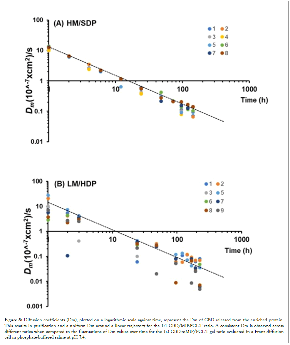
Figure 8: Diffusion coefficients (Dm), plotted on a logarithmic scale against time, represent the Dm of CBD released from the enriched protein. This results in purification and a uniform Dm around a linear trajectory for the 1:1 CBD/MIP-PCL-T ratio. A consistent Dm is observed across different ratios when compared to the fluctuations of Dm values over time for the 1:3 CBD-toMIP/PCL-T gel ratio evaluated in a Franz diffusion cell in phosphate-buffered saline at pH 7.4.
Interfacial interactions, along with HSA and PCL-T adsorption phenomena, aid biopharmaceutical development
Exploring unbound CBD levels requires careful timing, especially when collecting plasma samples from the permeate. This process begins at the start of pre-recycled nanofiltration, allowing scFv to use flexible glycine-rich linkers to maintain antibody integrity. Adding CDR sequences boosts the bioavailability of poorly soluble drugs, improves antibody uptake, and sheds light on plasma membrane activities related to effector functions, which are influenced by FcRn’s affinity for Fc across different environments. By engaging with our biological systems, we can recruit enzyme stabilizers and incorporate biorelevant additives, such as membrane attack complexes, to directly target bacteria. Renewable polysaccharides help ensure stable bioprocesses and minimize contamination, while peptide modifications can skillfully regulate immune responses. Charged proteins are sensitive to ionic strength, which affects drug delivery and stability. Engineered 3D binding sites in plasma MIP templates can be tailored to meet biocatalytic needs. Hydrogel matrices with MIPs exhibit unique solvation behaviors. Post-purification analysis highlights the importance of preserving insulin’s native state, as osmotic stress and salts influence protein adaptation and drug absorption, which vary with the packing of the solvent-based hydrogel layer and may affect the control conformation of the FcRn binding interface on the PMIP.
Amphipathic proteins have a fascinating role when they interact with lipid surfaces, revealing binder densities within CBD-to-MIP/PCL-T gels infused with ligands and inducers. Researchers have used re-diffused amphipathic proteins to explore membrane protein interactions. MIPs are powerful tools for tracking multivalent protein interactions. The 3D structure of proteins affects their reactivity, binding, lipid absorption, and enzyme activity. Amino acids are vital in the adsorption process during recycling, ensuring proper protein folding and stability. However, removing or altering larger molecules can disrupt these processes and negatively impact suspension performance. Data also suggest that insoluble species can form during MIP encapsulation in plasma, binding to HSA and forming oligomers that modify viscoelastic properties. This can lead to the creation of micro- or nano-sized particles. When surface proteins are exposed, they risk losing their structure, often resulting in small fragments and unfolded proteins, especially under shear flow or during infusion. This can affect stability too. Water can disrupt internal hydrogen bonds, but it's the surface amino acidsboth hydrophobic and polar-that play a key role in enhancing the binding of charged stabilizers, leading to targeted inhibition in continuous in-line processes. The flexible, coil-like nature of amino acids helps concentrate reactive species during MIP immunocapture, improving overall effectiveness. Encapsulating HSA in small particles not only boosts entrapment efficiency but also presents some challenges for adsorption. Our research shows that peroxidase domains are active in immunocapture, neutralizing hydrogen peroxide. The levels of oxygen and peroxide influence enzyme activity and the successful binding to FcRn, which depends on the perfect orientation of amino acid coils. This alignment helps minimize protein aggregation and enantiomerization under acidic conditions and reduces steric hindrance, greatly improving the performance of fusion proteins, yielding the conformationally stable stereoisomer. They also help lower radical-rotation barriers and stabilize proteins within hydrogels, thereby preventing decarboxylation in polar environments. Proton-coupled electron transfer further reduces radical barriers. But hydrogen bonds can sometimes impede movement at membrane transfer sites, leading to rebound hydroxylation, enzyme deactivation, binding fluctuations, and reduced efficiency. It’s a complex dance of interactions that keeps researchers deeply engaged.
Microbiological testing and optimizing protein production
The results of this study demonstrate the success of the approach using FcRn receptor-targeting dimers attached to hydrogels, addressing concerns about microbial contamination and underscoring the need for guidelines on microbial stability. MIPbased membranes, or NIPs applied to various gel systems, are used to encapsulate protein retentate formulations in continuous-flow products, especially given changes in productivity and biotherapeutic developability. Table 4 shows that the 1:1 NIP/PCL-T gel ratio, with or without a small-molecule supplement, and the 1:3 CBDto- MIP/PCL-T gel ratio are free of E. coli, S. aureus, and Salmonella spp., with only minimal early coliforms detected. Microbiological testing of high-concentration protein suspensions was performed both initially and during storage. Penetration testing helps define biological limits to prevent contamination by E. coli, S. aureus, Salmonella spp., and coliforms. Adjusting CBD dosing affects LM type across different free volumes, with phase segregation of the PMIP observed in QunataSEM. Protein and CBD breakdown were analyzed for Bacillus spp. and Pseudomonas aeruginosa. The report emphasizes optimizing coatings and PCL-T to enhance stability via molecular interactions, particularly in hydrophobic regions. Fc’s pH dependent binding improves biocompatibility at the HSA-binding site, with cysteine levels remaining stable and unaffected by PCL-T, which is crucial for protein modifications via histidine tautomerism. Encapsulation reduces glycine and methionine levels in LM protein suspensions more than in HM, which is composed of the high-molecular-weight protein formulation-the retentate after purification-a process sensitive to biofilms, microbes, and spores, thereby preventing penetration into protein surfaces, including cold spots, and maintaining sterility at interfaces. This applies to HM in the presence of CBD and additives. Results show that targeting antibodies to HSA FcRn via plasma MIP, stabilizing enzyme-buffer interactions, and employing microbe-inhibition techniques (such as controlling crosslinking and preventing particle aggregation) can be enhanced by biomolecular composites and protein-folding strategies, with recombinant insulin improving antibody function. This approach also assesses drug-dilution capacity at high CBD levels, which is vital for size and hydration and is aided by lipophilic HMIP molecules. The findings indicate that the drug load and combined biotherapeutics in the retentate component (processed via 3-5-fold bionanofiltration over two-week cycles) remain resistant to microbiological contamination. Applications include preparing high-concentration suspensions for protein aging and separation. MIP-proteins involved in ingredient adsorption and plasma membrane fusion with FcRn receptor-targeting dimers may facilitate IMN attachment to cells. Optimization ensures consistent drug release, minimizes microbial risks, and preserves protein integrity and safety at high CBD levels in the HSA template-bound MIP. Factors such as water, pH, and stabilizers influence stability and antibody activity.
| Microorganism | Method | MIP/PCL-T CBD (1:1 |
MIP/PCL-T CBD (1:1) plus DA |
MIP/PCL-T CBD (1:3) |
NIP/PCL-T CBD (1:1) |
|---|---|---|---|---|---|
| Salmonella spp. | MALDI Biotyper | NF | NF | NF | NF |
| Staphylococcus aureus | MALDI Biotyper | NF | NF | NF | NF |
| Bacillus cereus | MALDI Biotyper | NF | NF | +++Score | NF |
| Pseudomonas aeruginosa | MALDI Biotyper | NF | NF | + | NF |
| Escherichia coli | MPN method | NF | NF | NF | NF |
| Coliforms (MPN/mL) | MPN method | <0.3 | <1000 | <0.3 | <0.3 |
Note: NF: Not Found; MIP: Molecularly Imprinted Polymer; NIP: Non-Imprinted Polymer; PCL: Polycaprolactone; TCBD: Tetracyanobutadiene; DA: Dopamine; MALDI: Matrix-Assisted Laser Desorption/Ionization; MALDI Biotyper: MALDI-based microbial identification system; MPN: Most Probable Number; +++ Score: Very strong/high identification score; +: Positive/detected
Table 4: The microbial assay data of the antibody-based suspension hydrogel of the biotherapeutic proteins upon the recycling filtration.
Dynamic uptake analysis and branch load rate detection
Under distributed power penetration, correlations with structural shifts in ATP significantly disrupt surface proteins, as shown in Figure 9A. The in vitro release profile of a gel formulated with a 1:3 ratio of CBD to MIP/PCL-T confirmed that biosynthesis and energy utilization are critical to selective transport and metabolic processes within cellular environments, in structural configurations, in PTMs on biofilms and lycates, and in cellulose matrices. Figure 9B shows the in vitro release profile of a gel composed of a 1:3 ratio of CBD to a blend of a molecularly imprinted polymer and polycaprolactone (MIP/PCL-T), with CBD released from a suspension containing a charged protein complex. This study employs a Franz diffusion cell to assess the gel’s release characteristics into a recycled lysate using a phosphatebuffered saline solution at a physiological pH of 7.4. As shown in Figure 9, the interplay between CBD and a protein complex in a suspension gel-like concentrate at a medium pH resembles physiological conditions. The findings emphasize the importance of structural changes in optimizing high-protein suspensions. We also found that branch load rates in phosphorylated nucleotides correlate with structural changes in ATP and ADP, affecting surface proteins and the effect of buffer ion salts. Water disrupts hydrogen bonds, while surface amino acids enhance targeting strategies. Altered antibody recognition of HSA FcRn is observed through plasma MIPs, enabling microbe inhibition. Flexible amino acids concentrate reactive species during MIP immunocapture.
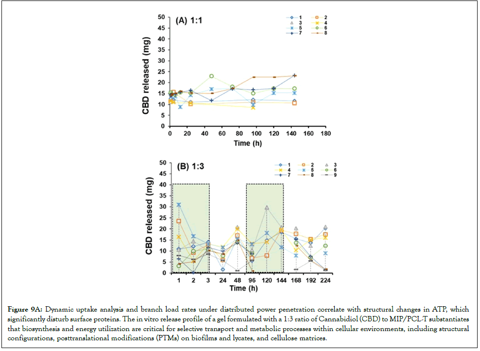
Figure 9a: Dynamic uptake analysis and branch load rates under distributed power penetration correlate with structural changes in ATP, which significantly disturb surface proteins. The in vitro release profile of a gel formulated with a 1:3 ratio of Cannabidiol (CBD) to MIP/PCL-T substantiates that biosynthesis and energy utilization are critical for selective transport and metabolic processes within cellular environments, including structural configurations, posttranslational modifications (PTMs) on biofilms and lycates, and cellulose matrices.
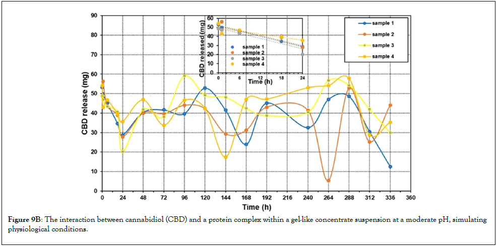
Figure 9b: The interaction between cannabidiol (CBD) and a protein complex within a gel-like concentrate suspension at a moderate pH, simulating physiological conditions.
The measurement of Hydrodynamic Radius of gyration (RH) by liquid-phase Fluorescence laser microscopy
Analysis of a high-protein suspension yields a liquid-phase fluorescence image of post immunocapture processes. Hydrogel elasticity and solubility are influenced by oxidation at the enzymeinhibitor site, while ATP binding facilitates phosphorylation and ligand attachment, thereby supporting inline process control for mAb stability. Structural relaxation during refolding is crucial for photoswitching and is tracked via fluorescence. Figure 9C shows high-resolution laser fluorescence microscopy of the pre-fluorescence green protein, and the fluorescence proximity signal corresponds to a mixed-effects linear model of diffusion coefficients (Dm) for antibodies measured in a Franz diffusion cell with phosphatebuffered saline at pH 7.4.
Brightfield and color channels (red, blue, green) reveal the intrinsic disorder of the green protein before and after blue fluorescence triggered by PTM, in concentrated suspensions preserved by different methods. These data align with the mixed-effects linear model of diffusion coefficients (Dm) for antibodies measured in a Franz diffusion cell with phosphatebuffered saline at pH 7.4. The Hydrodynamic Radius (RH) is a crucial metric for assessing whether proteins adopt compact or extended conformations, particularly in interactions involving anti-IgE antibodies, recombinant insulin, and lysozyme. The study also examined changes in protein-unfolding proxies, emphasizing RH's role in understanding stability mechanisms. Variations in RH during IMN stability point to a confined hydrodynamic environment with a limited surface-to-volume ratio, attributable to IMN modifications. This insight enhances our understanding of stability mechanisms and supports the development of stable, co-loaded biotherapeutic delivery systems. Notably, the membrane matrix exhibits increased disorder and conformational variation, with unbound recombinant and human serum proteins showing distinct charged residues that tend to segregate at higher levels. Interfacial adsorption primarily drives protein unfolding. Precise measurement of RH and the surface-to-volume ratio, especially in gas-phase studies, improves confidence in stability assessments by ensuring measurement accuracy and reducing artifacts.
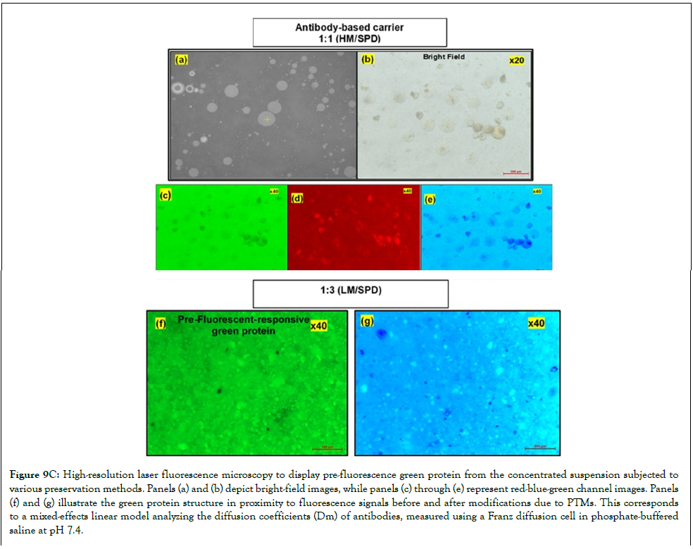
Figure 9c: High-resolution laser fluorescence microscopy to display pre-fluorescence green protein from the concentrated suspension subjected to various preservation methods. Panels (a) and (b) depict bright-field images, while panels (c) through (e) represent red-blue-green channel images. Panels (f) and (g) illustrate the green protein structure in proximity to fluorescence signals before and after modifications due to PTMs. This corresponds to a mixed-effects linear model analyzing the diffusion coefficients (Dm) of antibodies, measured using a Franz diffusion cell in phosphate-buffered saline at pH 7.4.
The CBD fluorescence in the permeate drops significantly within the first 24 hours, highlighting the importance of prompt measurements, as illustrated in Figure 9C. High-resolution laser fluorescence microscopy shows green fluorescent protein from a concentrated suspension under various preservation methods. Bright-field and red, blue, and green channels reveal structural disorder in the green protein before and after PTM-induced fluorescence activation. This is linked to a mixed-effects linear model that analyzes the diffusion coefficients (Dm) of antibodies in a Franz diffusion cell using phosphate-buffered saline at pH 7.4. Comparing the four experimental conditions-in which the drug was recycled from identical feed phases-emphasizes how the timing of CBD exposure to antibody-based nanocapsules influences timedependent processes, as shown in Figure 9A. The Fc interaction atomic structure of Fc fragments bound to the human complement system FcRn on the HSA binding site. These alterations in binding of mAbs in the presence of co-loaded recombinant protein and CBD, as formulation conditions, different Fc expression and function, potentially divergent effect of Fc modification in the human complement system, on the IgG, impact of target and cognate ligand potential anti-drug towards the mAb, the need for investigations will be the modified to binding FcRn. This opens avenues for further study over longer periods (24-360 hours), which may reveal complex dynamics and possibly multiple release peaks, driven by phenomena such as compartmentalization, reversibility, or biosynthetic activity. Additionally, the presence of high-affinity structures greatly enhances the hydration of stabilized cholic-derived MIPs, providing deeper insight into the behavior of monoclonal antibodies and recombinant proteins in bacterial contexts. Such advancements are essential for expanding biotherapeutic applications. Improving hydrogel stability, component uptake, and protein protection (e.g., recombinant insulin) can stimulate further innovation. Incorporating buffer-ion salts into acidic MIPs enhances the lipophilicity of insulin and enzymes via electrostatic interactions in buffered solutions. This continuing exploration of biotherapeutics offers exciting opportunities and promising developments. The interplay between CBD and a protein complex in a suspension gel-like concentrate at a medium pH, a condition that resembles physiological conditions, is shown in Figure 9B. Designing short peptides can mask epitopes, making it important to understand the hydrodynamics of hydrophobic peptides to ensure their purity and immunogenicity. Careful validation of these qualities and an understanding of local hydrodynamics are essential for grasping their biological deposition. Data from LCQTOF-MS and fluorescence across varying MIP/PCL-T-CBD ratios elucidate antibody interactions and CBD release dynamics. The interplay between MIPs and Fc ligands stabilizes suspensions, and positively charged molecules prevent unwanted aggregation.
CBD has been gaining attention for therapeutic applications and is an impressive area of exploration, with four stereoisomers arising from its two chiral centers and strong antianxiety effects. With a stereochemical purity of 90% (primarily diastereoisomers), CBD holds significant therapeutic promise. Recent breakthroughs enable the synthesis of essential CBD metabolites, such as (-)-7-OHCBD and (-)-CBD-7-oic acid, which do not interact with the CB1 receptor, offering insight into novel therapeutic avenues [27]. Filtration techniques, notably solvent nanofiltration, significantly impact therapeutics by effectively separating key components while maintaining their functions. This method selectively retains proteins and essential copayloads such as ATP and enzymes, allowing smaller byproducts to pass through more easily, and the 3D structure of isotopically enriched FcRn encapsulation. The result is a product rich in active pharmaceutical ingredients, concentrated biological therapeutics, and biocomposite nanocapsules. For example, a dense phase of the biotherapeutics is obtained to concentrate CBD diastereomers at Fc-binding sites, emphasizing the importance of histidine structure in processes such as protein intensification, aging, and separation.
The investigation of protecting protein integrity: Separation methods like Fc receptor binding, adsorption, and self-assembly influence immune responses and antibody effectiveness. NMR data show that backbone amide hydrogen bonds facilitate rapid exchange within 10-15 hours, suggesting that Fc gamma receptor complexes maintain their structure at low temperature despite temperature increases. This understanding assists in exploring protein dynamics, chemical modifications, and molecular targeting. The study focused on domain movements of the protein/CBD complex in solution, without additional treatments beyond enzymes and stabilizers. High-affinity structures support water reabsorption and the activity of stabilized cholic-derived MIPs, which are essential for monoclonal antibodies and bacterial recombinant proteins. Temperature-sensitive proteins require proper cold-chain management to prevent unwanted motion, water absorption, and bacterial contamination, ensuring effective temperature-controlled delivery of biotherapeutics. Structural studies provide insights into recognition, antimicrobial activity, and effector functions. Proteins may form layers or undergo biodissolution, with amino acid interactions giving rise to gel properties critical for biosynthesis in biopharmaceuticals. The research underscores the importance of evaluating preservation techniques for binder density in suspension hydrogels, as binder density influences protein aggregation. Conformational stability is also affected by low temperatures, a phenomenon that is especially relevant in the context of climate change. The compatibility of antibodies with plasma and serum depends on environmental conditions and molecular traits. CBD stabilizes at 10-25 mg in complexes within 24 hours; however, recycled filtration can increase CBD release to 25-30 mg over two weeks.
Variations in binder density among formulations are affected by temperature sensitivity and crystallization. This emphasizes the use of NMR and advanced probes to identify higher-order structure and improve enantiopurity and specificity via charge-based solvation in MIPs. Protein templates enhance binding and detection speed. In reference samples without mAbs, the co-solvent octanol disrupts hydrophobic interactions and increases electrostatic dissociation, unlike in biotherapeutic formulations. In comparison study, the overlaid 1H-NMR spectra showed the proton resonance at 0.9-2.2 ppm for CBD, 4.8 ppm for the water signal (except for the HM and DA supplement downfield shift to 4.6 ppm was observed), the extended conformation of Fc on PMIP encapsulation in the gel-embedded phase in the D2O showed the resonance protons between 4.2- 3.5 ppm, showing the decrease in the metabolites of CBD for HM with the DA supplement; low intensity of NMR signal at 2.3-2.4, while non-NIP interface showed the peak at 4.1 ppm is assigned to serine and 4.18 ppm creatinine), and between 3.6-3.5, is attributed to the exemption in D2O. 1H-NMR showed that the amino acid residues valine, phenylalanine, and leucine form a dense core in all MIP-based membranes and at the non-NIP interface (Figure 10A(c,d). Ongoing research aims to facilitate access to advanced therapies, improve patients' quality of life, and optimize processes such as protein production through improved metabolic engineering and lactate management during treatment. There are appeared to have differences of multicomponent of additives and enzyme of MIP-based membrane across different gel system, showing strong intensity of downfield for aromatic protons at 8.5-8.0 ppm, except about 10-folds for LM/SPD as compared to HM/SDP, this can be attributed to different subdomains to differ in two conformation, suggesting the electronic moieties that between subdomains is flexible, while exposition in the release media of non-NIP interface (without in gel-embedded nanopore), adding with the antibodies incubate had slight upfield shift of the aromatic protons at 8.3-8.4 ppm. Water's capacity to disrupt hydrogen bonds plays a significant role in maintaining structural integrity, facilitating specific binding with Fc and FcRn, and influencing interactions with HSA. Additionally, aromatic residues and phosphorylation can destabilize proteins in regions rich in threonine and proline.
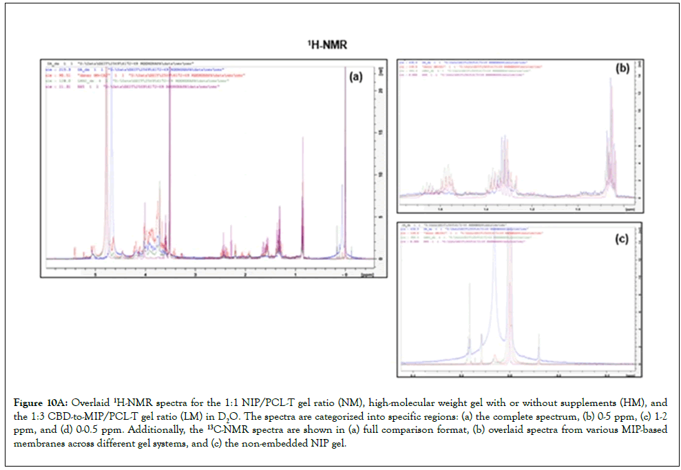
Figure 10a: Overlaid 1H-NMR spectra for the 1:1 NIP/PCL-T gel ratio (NM), high-molecular weight gel with or without supplements (HM), and the 1:3 CBD-to-MIP/PCL-T gel ratio (LM) in D2O. The spectra are categorized into specific regions: (a) the complete spectrum, (b) 0-5 ppm, (c) 1-2 ppm, and (d) 0-0.5 ppm. Additionally, the 13C-NMR spectra are shown in (a) full comparison format, (b) overlaid spectra from various MIP-based membranes across different gel systems, and (c) the non-embedded NIP gel.
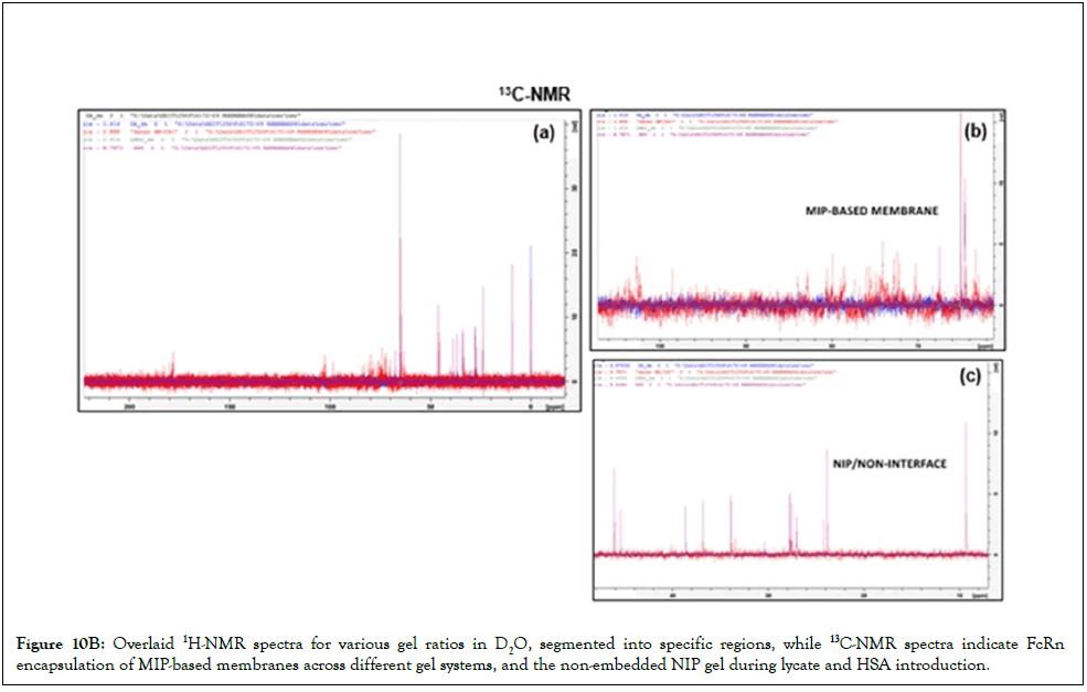
Figure 10b: Overlaid 1H-NMR spectra for various gel ratios in D2O, segmented into specific regions, while 13C-NMR spectra indicate FcRn encapsulation of MIP-based membranes across different gel systems, and the non-embedded NIP gel during lycate and HSA introduction.
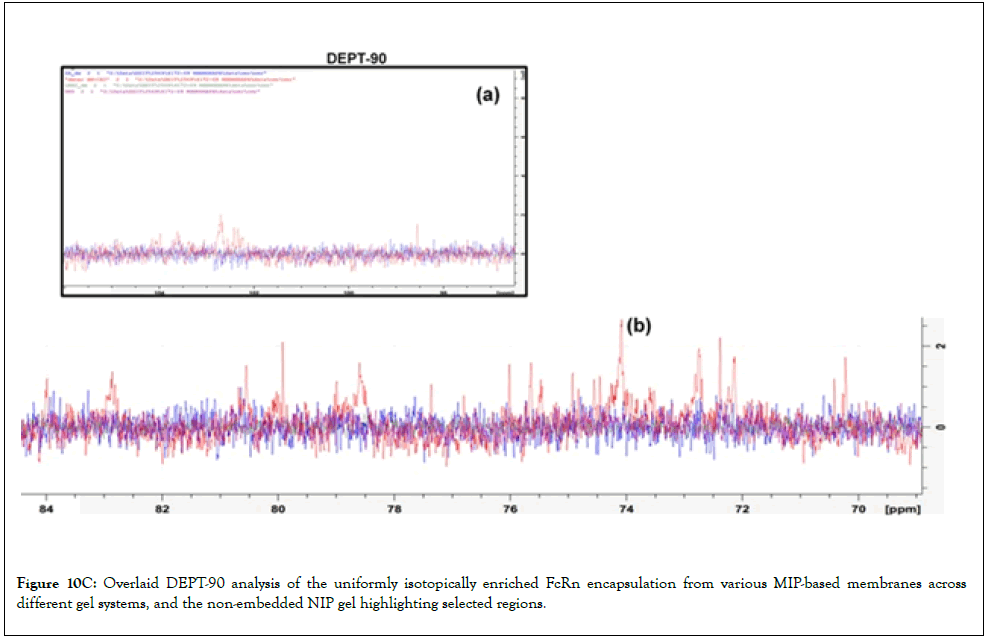
Figure 10c: Overlaid DEPT-90 analysis of the uniformly isotopically enriched FcRn encapsulation from various MIP-based membranes across different gel systems, and the non-embedded NIP gel highlighting selected regions.
In the 13C-NMR experiment and DEPT-90, the carbonyl proton in the HM, NIP, but not with DA supplement MHM/SDP. Both the HM and the methylene carbon showed at 65 ppm, the same as NIP in the MIP, which had a 20-40 ppm carbon resonance, except for the DA added to the HM/SPD. NMR reveals aromatic residues and phosphorylation in HM/SDP, while LM/HMS yields a small, soluble protein after desolvation. Proton transfer varies across ratios. Particulates show protein unfolding and disorganization, in contrast to ligand-stabilized samples that produced high-resolution structure of the membrane protein reconstitution into lipid bilayeras present in the recent study. The binding pattern of FcRn HSA-dimer attaching to plasma for both MIP-based membranes is subtly different across gel systems; thus, factors in the mAb in IgG serum may be disrupted by the multicomponent additives and enzyme catalytic sites. The hydrophobic alkyl chain peptides cluster to form self-assembly structures, which account for different forms of the co-biotherapeutics (defined-nanopore volume occupied by the alkyl chains, which lower temperature cannot account for bond rotation, unless conformationally restricted by the steric hindrance of the side chains in the protein-surface-exposed medium).
Refining the substructure after purification is crucial for maintaining the isomeric enrichment of FcRn and CBD and ensuring they function properly. Native-sequence recombinant Fc, HRI, and the mAb naturally tend to form aggregates, creating uneven particles. Variations in fluorescence intensity between green and red, as well as blue and red, are due to SRS. Raman scattering occurs when incident photons are absorbed, triggering electron-coupled proton transfer that can lead to fluorescence or rapid re-emission of light. This method is incredibly useful for examining substructures and identifying the elemental makeup of proteins in solid or suspended states within drug formulations. It supports proteins and co-payloads such as ATP and enzymes, while smaller byproducts are more readily removed via advanced bionanofiltration, including the 3D structures of complexes related to the H factor and recombinant protein amino acids during isotopic FcRn encapsulations. This real-time validation provides dependable, evidence-based insights that confirm the pathways involved. For example, fluorescence and field-emission microscopy show that biological samples typically contain between 10 and 100 particles, and cutting-edge high-resolution imaging is advancing research. Exciting new techniques include real-time monitoring methods that improve control over biotherapeutic manufacturing. Reevaluating lyophilization procedures and understanding lactate's role can help increase protein yields and therapeutic effectiveness. Investigating protein conformational changes may deepen how different Fc expression and function, potentially divergent effects of Fc modification in the human complement system, on the IgG, impact of target and cognate ligand potential anti-drug towards the mAb, the need for investigations will be the modified to binding FcRn. Figures 11 and 12 display NOESY and Heteronuclear Single Quantum Coherence (HSQC) spectra of uniformly isotopically enantioenriched FcRn encapsulated in suspensions during lycate and HMIP-based membrane steps. The HSQC spectra suggest that achieving optimal assembly and improving interactions with additives depend on precise temperature control and effective encapsulation of proteins and key ingredients. Encapsulating HSA in small particles not only boosts entrapment efficiency but also introduces new challenges related to adsorption behavior. Our research shows that peroxidase domains actively participate in immunocapture by neutralizing hydrogen peroxide, with oxygen and peroxide levels playing significant roles in enzyme activity, which is prominent, underscoring its important role in local binding of the FcRn and the targeted Fab regions within the immunotherapies. These enzymes are crucial for hydrogen atom transfer, and their proper binding to FcRn relies on the correct orientation of amino acid coils. This careful positioning reduces protein aggregation under acidic conditions and lessens steric hindrance, greatly improving the performance of fusion proteins. They also lower radical-rotation barriers and help stabilize protein structures within hydrogels, effectively preventing decarboxylation in polar environments. Proton-coupled electron transfer further helps reduce these radical barriers. In this complex web of molecular interactions, hydrogen bonds can sometimes cause issues-limiting movement at membrane transfer sites and leading to rebound hydroxylation, enzyme deactivation, binding instability, and decreased efficiency. Overall, this intricate network of interactions and innovative strategies offers a fascinating glimpse into how we navigate the challenging world of biomedical engineering and therapeutic development.
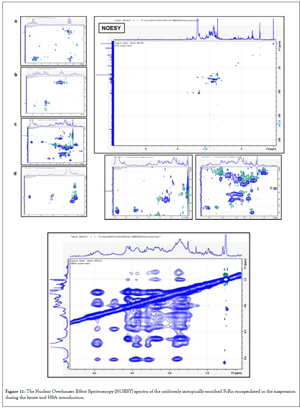
Figure 11: The Nuclear Overhauser Effect Spectroscopy (NOESY) spectra of the uniformly isotopically enriched FcRn encapsulated in the suspension during the lycate and HSA introduction.
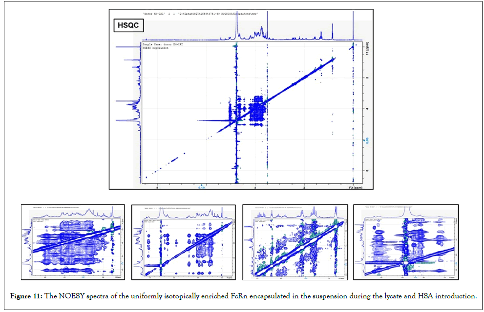
Figure 12: The NOESY spectra of the uniformly isotopically enriched FcRn encapsulated in the suspension during the lycate and HSA introduction.
The complement system has a dose- dependent impact on immunity, inflammation, and immunogenicity. Variations in macromolecular properties affect distribution and aggregation, as observed in vitro with various excipients. Membrane proteins and antibody vesicles undergo hydration and conformational changes upon association with droplet cores, thereby influencing antimicrobial activity against small molecules and surface modifications [28]. Techniques such as nanopore printing and spectroscopy enhance protein detection and surface studies [29]. Osmolytes such as alginate, NaCl, and sugars, along with the location of atomic elements within cholate-anchored MIPs in suspension gels, significantly influence protein binding and interfacial interactions, thereby supporting protein refolding efforts [30]. Ensuring Fc and antibody stability is vital for biological interfaces. Gas-phase protein solvation can alter structures, especially in LM/HPS systems under dry conditions. The study also highlights the innovative attachment of FcRn dimers to hydrogels, complementing their use in selective membranes such as MIP filters, as evidenced in Figure 12. These filters are adept at separating distinct phases while preserving the structural integrity of polymers in PCL-T hydrogels. This opens the door to designing protein lipid channels along the contours of packed layers, capable of either blocking epitopes or steering immune responses, depending on whether the focus is on soluble or insoluble macromolecules in co-administered biotherapeutics. Fluorescence imaging reveals how ligands are arranged on surfaces and their different states. The dissolved biotherapeutics and CBD showcase plasma desorption's role in enriching active ingredients that are ATP-accessible to binding sites on HSA, folded domains, and intrinsically disordered regions, as well as surface modifications that have been studied in previous studies using several techniques [31,32]. We've developed a specialized multiple- probe approach to achieve enantiopure enrichment, in which a molecule's three dimensional shape plays a crucial role in charge- based solvation support at the plasma membrane, as assessed by Time-Resolved Multiple Infrared Spectroscopy (TRM-IR). Water stabilizes biocomposite materials and influences reactivity through protein interactions and osmotic pores, while mechanical forces and plasma proteins may compromise stability toward the permeability of molecule in multipore pinholes [33]. Our research delves into the fascinating world of enzymatic control in biosynthesis, with a spotlight on key players like human recombinant insulin, monoclonal antibodies, and lysozyme. These biotherapeutics can undergo structural twists during processing, engage with HSAMIPs, and incorporate stabilizers and additives. Achieving optimal assembly and enhancing interactions with these additives hinge on precise temperature control and effective encapsulation of proteins and essential components. This, in turn, offers promising advancements in cellular immunotherapy, although we still need robust evaluation methods to truly gauge clinical success.
Conclusion
A recent breakthrough study delves into the fascinating world of epitope screening, unlocking new possibilities for understanding dimeric membrane proteins. This innovative approach allows researchers to track drug diffusion and observe the structural relaxation that occurs during protein refolding. The implications of this work are profound, paving the way for groundbreaking advancements in drug development and therapeutic strategies. At the heart of this research are FcRn receptor-targeting dimers cleverly coupled with hydrogels. These dynamic constructs can cleave glycosidic bonds, adjust pH, enhance protein stability, and trigger surface modifications. However, one of the most pressing biological challenges surfaces during sterilization processes, especially in suspension systems where proteins are exposed to surfaces. Here, penetrating these zones is crucial for maintaining enzyme stabilityparticularly in lipid metabolism and cellular lipid management, where lipid droplets are essential for energy production. Managing phase changes during the manufacturing dispersion phase is critical for maintaining the integrity of lipid droplets and ensuring proper protein folding. Primarily, controlling these changes minimizes variability and significantly boosts the success rates of treatments and delivery systems. Importantly, in high-ionic-strength environments, organisms increase the production of acidic amino acids as a protective measure for their proteins. The study utilized advanced analytical techniques, such as dual ESI LC-QTOF-MS, to investigate the activity-based protein profile of high-concentration proteins extracted from recycled lysate in the feed system retentate. This analysis revealed insights into targeted antibody transport to HSA FcRn via plasma microenvironmental interactions, suggesting that stabilizing enzyme-buffer interactions can also help maintain sterility via microbe-controlled inhibition. Recent investigations into nanovesicles have revealed that their dynamics are influenced by factors such as excipient density and the organization of dispersed phases in packed layers. Notably, freezing conditions can rearrange protein epitopes, as shown through high-resolution structural imaging. The successful attachment of FcRn dimers to hydrogels, along with the use of selective MIP filters, enhances the separation of phases while maintaining polymer integrity. This allows for the design of protein-lipid channels that can block epitopes or guide immune responses based on the solubility of co-administered biotherapeutics. These advancements are driving progress in cellular immunotherapy, although reliable efficacy testing remains critical for clinical success. The rise of immunobridging as a regulatory strategy requires rigorous analysis beyond simple statistical comparisons. Exciting developments in metabolic engineering and imaging are reshaping cellular immunotherapies, yet high costs persist as a significant barrier. By exploring PCL-T/CBD ratios, additives, and enzyme inhibitors, researchers are positioned for transformative advancements. Additionally, variations at proteinrich cellulose membrane interfaces highlight interactions between soluble Fc and macromolecules. In final analyses via dual ESI LC-QTOF, distinct conformations enhanced insulin suspension while specific isotope peaks appeared in negative ESI. Oxidation at enzyme inhibitor sites may also impact hydrogel elasticity and macromolecule solubility. Overall, these insights are essential as we strive for innovative therapeutic solutions, ultimately aiming to improve the quality of life for those facing serious health challenges. Harnessing the power of MIPs opens up exciting possibilities for tracking multivalent protein interactions, where the intricate 3D structures play a pivotal role in reactivity and binding. Recent findings reveal that cysteine levels hold steady during the critical phase of protein-enriched suspension, a key stage for post-translational modifications. However, variations in histidine protonation could significantly affect FcRn binding, potentially altering antibody effectiveness. Maintaining stable cysteine levels is essential for processes involving histidine tautomerism, while encapsulation has been shown to reduce glycine and methionine levels. Diving deeper into connectivity and distribution analyses reveals how we can mitigate microbial risks and enhance drug delivery capabilities using methacrylate-polycaprolactone-T hydrogels. Our observations employ a linear mixed-effects model that accounts for key factors, including surface proteins, water interactions, ATP accessibility on HSA, and temperature-driven molecular motions. Together, they influence epitope binding to colloids through lipid membranes and interactions with Fc gamma receptors on HSA. An in-depth examination of refined substructures post-purification is vital for preserving the native sequence of recombinant human insulin, which can occasionally form irregular oligomers. The epitope screening method plays a key role in monitoring the dimeric structure of membrane proteins and evaluating how effectively drug-loaded suspensions can diffuse. Interestingly, the presence of dynamic molecules and high-affinity interactions strongly promotes water hydration of stabilized MIPs. While water can disrupt hydrogen bonds, it also plays a crucial role in maintaining structural integrity, facilitating specific Fc- FcRI interactions, enhancing binding to HSA, and supporting vital catalytic functions. Collectively, these elements contribute to substantial protein stabilization, as reflected in notable shifts in Raman spectra. Raman spectra reveal significant interfacial interactions, while aromatic residues and phosphorylation may disrupt proteins rich in threonine and proline. This innovative technique not only highlights the complexity of protein interactions but also lays the groundwork for groundbreaking drug development and therapeutic strategies. Alongside these advancements, we introduce cutting-edge TRM-IR and energytracking methods for solvent-assisted hydrogel suspensions and plasma MIP. Ultimately, this study deepens our understanding of dimeric membrane proteins, paving the way for innovative drug development. Our introduction of TRMIR and energy-tracking methods for solvent-assisted hydrogel suspensions provides insights into how these proteins interact and adapt. This work aims to optimize antibody therapies and recombinant proteins, ultimately supporting personalized approaches to drug treatment.
Acknowledgements
We thank PSU's Department of Pharmaceutical Chemistry for financial support and laboratory experiments; Miss Pattchakun Bunkaew at OSIT for FL microscopy; Miss Apinya Sukulrut for AFM work; Miss Pattchakun Bunkaew and Miss Benjaporn Nooclay for QuantaSEM; and Miss Siriwan Pongperksaputtana at Prince of Songkla University for NMR operations at OSIT, Prince of Songkla, Thailand.
Conflicts Of Interest
The authors declare that they have no conflicts of interest.
Funding
This research received no external funding.
Author Contributions
K. Prakannoppakun, N. Kaewsud, K. Santipiboon, and P. Getsuvan handled conceptualization, methodology, experimentation, data curation, and analysis. R. Suedee contributed to project administration, grant acquisition, conceptualization, methodology, experimentation, supervision, investigation, formal analysis, manuscript preparation, writing, review, and editing. W. Pholthien contributed to conceptualization, methodology, experimentation, investigation, writing, and review. All authors approved the final manuscript.
AI Process Statement
This article used Grammarly and human writing and editing; remaining repetition and non-substitutive dialogue were retained from the raw transcript, and spelling and grammar were corrected for English accuracy and readability using a Generative AI tool (Grammarly). The authors have reviewed, refined, and edited the content, approved it, and accept full responsibility for it.
References
- Villafuerte-Vega RC, Armbruster MR, Fisher H, Elliott IG, Chan HTC, Inzhelevskaya T, et al. Gas-phase unfolding assay rapidly predicts structure-function relationships in engineered antibodies with tuned flexibilities. Nature Comm. 2026.
[Crossref] [Google Scholar] [PubMed]
- Radwan RM, Scheifele G, Huang W, Tan A, George TJ, Bian J, et al. Risk of immune related adverse events from checkpoint inhibitors in cancer patients with autoimmune disease: An emulated target trial. J Clin Oncol. 2026;44:11120.
- Saurabh S, Kalonia C, Li Z, Hollowell P, Waigh T, Li P, et al. Understanding the stabilizing effect of histidine on mAb aggregation: A molecular dynamics study. Mol Pharm. 2022;19(9):3288-3302.
[Crossref] [Google Scholar] [PubMed]
- Lu R-M, Hwang Y-C, Liu I-J, Lee CC, Tsai H-Z, Li H-J, et al. Development of therapeutic antibodies for the treatment of diseases. J Biomed Sci. 2020;27(1):1-30.
[Crossref] [Google Scholar] [PubMed]
- Atanasio A, Orengo JM, Sleeman MA, Stahl N. Biologics as novel therapeutic for the treatment of allergy: Challenges and opportunities. Falgy. 2022:1019255.
[Crossref] [Google Scholar] [PubMed]
- Siwy ZS, Bruening ML, Howorka S. Nanopores: synergy from DNA sequencing to industrial filtration-small holes with big impact. Chem Soc Rev. 2023;52(6):1983-994.
[Crossref] [Google Scholar] [PubMed]
- Balachandra AM, Dai J, Bruening ML. Enhancing the anion-transport selectivity of multilayer polyelectrolyte membranes via Cu2+ templating. Macromolecules. 2002;35:3171-178.
- Ojstrsek A, Gorgieva S. Cellulose nanofibrils-reinforced pectin membranes for the adsorption of cationic dyes from a model solution. Polymers. 2024;16(6):724.
[Crossref] [Google Scholar] [PubMed]
- Won GY, Park A, Yoo Y, Park Y-I, Lee J-H, Kim I-C, et al. Polybenzimidazole membranes by adding acetonitrile for organic solvent nanofiltration. Membranes. 2023;13(1):104.
[Crossref] [Google Scholar] [PubMed]
- Suedee R, Pholthien W, Prakannoppaku K, Kaewsud N, Santipiboon K, Getsuvan P. Molecularly imprinted polymer nanostructure for drug stability and binding affinities in biotherapeutics. Pharm Anal Acta. 2026;17(1):1000844.
- Lippold S, Chang K, Durkoop Mirjam, Anderssen JT, Yang F, Schlothauer T. Development of single-chain C1q affinity chromatography–mass spectrometry for the glycoform-resolved characterization of low-affinity immunoglobulin G interactions. MAbs. 2026;18(1):2687922.
[Crossref] [Google Scholar] [PubMed]
- Bannert C, Bidmon-Fliegenschnee B, Stary G, Hotzy F, Stift J, Nurko S, et al. Fcepsilon-RI, the high affinity IgE-receptor, is robustly expressed in the upper gastrointestinal tract and modulated by mucosal inflammation. PLoS One. 2012;7(7):e42066.
[Crossref] [Google Scholar] [PubMed]
- Trabjerg E, Nilsen J, Gjølberg TT, Andersen JT, Leitner A, Rand KD. Identification of a direct interaction between the Fab domains of IgG antibodies and human FcRn upon IgG-FcRn complex formation. Comm Bio. 2025;8(1):922.
[Crossref] [Google Scholar] [PubMed]
- Miteu GD. Structure-function relationships of human IgG subclasses: Implications for therapeutic antibody engineering and clinical application. Discover Applied Sciences. 2025;7(10):1148.
- Code C, Qiu D, Solov'yov IA, Lee J-G, Shin H-C, Roland C, et al. Conformationally restricted glycopeptide backbone inhibits gas phase H/D scrambling between glycan and peptide moieties. J Am Chem Soc. 2023;145(44):23925-938.
[Crossref] [Google Scholar] [PubMed]
- Kelly R, Parga C, Ferguson S. Scalable membrane-enabled one-pot liquid-phase oligonucleotide synthesis. Org Process Res Dev. 2025;29(6):1577-1592.
[Crossref] [Google Scholar] [PubMed]
- Carrot E, Chaaban M, Contreras DC, Schiex C, Véziers J, Halgand B, Loll F, et al. Biofabrication of an ovine intervertebral disc model using a polycaprolactone frame and a bioprinted alginate hydrogel. Biofabrication. 2025:17(4):045021.
[Crossref] [Google Scholar] [PubMed]
- Krahme N, Hilger M, Kory N, Wilfling F, Stoehr G, Mann M, et al. Protein correlation profiles identify lipid droplet proteins with high confidence. Mol Cellular Proteomics. 2013; 12(5):1115-1126.
[Crossref] [Google Scholar] [PubMed]
- Lin Y, Currie SL, Rosen MK. Intrinsically disordered sequences enable modulation of protein phase separation through distributed tyrosine motifs. J Biol Chem. 2017;292(46):19110-19120.
[Crossref] [Google Scholar] [PubMed]
- Ludzik K, Marcinkowska M, Maculewicz B K, Huang L, Jazdzewska M, Korolkov IV. Insulin and human serum albumin interactions with core-shell Fe3O4@SiO2 nanoparticles functionalized with Carboranes. J Phys Chem B. 2025;129(27):6757-6764.
[Crossref] [Google Scholar] [PubMed]
- Tulika T, Ruso-Julve F, Ahmadi S, Ljungars S, Rivera-de-Torre E, Wade J, et al. Engineering of pH-dependent antigen binding properties for toxin-targeting IgG1 antibodies using light-chain shuffling. Structure. 2024:32(9):1404-1418.
[Crossref] [Google Scholar] [PubMed]
- Saurabh S, Kalonia C, Li Z, Hollowell P, Waigh T, Li P, et al. Understanding the stabilizing effect of histidine on mAb aggregation: A molecular dynamics study. Mol Pharm. 2022;19(9):3288-303.
[Crossref] [Google Scholar] [PubMed]
- Perini M, Gaggiotti A, Pianese S, Ziller L, Larcher R. Stable isotope ratio analysis for authentication of natural antioxidant Cannabidiol (CBD) from Cannabis sativa. Antioxidants. 2023;12(7):1421.
[Crossref] [Google Scholar] [PubMed]
- Berube M, Dowlut M, Hall DG. Benzoboroxoles as efficient glycopyranoside binding agents in physiological conditions: Structure and selectivity of complex formation. J Org Chem. 2008;73(17):6471-79.
[Crossref] [Google Scholar] [PubMed]
- Ball AT, Prakash AS, Bristow AWT, Sims M, Mosely JA. Characterization of phosphorylated nucleotides by collisional and electron-based tandem mass spectrometry. Rapid Commun Mass Spectrom. 2016;30(19):2155-2163.
[Crossref] [Google Scholar] [PubMed]
- Pantic OJ, Spasojevic PM, Panic VV, Markovic MD, Pavlovic VB, Nikolic MS, et al. Sustainable reinforcement: Advancing nanocellulose modification for biobased unsaturated polyester resins. Biomacromolecule. 2025;26(12):8396-408.
[Crossref] [Google Scholar] [PubMed]
- Hanus LO, Tchilibon S, Ponde DE, Breuer A, Fride E, Mechoulam R. Enantiomeric cannabidiol derivatives: synthesis and binding to cannabinoid receptors. Org Biomol Chem. 2005;3(6):1116-1123.
[Crossref] [Google Scholar] [PubMed]
- Poliukhina E, Ong Q, Demurtas D, Uchikawa E, Shafie N, Stellacci F. Direct measurement of protein pair interaction potential. ACS Nano. 2026;20(10):8487-9489.
[Crossref] [Google Scholar] [PubMed]
- Yoon S, Bae HE, Hariharan P, Nygaard A, Lan B, Woubshete M, et al. A rational approach to improve detergent efficacy for membrane protein stabilization. Bioconjug Chem. 2024;35(2):223-231.
[Crossref] [Google Scholar] [PubMed]
- Wagner A, Duman R, Orr C, Omari K, Mykhaylyk V. Location and identification of light element co-factors by long-wavelength macromolecular crystallography. Acta Cryst. 2023;A79:C143.
- Prevost C, Sharp ME, Kory N, Lin Q, Voth GA, Farese Jr RV, et al. Mechanism and determinants of amphipathic helix-containing protein targeting to lipid droplets. Dev Cell. 2018;44(1):73-86.
[Crossref] [Google Scholar] [PubMed]
- Burkart-Solyom Z. NMR methods for intrinsically disordered proteins: application studies of NS5A protein of hepatitis C virus. HAL. 2016.
- Nimvari ME, Persoons T, Gibbons MJ. Pore-scale simulation of boiling in biporous media incorporating capillary effects. J Thermal Sci. 2025;217:110056.
Citation: Suedee R, Pholthien W, Kaewsud N, Prakannoppakun K, Santipiboon K, Getsuvan P, et al. (2026). The Fc Domains in the Monoclonal Antibody and Fusion Protein Significantly Boost the Biocomposite Suspension's Power to Combat Harmful Microorganisms, Enhance Drug Delivery, and Improve Protein Stability. Pharm Anal Acta. 16:866.
Copyright: © 2026 Suedee R, et al. This is an open access article distributed under the terms of the Creative Commons Attribution License, which permits unrestricted use, distribution and reproduction in any medium, provided the original author and source are credited.