Indexed In
- RefSeek
- Hamdard University
- EBSCO A-Z
- Geneva Foundation for Medical Education and Research
- Euro Pub
- Google Scholar
Useful Links
Share This Page
Journal Flyer
Open Access Journals
- Agri and Aquaculture
- Biochemistry
- Bioinformatics & Systems Biology
- Business & Management
- Chemistry
- Clinical Sciences
- Engineering
- Food & Nutrition
- General Science
- Genetics & Molecular Biology
- Immunology & Microbiology
- Medical Sciences
- Neuroscience & Psychology
- Nursing & Health Care
- Pharmaceutical Sciences
Mini Review - (2024) Volume 9, Issue 1
Retinopathy and their Mechanism
Ifrah Saroosh1, Aisha Shakir1, Tehseen Faiz2, Haim Sajid1, Muhammad Aetesam Nasir1, Maham Ifikhar1, Muhammad Adil Umar1 and Fatima Mazhar3*2Department of Ophthalmology, HITEC-Institute of Medical Sciences, Taxila, Pakistan
3Department of Microbiology, Muhammad Nawaz Sharif University of Agriculture, Multan, Pakistan
Received: 04-Mar-2024, Manuscript No. JEDD-24-25090; Editor assigned: 06-Mar-2024, Pre QC No. JEDD-24-25090 (PQ); Reviewed: 20-Mar-2024, QC No. JEDD-24-25090; Revised: 27-Mar-2024, Manuscript No. JEDD-24-25090 (R); Published: 03-Apr-2024, DOI: 10.35248/2684-1622.24.9.228
Abstract
Retinopathy is the emerging disease in Pakistan over the age of 30 in diabetic patients. In diabetic patients this disease causes blindness, visual loss, and hazy eyesight. Several suggested metabolic mechanisms that connect micro vascular problems to hyperglycemia. Activation of Protein Kinase C (PKC), oxidative stress, polyol buildup, and the production of Advanced Glycation End Products (AGEs) are a few of these. The anti- Vascular Endothelial Growth Factor (VEGF) drugs, laser, eye surgery is the treatment of Retinopathy. The patient HBA1C lab test below than 6.5% will be preferred for surgery. The Diabetic patient should control their blood glucose level to reduce the complexity of the disease.
Keywords
Retinopathy; Diabetic macular edema; Neo vascular glaucoma; Retinal detachment
Introduction
Retinopathy is a disease that can cause blindness and vision loss in aged peoples. It mostly occur in diabetic patients so it called Diabetic Retinopathy [1]. Worldwide, the number of people with diabetes is more than 135 million. Over 18.2 million Americans, or 6.3% of the population, have diabetes, and 800,000 new instances of type 2 diabetes are diagnosed in the country each year [2]. It affects the blood vessels in the light sensitive layer of tissues of the retina. In early stages of this disease have no symptoms. But some people face problems in reading and seeing faraway objects. In the later stage of disease, the bleeding from blood vessels of retina in the form of gel like fluid and patient will see dark, floating spots like cobwebs. If the treatment on time it will benefit the patient but after time without time it become worse [3].
Review of Literature
Diabetic Macular Edema (DME)
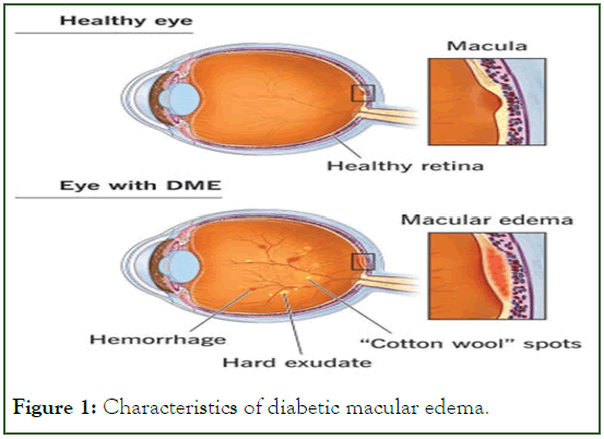
Approximately 1 in 15 diabetics will eventually develop DME. DME occurs when fluid leaks from retinal blood vessels into the macula, a region of the retina required for crisp, center vision. This results in hazy eyesight (Figure 1) [4].

Figure 1: Characteristics of diabetic macular edema.
Neo vascular glaucoma
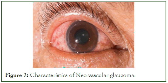
Unusual blood vessels that emerge from the retina as a result of diabetic retinopathy may obstruct the flow of fluid out of the eye. One kind of glaucoma, which is a collection of eye conditions that can result in blindness and visual loss, is brought on by this (Figure 2) [5].

Figure 2: Characteristics of Neo vascular glaucoma.
Glaucoma
A class of eye conditions known as glaucoma can result in blindness and visual loss by harming the optic nerve, a nerve located at the back of the eye. patient might not notice the symptoms at first since they might appear so slowly [6]. A thorough dilated eye exam is the only method to determine if patient have glaucoma. While there is no known cure for glaucoma, vision protection and damage may frequently be stopped with early intervention.
Retinal detachment
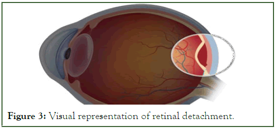
The back of patient’s eye may develop scars as a result of diabetic retinopathy. Tractional retinal detachment refers to the condition when your retina pulls away from the back of your eye due to scarring (Figure 3) [7].

Figure 3: Visual representation of retinal detachment.
Pathophysiology
There are several suggested metabolic mechanisms that connect micro vascular problems to hyperglycemia. Activation of protein kinase C (PKC), oxidative stress, polyol buildup, and the production of Advanced Glycation End products (AGEs) are a few of these. Through their influence on cellular metabolism, signaling, and growth factors, these mechanisms are hypothesized to modify the disease process [8].
Polyol accumulation of polyol occurs in experimental hyperglycemia, which in rats and dogs is associated with the development of basement thickening, pericyte loss, and micro aneurysm formation [9]. High concentrations of glucose increase flux through the polyol pathway with the enzymatic activity of aldosereductase, leading to an elevation of intracellular sorbitol concentrations. This rise in intracellular sorbitol accumulation has been hypothesized to cause osmotic damage to vascular cells [10].
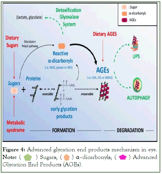
Aldose Reductase Inhibitors (ARIs) have been evaluated for the prevention of retinal and neural damage in diabetes. However, three clinical trials of ARIs in humans have not shown efficacy in preventing the incidence or progression of retinopathy. The efficacy of new, more potent ARIs remains to be evaluated in clinical trials [11]. AGEs another wellcharacterized pathway is damage resulting from accumulation of AGEs. High serumglucose can lead to non-enzymatic binding of glucose to protein side chains, resulting in the formation of compounds termed AGEs. After 26 weeks of induced hyperglycemia, the retinal capillaries of diabetic rats have marked accumulation of AGEs as well as a loss of pericytes [12]. Furthermore, diabetic rats treated with amino guanidine (AGE formation inhibitor) have reduced AGE accumulation and reduced histological changes, including micro aneurysm formation and pericyte loss. An ongoing clinical trial is investigating the effect of amino guanidine in humans. Preliminary results suggest that amino guanidine reduces the progression of retinopathy but is associated with anemia (Figure 4).

Figure 4: Advanced glycation end products mechanism in eye.
Note:  Advanced Glycation End Products (AGEs).
Polyol accumulation
In rats and dogs, experimental hyperglycemia leads to the accumulation of polyol and is linked to the development of micro aneurysm formation, pericyte loss, and basement thickening. Elevated intracellular sorbitol concentrations result from high glucose concentrations increasing flow via the polyol pathway with the enzymatic activity of aldose reductase [9]. It has been proposed that vascular cells may sustain osmotic injury as a result of this increase in intracellular sorbitol buildup. Aldosereductase Inhibitors (ARIs) have been studied in relation to preventing diabetic nerve and retinal damage. Nonetheless, according to three human clinical trials, ARIs are ineffective at stopping the onset or development of retinopathy. Clinical studies need to be conducted to determine the effectiveness of new, more powerful ARIs [13].
Advanced Glycation End Products (AGEs)
Damage brought on by a buildup of AGEs is another well-established route. Elevated blood glucose levels have the potential to cause non enzymatic glucose binding to side chains of proteins, which can lead to the creation of substances known as AGEs. Following 26 weeks of induced hyperglycemia, diabetic rats' retinal capillaries show a significant buildup of AGEs and a pericyte loss [14]. Additionally, diabetic rats given amino guanidine, an inhibitor of AGE synthesis, showed decreased buildup of AGE and decreased histological alterations, such as the development of micro aneurysms and the loss of pericytes [15]. The effects of amino guanidine in humans are being studied in a clinical study that is now underway. According to preliminary findings, amino guanidine slows the development of retinopathy but is linked to anaemia.
Conclusion
In Pakistan, over the age of 30 the retinopathy become leading disease in diabetic patient. Several mechanisms like PKC, AGES play role in this disease. In the patient the diseases causes color blindness, overall blindness, hazy eyesight, and visual loss. The doctor suggest the patient towards surgery and maintain hemoglobin A1C tests below than 6.5. The Laser and VEGF are alternative treatment against surgery in this disease. After surgery patient have care from sunlight exposure.
Author Contributions
All authors contribute equally
Acknowledgements
I am grateful to all of those with whom I have had the pleasure to work during this and other related projects.
Funding
No funding for this research.
Conflict of Interest
The authors have no competing interest.
References
- Shah CA. Diabeticretinopathy: A comprehensive review. Indian J Med Sci. 2008; 62(12):500-519.
[Crossref]
- Jonnalagadda SS. Effectiveness of medical nutrition therapy: Importance of documenting and monitoring nutrition outcomes. J Am Diet Assoc. 2004;104(12):1788-1792.
- Sayres R, Taly A, Rahimy E, Blumer K, Coz D, Hammel N, et al. Using a deep learning algorithm and integrated gradients explanation to assist grading for diabetic retinopathy. Ophthalmology. 2019;126(4):552-564.
[Crossref] [Google Scholar] [PubMed]
- Holekamp NM. Overview of diabetic macular edema. Am J Manag Care. 2016;22(10 suppl):s284-291.
[Google Scholar] [PubMed]
- Hu CX, Zangalli C, Hsieh M, Gupta L, Williams AL, Richman J, et al. What do patients with glaucoma see? Visual symptoms reported by patients with glaucoma. Am J Med Sci. 2014;348(5):403-409.
[Crossref] [Google Scholar] [PubMed]
- Bhowmik D, Kumar KS, Deb L, Paswan S, Dutta AS. Glaucoma-A eye disorder its causes, risk factor, prevention and medication. The Pharma Innovation. 2012;1(1, Part A):66.
- Peate I. Retinal detachment. British Journal of Healthcare Assistants. 2022. 16(5):236-241.
[Crossref]
- González P, Lozano P, Ros G, Solano F. Hyperglycemia and oxidative stress: An integral, updated and critical overview of their metabolic interconnections. Int J Mol Sci. 2023;24(11):9352.
[Crossref] [Google Scholar] [PubMed]
- G Obrosova I, F Kador P. Aldose reductase/polyol inhibitors for diabetic retinopathy. Curr Pharm Biotechnol. 2011;12(3):373-385.
[Crossref] [Google Scholar] [PubMed]
- Garg SS, Gupta J. Polyol pathway and redox balance in diabetes. Pharmacol Res. 2022;182:106326.
[Crossref] [Google Scholar] [PubMed]
- Mara L, Oates PJ. The polyol pathway and diabetic retinopathy. Diabetic Retinopathy. 2008:159-186.
- Smit AJ, Lutgers HL. The clinical relevance of Advanced Glycation Endproducts (AGE) and recent developments in pharmaceutics to reduce AGE accumulation. Curr Med Chem. 2004;11(20):2767-2784.
[Crossref] [Google Scholar] [PubMed]
- Zhu C. Aldose reductase inhibitors as potential therapeutic drugs of diabetic complications. Diabetes mellitus-insights and perspectives. 2013:17-46.
- Chilelli NC, Burlina S, Lapolla A. AGEs, rather than hyperglycemia, are responsible for microvascular complications in diabetes: A “glycoxidation-centric” point of view. Nutr Metab Cardiovasc Dis. 2013;23(10):913-939.
[Crossref] [Google Scholar] [PubMed]
- Luo D, Fan Y, Xu X. The effects of aminoguanidine on retinopathy in STZ-induced diabetic rats. Bioorg Med Chem Lett. 2012;22(13):4386-4390.
[Crossref] [Google Scholar] [PubMed]
Citation: Saroosh I, Shakir A, Faiz T, Sajid H, Nasir MA, Ifikhar M, et al (2024) Retinopathy and their Mechanism. J Eye Dis Disord. 9:228.
Copyright: © 2024 Saroosh I, et al. This is an open access article distributed under the terms of the Creative Commons Attribution License, which permits unrestricted use, distribution, and reproduction in any medium, provided the original author and source are credited.
Sources of funding : No funding for this research.
