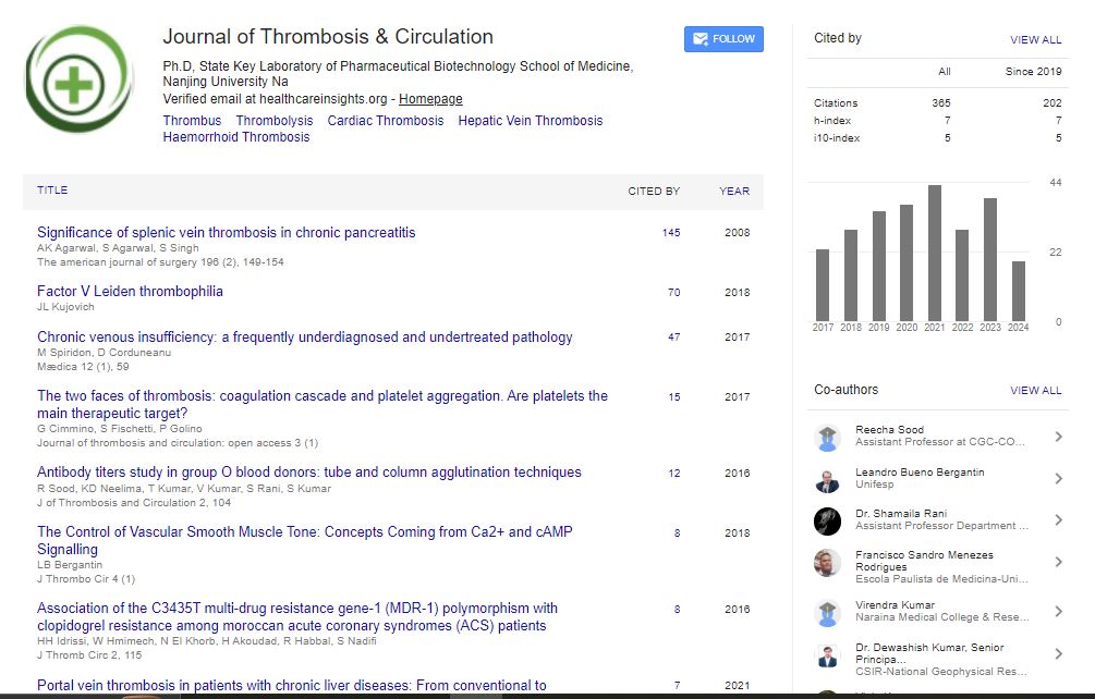

Indexed In
- RefSeek
- Hamdard University
- EBSCO A-Z
- Publons
- Google Scholar
Useful Links
Share This Page
Journal Flyer
Open Access Journals
- Agri and Aquaculture
- Biochemistry
- Bioinformatics & Systems Biology
- Business & Management
- Chemistry
- Clinical Sciences
- Engineering
- Food & Nutrition
- General Science
- Genetics & Molecular Biology
- Immunology & Microbiology
- Medical Sciences
- Neuroscience & Psychology
- Nursing & Health Care
- Pharmaceutical Sciences
Case Report - (2021) Volume 7, Issue 5
Retinal Vein Apoplexy: Pathogenesis, Risk Factors Hematological Disorders and Treatment
Shiva Varma*Received: 06-Sep-2021 Published: 27-Sep-2021, DOI: 10.35248/2572-9462.21.7.166
Abstract
Retinal vein impediment (RVO) is the most common retinal vascular sickness after diabetic retinopathy. Attributable to its multifactorial nature, notwithstanding, the board of this condition remains a test. Of the two fundamental kinds of RVO, branch retinal vein impediment (BRVO) is more pervasive than central retinal vein impediment (CRVO). Most patients create the disease at an older age, and the greater parts of them have associated foundational messes (for example hypertension, hyperlipidemia and/or diabetes mellitus). There is no proof to suggest routine testing for heritable thrombophilia’s in patients with RVO. The fundamental driver of the visual debilitation is macular edema, while neovascularization of the retina and optic plate are the most genuine inconveniences prompting glassy hemorrhage, retinal separation and neovascular glaucoma. Macular grid laser photocoagulation is an effective treatment for macularedema in patients with BRVO and a visual keenness of 20/40 or less. Other treatment choices for decreasing the edema are intra vitreal steroids, against VEGF medications and vitrectomy. The recently presented intra vitreal use of steroids and against VEGF medications might end up being a superior methodology for improving visual keenness. At long last, dissipate pan retinal laserphotocoagulationcan effectively treat neovascularization and its secondary complications.
Introduction
Of the two main kinds of RVO, focal retinal vein impediment (CRVO) and branch retinal vein impediment (BRVO), the last is more prevalent. BRVO, nonetheless, regularly happens at an arteriovenous crossing, where, as a rule, the course passes anterior to the vein. Another kind of RVO is hemi-vein occlusion, presenting as impediment of only one trunk of the focal retinal vein. This audit centers around the pathogenesis of RVO, with specific accentuation on the relationship with cardiovascular danger factors and thrombophilia messes. Key visual signs and treatment the board are likewise illustrated. The most as of late distributed examination summing up the pre-valence of RVO as announced in investigations from the United States, Europe, Asia and Australia has shown an age-and sex-normalized commonness of 5.20 per 1000 (95% confidence Correspondence stretch for any type of RVO [1].
Systemic risk factors
RVO presents chiefly in more established people, more than half of cases happening in people more seasoned than 65. There is solid proof from enormous investigations of expanded danger of this diseasing patient with fundamental arteriosclerotic vascular infection. An expanded danger of RVO has been displayed in patients with hypertension, diabetes mellitus, dyslipidemia, high weight list and smoking. Others hazard factors are various types of vasculitis, neoplasia and medication consumption.
Degenerative changes of vessel wall
The common confinement of BRVO is an arteriovenous (AV) crossing. The histological changes in the vessel divider at the A/V intersection have been explored in a few examinations, notwithstanding, showed that there was no histological proof for the normal venous pressure at the intersection. Seitz portrayed the clinical-histological sequelae in one eye with BRVO a couple of hours after beginning. There was no blood-clots destroying the venous lumen at the A/V intersection yet the endothelium was harmed. Mechanical pressure of the vein through the inflexible course in the A/V intersection might bring about fierce blood flow, delivering injury to venous endothelium that prompts impediment of the vein.
Hematological disorders
A few investigations have uncovered a relationship among RVO and hyperviscosity because of high hematocrit. Higher blood consistency expands erythrocyte accumulation under states of low blood flow. Improvement of the miniature flow through diminished blood consistency is the system of activity of hemo dilution, which is talked about underneath. The writing on the relationship between thrombophilic components and RVO is exceptionally conflicting. One significant explanation is the set number of researched patients. No single presently distributed examination has accomplished the necessary example size for distinguishing the relationship among RVO and thrombophilia with sufficient measurable force. To date no multi focus imminent examination has been finished. Henceforth, to explain the job of thrombophilic messes in patients with RVO, a meta-examination of all distributed investigations stays the solitary choice. Five meta-examinations researching the relationship among RVO and thrombophilia have been distributed to date. The thrombophilic messes that are suspected to be hazard factors for RVO.
Symptoms and clinical signs
Patients with CRVO generally present with unexpected, effortless, one-sided loss of vision. Notwithstanding, patients with BRVO can be asymptomatic, particularly in case BRVO is in nasal retinal quadrants, or they might give obscured vision, typically including the area of visual field relating to the space of the impeded venous branch. By and large, macular edema is the main source of decreased vision in patients with RVO. The fundus appearance in RVO can change as indicated by seriousness of the venous impediment. Table 3 presents clinical findings average for the intense and late phases of RVO [2].
Treatment
Various treatment modalities have been upheld for the administration of RVO. Until now, in any case, no causal treatment has been demonstrated in enormous randomized investigations to be compelling. Therefore, current medicines center around the sequelae of RVO, for example, macular edema and retinal neovascularization. Most examinations that have researched the mediations for RVO experience the ill effects of methodological limits (for example insufficient factual force, nonappearance of a benchmark group, and consideration of patients with various kinds of RVO that have an alternate regular history. A large portion of the accessible investigations have a review plan or depict little case series. Every one of these elements are answerable for extremely restricted proof and result in disarray in the clinical proposals. In future, further randomized clinical preliminaries with severe stratification of patient in clusio measures are required [3].
Laser photocoagulation
Laser treatment is a set up technique for use in patients with RVO. Two fundamental laser procedures can be utilized: macular network photocoagulation for treatment of macular edema, and container retinal (disperse) laser photocoagulation for treatment of retinal and additionally circle neovascularization. The consequences of imminent, randomized clinical preliminaries assessing the efficacy of these strategies over just about 20 years address to date the most significant level of proof corresponding to treatment of RVO. Surgical treatment Careful methods de-pressurize the retinal vein in the space of the impediment. In spiral optic neurotomy (RON) a cut of the lamina cribrosa at the edge of the optic nerve is made and the tension on the focal vein is eased. Sheathotomy, a method for BRVO, makes a cut in the adventitial sheath at the reason behind A/V intersection and isolates the vein from the overlying supply route. In both of these methods the chief advance is a standards plana vitrectomy. Despite the fact that the information is dubious, generally speaking, most of the reports have proposed that these medicines further develop VA. Notwithstanding, a few investigations contrasting vitrectomy alone and vitrectomy joined with sheathotomy and additionally RON have called attention to the shortfall of any distinction in clinical result. It is proposed that the critical job in this a medical procedure is played by straightforward vitrectomy and potential components of activity incorporate worked on retinal oxygenation, and evacuation of vitreomacular footing and cytokines answerable for the vascular penetrability. Overall, given the solitary moderate visual result and conceivable sight-undermining inconveniences, careful methods are not by and large suggested [4].
Surgical treatment
Careful methods de-pressurize the retinal vein in the space of the impediment. In spiral optic neurotomy (RON) a cut of the lamina cribrosa at the edge of the optic nerve is made and the tension on the focal vein is eased. Sheathotomy, a method for BRVO, makes a cut in the adventitial sheath at the reason behind A/V intersection and isolates the vein from the overlying supply route. In both of these methods the chief advance is a standards plana vitrectomy. Despite the fact that the information is dubious, generally speaking, most of the reports have proposed that these medicines further develop VA. Notwithstanding, a few investigations contrasting vitrectomy alone and vitrectomy joined with sheathotomy and additionally RON have called attention to the shortfall of any distinction in clinical result. It is proposed that the critical job in this a medical procedure is played by straightforward vitrectomy and potential components of activity incorporate worked on retinal oxygenation, and evacuation of vitreomacular footing and cytokines answerable for the vascular penetrability. Overall, given the solitary moderate visual result and conceivable sight-undermining inconveniences, careful methods are not by and large suggested [4].
Conclusion
RVO is a perplexing retinal vascular issue that regularly prompts extreme visual impedance. Enormous levels of patients with RVO have a background marked by cardiovascular illness, hypertension, hyperlipidemia as well as diabetes mellitus. Assessment of another patient with RVO should incorporate as a first line, assessment of circulatory strain, lipid profile and glucose levels in light of the fact that RVO might be a show of significant vascular grimness. Analysis of thrombophilia doesn't change the administration of RVO. Given the absence of remedial result, evaluating for heritable thrombophilia in patients with RVO isn't needed.
REFERENCES
- Brunette I, Boghen D. Central retinal vein occlusion complicating spontaneous carotid-cavernous fistula. Case report. Arch Ophthalmol. 1987;105:464-465
- Feman SS, Stein RS. Waldenstrom's macroglobulinemia, a hyperviscosity manifestation of venous stasis retinopathy. Int Ophthal. 1981;4(1):107-12.
- Yau JW, Lee P, Wong TY, Best J, Jenkins A. Retinal vein occlusion: an approach to diagnosis, systemic risk factors and management. Intern Med J. 2008;38(12):904-10.
- David R, Zangwill L, Badarna M, Yassur Y. Epidemiology of retinal vein occlusion and its association with glaucoma and increased intraocular pressure. Ophthalmol. 1988;197(2):69-74
Citation: Varma S (2021) Retinal Vein Apoplexy: Pathogenesis, Risk Factors Hematological Disorders and Treatment. J Thromb Circ. 7:166
Copyright: © 2020 Varma S. This is an open-access article distributed under the terms of the Creative Commons Attribution License, which permits unrestricted use, distribution, and reproduction in any medium, provided the original author and source are credited.