Indexed In
- The Global Impact Factor (GIF)
- CiteFactor
- Electronic Journals Library
- RefSeek
- Hamdard University
- EBSCO A-Z
- Virtual Library of Biology (vifabio)
- International committee of medical journals editors (ICMJE)
- Google Scholar
Useful Links
Share This Page
Journal Flyer

Open Access Journals
- Agri and Aquaculture
- Biochemistry
- Bioinformatics & Systems Biology
- Business & Management
- Chemistry
- Clinical Sciences
- Engineering
- Food & Nutrition
- General Science
- Genetics & Molecular Biology
- Immunology & Microbiology
- Medical Sciences
- Neuroscience & Psychology
- Nursing & Health Care
- Pharmaceutical Sciences
Image Article - (2021) Volume 20, Issue 6
Occlusal Contacts for Implant Dentistry
Philip L. Millstein1, Carlos Eduardo Sabrosa2, Wai Yung3 and Karen Geber42Department of Integrated Procedures, University of the State of Rio de Janeiro, Rio de Janeiro, Brazil
3Department of Engineering, Johnson and Wales University, Providence, Rhode Island, USA
4Department of Dentistry, Private practice, Rio de Janeiro, Brazil
, DOI: 10.35248/2247-2452.21.20.1140
Abstract
This study uses a computerized measuring system to observe predictable light and heavy occlusal contact. The method presented offers a means by which the occlusion of a restoration can be adjusted before delivery, on insertion, and monitored overtime.
Keywords
Dental, Implant dentistry, Dental management.
About the Study
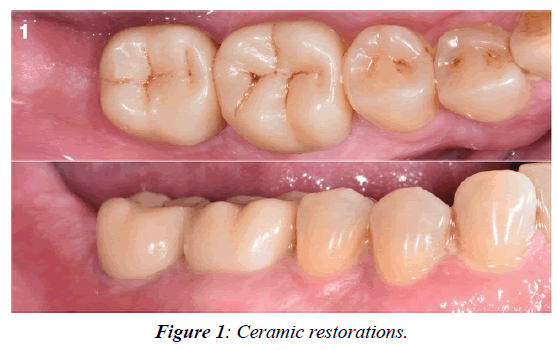
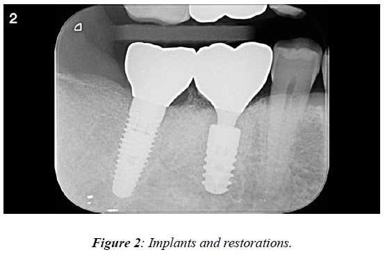
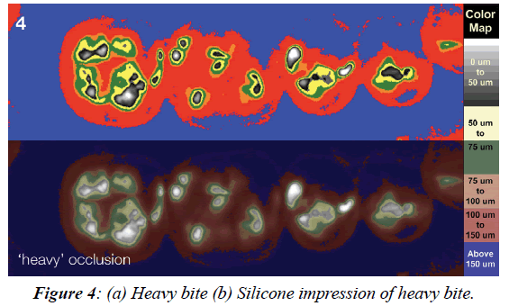
Implant restorations differ from conventional restorations because they lack a periodontal housing which among other purposes is used to absorb the everyday forces from eating and grinding [1]. Implant restorations function the same but the physiological demands differ [2]. To accommodate this lack of periodontal support it is advocated that both a light and heavy bite be made [3]. We use an occlusal scan to record the bites and to incorporate them into our restorations [4]. The procedure is as follows: For single and or multiple crowns we first establish the existing occlusion by closing commands and recording the occlusal position [5]. This is accomplished by positioning the wand of the scanning system close to the restoration to be constructed using cerec (dentsply, sirona). The completed restoration is inserted. At this time a silicone bite in a triple tray (premier, plymouth meeting, PA) is processed and shown on the computer screen. Using the same tray and a non-set impression material a second bite is recorded by biting hard [6]. This bite is compared to the previous bite on the screen (Figures 1-4). If the light bite is almost in contact and the heavy bite is in occlusal contact, the restoration is approved. [7] If not, the ceramic restoration is adjusted by adding or subtracting ceramic material.

Figure 1: Ceramic restorations.
Figure 2: Implants and restorations.
Figure 3: (a) Light bite, (b) Silicone impression of light bite.
Figure 4: (a) Heavy bite (b) Silicone impression of heavy bite.
The scanning system uses a DC light box, a camera and an analyzer (image J freeware) that processes the optical and computer images. The triple trays are market available as is the instant impression material. The total impression processing time is less than a minute with this procedue. Intensity of closure can be gauged by studying the optical patterns. A permanent record can then be stored along with routine radiographs.
Conclusion
A method is presented whereby the occlusion of a restoration can be adjusted before delivery, on insertion, and monitored overtime.
REFERENCES
- Fuselier B, Wei-en W, Loughner B. The problem with bruxism. Decisions in Dentistry. 2020;6:17-21.
- Korh A, Kowalsky M. Translating touch. Science News. 2021;199(8):1721.
- Millstein PL, Merrill EW, inventors. Method and apparatus for occlusal position measurement and recording. 2013;529(8):254.
- Millstein PL. A method to determine occlusal contact and noncontact areas: Preliminary report. J Prosthet Dent. 1984;52(1):106-110.
- Millstein PL, Sabrosa CE, Florencio S, Geber K. Consistency and Repeatability of Digitized Occlusal Records. Int J Dentistry. 2020;3(3):24-29.
- Krahenbuhl JT, Cho SH, Irelan J, Bansal NK. Accuracy and precision of occlusal contacts of stereolithographic casts mounted by digital interocclusal registrations. J Prosthet Dent. 2016;116(2):231-236
- Edher F, Hannam AG, Tobias DL, Wyatt CC. The accuracy of virtual interocclusal registration during intraoral scanning. J Prosthet Dent. 2018;120(6):904-912.


