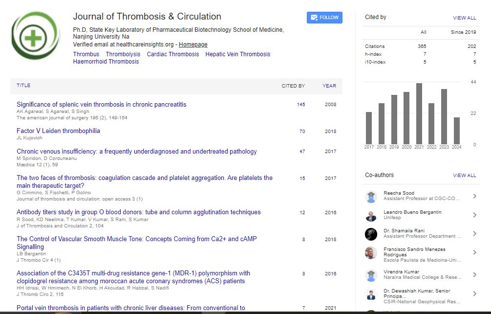

Indexed In
- RefSeek
- Hamdard University
- EBSCO A-Z
- Publons
- Google Scholar
Useful Links
Share This Page
Journal Flyer
Open Access Journals
- Agri and Aquaculture
- Biochemistry
- Bioinformatics & Systems Biology
- Business & Management
- Chemistry
- Clinical Sciences
- Engineering
- Food & Nutrition
- General Science
- Genetics & Molecular Biology
- Immunology & Microbiology
- Medical Sciences
- Neuroscience & Psychology
- Nursing & Health Care
- Pharmaceutical Sciences
Short Communication - (2024) Volume 10, Issue 1
Novel Biomarkers and Imaging Methods for Thromboembolism Early Identification
Xijing Xia*Received: 01-Jan-2024, Manuscript No. JTCOA-24-25345; Editor assigned: 03-Jan-2024, Pre QC No. JTCOA-24-25345 (PQ); Reviewed: 17-Jan-2024, QC No. JTCOA-24-25345; Revised: 24-Jan-2024, Manuscript No. JTCOA-24-25345 (R); Published: 31-Jan-2024, DOI: 10.35248/2572-9462.24.10.264
Description
Thromboembolism, surround conditions such as Deep Vein Thrombosis (DVT) and Pulmonary Embolism (PE), represents a major cause of morbidity and mortality worldwide [1,2]. Early identification and prompt treatment of thromboembolism are essential for preventing complications and improving patient outcomes. In recent years, advances in biomarker research and imaging techniques have provided novel approaches for the early identification and risk stratification of thromboembolism.
Biomarkers for thromboembolism detection
Biomarkers are measurable indicators of biological processes or disease states and play an important role in the early detection and risk stratification of thromboembolism [3]. Traditional biomarkers such as D-dimer, a degradation product of crosslinked fibrin, have been widely used in clinical practice for the diagnosis of thromboembolic events. However, their diagnostic accuracy may be limited by factors such as sensitivity, specificity, and cutoff values.
Recent research efforts have focused on identifying novel biomarkers that offer improved diagnostic performance and prognostic value in thromboembolism. For example, microRNA (miRNA) profiling has shown as a potential approach for identifying dysregulated miRNAs associated with thrombotic risk [4]. Specific miRNA signatures in plasma or circulating extracellular vesicles have been implicated in thromboembolism pathogenesis and could serve as biomarkers for early detection and risk stratification [5]. Furthermore, proteomic and metabolomics profiling techniques enable comprehensive analysis of protein and metabolite signatures associated with thromboembolic events. By identifying unique biomarker profiles in patients with thromboembolism, these omics approaches offer insights into underlying pathophysiological mechanisms and potential targets for intervention.
Imaging methods for thromboembolism visualization
Imaging techniques play an essential role in the diagnosis and localization of thromboembolic events, enabling accurate visualization of thrombi within blood vessels. Conventional imaging modalities such as ultrasound, Computed Tomography (CT), and Magnetic Resonance Imaging (MRI) are routinely used in clinical practice for the detection of DVT, PE, and other thromboembolic conditions [6].
Recent advances in imaging technology have led to the development of novel techniques with enhanced sensitivity, specificity, and spatial resolution for thromboembolism visualization. For example, Contrast-Enhanced Ultrasound (CEUS) employs micro bubble contrast agents to improve the detection of peripheral thrombi and assess thrombus burden in real time [7]. CEUS offers advantages such as portability, non-invasiveness, and lack of ionizing radiation, making it an attractive option for point-of-care imaging in thromboembolism diagnosis.
Moreover, molecular imaging approaches, such as Positron Emission Tomography (PET) and single-photon emission computed tomography (SPECT), enable non-invasive visualization of thrombus-specific molecular targets [8]. Targeted molecular imaging agents, including radiolabeled peptides, antibodies, and nanoparticles, selectively bind to thrombus associated biomarkers or cellular receptors, allowing precise localization and characterization of thrombi in vivo.
Integration of biomarkers and imaging techniques offers a comprehensive approach to thromboembolism detection and risk stratification, combining the strengths of both modalities for improved diagnostic accuracy and patient management. Biomarker-based risk scores, incorporating multiple biomarkers associated with thromboembolic events, can complement imaging findings and provide additional prognostic information.
For example, combining D-dimer levels with imaging results from CT Pulmonary Angiography (CTPA) in patients suspected of having PE can enhance diagnostic accuracy and risk stratification [9]. Similarly, multimodal molecular imaging approaches, such as combining PET imaging with MRI or CT, enable simultaneous visualization of thrombus anatomy and molecular targets, offering insights into thrombus composition and vulnerability [10].
Despite the potential of novel biomarkers and imaging methods for thromboembolism early identification, several challenges remain to be addressed. These include standardization of biomarker assays, validation of imaging techniques in diverse patient populations, and integration of multimodal imaging data for clinical decision-making. Furthermore, translation of research findings into clinical practice requires strong evidence from large-scale prospective studies and validation in real-world settings. Collaborative efforts among researchers, clinicians, and regulatory agencies are essential for advancing biomarker and imaging research in thromboembolism and implementing these technologies into routine clinical care.
Conclusion
In conclusion, novel biomarkers and imaging methods offer potential approaches for the early identification and risk stratification of thromboembolism, enhancing diagnostic accuracy and patient management. Continued research efforts and technological advancements in biomarker discovery and imaging technology are needed to address existing challenges and realize the full potential of these innovative approaches in thromboembolism diagnosis and treatment.
References
- Stubbs MJ, Mouyis M, Thomas M. Deep vein thrombosis. BMJ. 2018;360:k351.
[Crossref] [Google Scholar] (All versions) [PubMed]
- Doherty S. Pulmonary embolism: An update. Australian family physician. 2017;46(11):816-20.
[Google Scholar] (All versions) [PubMed]
- Lip GY, Nieuwlaat R, Pisters R, Lane DA, Crijns HJ. Refining clinical risk stratification for predicting stroke and thromboembolism in atrial fibrillation using a novel risk factor-based approach: The euro heart survey on atrial fibrillation. Chest. 2010;137(2):263-72.
[Crossref] [Google Scholar] [PubMed]
- Pritchard CC, Cheng HH, Tewari M. MicroRNA profiling: Approaches and considerations. Nature Reviews Genetics. 201;13(5):358-69.
[Crossref] [Google Scholar] [PubMed]
- Lee YT, Fujiwara N, Yang JD, Hoshida Y. Risk stratification and early detection biomarkers for precision HCC screening. Hepatology. 2023;78(1):319-362.
[Crossref] [Google Scholar] [PubMed]
- Zlatareva DK, Traykova NI. Modern imaging modalities in the assessment of acute stroke. Folia Med (Plovdiv). 2014;56(2).
[Google Scholar] [PubMed]
- Kutty S, Biko DM, Goldberg AB, Quartermain MD, Feinstein SB. Contrast-enhanced ultrasound in pediatric echocardiography. Pediatr Radiol. 2021;51:2408-17.
[Crossref] [Google Scholar] [PubMed]
- Kowalewska B, Drozdz W, Kowalewski L. Positron Emission Tomography (PET) and Single-Photon Emission Computed Tomography (SPECT) in autism research: Literature review. Ir J Psychol Med. 2022;39(3):272-86.
[Crossref] [Google Scholar] (All versions) [PubMed]
- Zantonelli G, Cozzi D, Bindi A, Cavigli E, Moroni C, Luvarà S, et al. Acute pulmonary embolism: Prognostic role of Computed Tomography Pulmonary Angiography (CTPA). Tomography. 2022;8(1): 529-539.
[Crossref] [Google Scholar] [PubMed]
- Zhang L, Li X, Lyu Q, Shi G. Imaging diagnosis and research progress of carotid plaque vulnerability. J Clin Ultrasound. 2022;50(7):905-12.
[Crossref] [Google Scholar] [PubMed]
Citation: Xia X (2024) Novel Biomarkers and Imaging Methods for Thromboembolism Early Identification. J Thrombo Cir. 10:264.
Copyright: © 2024 Xia X. This is an open-access article distributed under the terms of the Creative Commons Attribution License, which permits unrestricted use, distribution, and reproduction in any medium, provided the original author and source are credited.