Indexed In
- Euro Pub
- Google Scholar
Useful Links
Share This Page
Journal Flyer

Open Access Journals
- Agri and Aquaculture
- Biochemistry
- Bioinformatics & Systems Biology
- Business & Management
- Chemistry
- Clinical Sciences
- Engineering
- Food & Nutrition
- General Science
- Genetics & Molecular Biology
- Immunology & Microbiology
- Medical Sciences
- Neuroscience & Psychology
- Nursing & Health Care
- Pharmaceutical Sciences
Research Article - (2023) Volume 7, Issue 4
Motion Sickness Medication Cinnarizine could Impair Hippocampal Morphology, Memory and Learning in Wistar Rat Models
Gabriel Godson Akunna1*, Sule Sunday Obagu1, Lucyann C.A2 and Saalu L.C12Department of Health Economics, Bowen University, Iwo Osun State, Nigeria
Received: 28-Jul-2023, Manuscript No. JCMS-23-22367; Editor assigned: 31-Jul-2023, Pre QC No. JCMS-23-22367(PQ); Reviewed: 15-Aug-2023, QC No. JCMS-23-22367; Revised: 22-Aug-2023, Manuscript No. JCMS-23-22367(R); Published: 30-Aug-2023, DOI: 10.35248/2593-9947.23.7.239
Abstract
Traveling is an integral part of human existence and the need to be very comfortable while traveling is of outmost importance to humans but most people suffer from motion sickness while traveling and depend on medications like cinnarizine to help them with the nausea and sickness they feel while traveling. Cinnarizine an anti-histamine which has been approved for the treatment of motion sickness, vomiting, nausea, inner ear disorders and vertigo, it does this by reducing blood viscosity, reducing nystagmus in the labyrinth and by carrying out anti-vasoconstrictor activity. This study is aimed to see if cinnarizine given at different doses at different duration will have effect on the hippocampus and the learning and memory ability of persons who use it to treat motion sickness using Adult Wistar rats as model for this study.
Cinnarizine (10 mg/kg and 20 mg/kg) was administered orally using orogastric cannula and the memory and learning ability of the rats were tested using Morris water maze test after 21 days of treatment with cinnarizine, the rats were euthanized 21 days after oral administration. Tumor Necrosis Factor (TNF-α), Superoxide Dismutase (SOD) and Malondialdehyde (MDA) were measured. Groups treated with high dose of 20 mg/kg showed significantly shorter latencies when compared with the control group treated with normal saline which showed that learning has occurred. Groups administered with low dose of cinnarizine showed increase in MDA level and decrease in SOD level while those administered with high dose show high level of SOD and decrease in MDA level, TNF-α also showed same results. With this Data it can be concluded that cinnarizine improved learning and memory ability of treated rats and also possess anti-inflammatory properties.
Keywords
Cinnarizine; Water maze; Memory; Learning; Motion sickness
Abbreviations
Superoxide Dismutase (SOD); Malondialdehyde (MDA); Enzyme-Linked Immunosorbent Assay (ELISA); Nitro-Blue Tetrazolium (NBT); Dentate Gyrus (DG); Tumor Necrosis Factor-Alpha (TNF-α); Reactive Oxygen Species (ROS); Analysis of Variance (ANOVA); Statistical Package for the Social Sciences software (SPSS); Thiobarbituric Acid reaction (TBA); Thiobarbituric Acid Reactive Substances (TBARS); National Agency for Food and Drug Administration and Control (NAFDAC)
Introduction
The hippocampus is an extension of the cerebral cortex's temporal region [1]. The limbic lobe of the temporal lobe, which is where it is located, is distinguished externally as a layer that is tightly packed with neurons and creates an S-shaped curl structure [2]. The limbic system, which is a portion of the hippocampus and is thought of as a primitive brain, has issues with appetite, motivation, sex drive, mood, pain, and memory among other things [1]. Despite the fact that it is located sub-cortically, it is not thought to be a sub-cortical structure. The hippocampus is the area of the brain that has been studied the most, and clinical repercussions can result from hippocampal atrophy [3]. The hippocampus is the brain structure that is most seriously impacted in situations of neuropsychiatric illnesses including epilepsy, Alzheimer's disease, epilepsy, etc. Studies have shown that the hippocampus, which is part of the moral brain, is also involved in learning, memory, and spatial navigation [4,5].
According to Paillard motion sickness is a disorder brought on by discordant sensory perceptions while moving, and it manifests as a variety of autonomic symptoms including cold sweats, pallor, nausea, and vomiting. According to Diels, more than two thirds of passengers experience motion sickness, particularly those who sit in the back seat of moving vehicles. When the brain gets inconsistent data regarding actual bodily movement from several sensors, motion sickness frequently results [6].
According to the vestibular cortex and the brain are in charge of storing the experiences that were had under normal circumstances and are the sources of the contradicting information [6]. According to the degree of the incongruous motion-sensitive input signals and the capacity of the individual to adjust to this aberrant environment determine how severe motion sickness is [7].
The anti-vertigo medication cinnarizine was first created as an anti-histamine [8]. It has been approved for nausea, vomiting, motion sickness, vertigo, tinnitus associated with Meniere's disease and other middle ear disorders, as a nootropic drug (memory and cognitive function enhancer), and as an adjunct therapy for peripheral arterial disease (National Formulary of India, 2011) [9]. It is the main treatment for vestibular disorders [10]. Vertigo can also be treated with dimenhydrinate, prochlorperazine, and betahistine. Cinnarizine is authorized in India for the treatment of vertigo alone or in combination with other drugs (Drugs Controller General of India, 2010) [11].
By decreasing the activity of the vestibular hair cells that convey movement signals, it performs its role by interfering with the signal transmission between the vestibular apparatus of the inner ear and the vomiting center of the hypothalamus [12]. This procedure eliminates the discrepancies in signal processing between the inner ear motion receptor and the visual senses, which reduces uncertainty regarding whether the person is moving or standing in the brain [13]. This is how cinnarizine works to lessen motion sickness symptoms like nausea.
Therefore, the current study's objectives were to; examine the impact of cinnarizine on adult wistar rats body weight; examine the impact of cinnarizine on the levels of oxidative stress makers (SOD and MDA) and Tumor Necrosis Factor (TNF); and examine the impact of cinnarizine on the working memory version of the Morris water maze test and the hippocampal histology.
Materials and Methods
Animals
A total of twenty-five adult wistar rats (Ratus norvegicus) with an average weight of 222.12 g were purchased from the Animal House of the College of Health Sciences, Benue State University. The animals were housed in plastic cages at the Animal House, College of Health Sciences, Benue State University; under ambient temperature (25 ± 3°C) and 12-hour light and dark periodicity. The animals were fed on standard vital growers feed and tap water. All the animals were handled in this study according to Institutional guidelines describing the use of laboratory animals; and in accordance with the American Physiological Society guiding principles for research involving animals and human being (American Physiological Society, 1994). In addition, proper care was taken as per the ethical rules and regulation of the concerned committee of the Benue State University, Makurdi, Nigeria.
Cognitive test
The maze consisted of a plastic tank narrowed to 17 cm wide, 20 cm in height, 40 cm in length filled to a depth of about 18 cm with the temperature of the water maintained at 26°C the block escape platform was hidden from the sight of the rats [14]. The rats were treated for 21 days, two groups treated for 14 days and trained for 3 days and tested while the remaining two groups and control group was treated for 21 days and trained for 3 days and tested, during training and testing the rats were subjected to 3 trials and after each trial they were dried and taken for another trial and the time is been recorded using a stop watch with the average escape time calculated for each rat and recorded in seconds.
Biochemical analysis
The rats were euthanized 21 days after starting administration of cinnarizine they were first weighed and then anaesthetized by placing them in a closed jar containing cotton wool sucked with chloroform anesthesia 24 hours after the last treatment. Blood was collected directly from the heart through cardiac puncture using a syringe and needle [15]. The brain was dissected and the hippocampus was obtained.
Assay of inflammatory cytokine (TNF)
The concentration of TNF- as inflammation marker was determined by an Enzyme-Linked Immunosorbent Assay (ELISA) in 450 nm wave length using commercial rat TNF- assay kit (eBioscience, San Diego, CA, USA). The assays were carried out according to the manufacturer’s instructions.
Determination of lipid peroxidation
Wound tissue was collected and tissue homogenates were prepared for the measurement of lipid peroxidation rate by measuring the level of Malondialdehyde (MDA) using the Thiobarbituric Acid reaction (TBA). The homogenization mixture, 50 mmol/L Tris- HCL reagent (pH 7.4) were prepared using 10 mmol/L EDTA, 0.02% butylated hydroxytoluene and 180 mmol/L KCl. The tissue homogenate (0.2 ml) was added with 1.5 ml of 20% acetic acid, 0.2 ml of 8.1% sodium 1.5 ml of 0.95% thiobarbituric acid, dodecyl sulphate and 0.6 ml of distilled water and mixed well. The reaction mixture was then kept in a water bath at 95°c for 1 h. Later, mixture of 1.0 ml of distilled water and 5.0 ml of butanol/pyridine (15:1, v/v) were added and vortexed. The vortexed mixture was centrifuged (10,000 g) for 10 min, to obtain absorbance reading at 532 nm. The lipid peroxidation concentration was calculated using 1, 1, 3, 3-tetraethoxypropane as a standard and presented as nanomole TBARS per mg of protein.
Assay of Superoxide Dismutase (SOD) activity
Superoxide dismutase activity was measured according to the method as described by Rukmini [16,17]. The principle of the assay was based on the ability of SOD to inhibit the reduction of Nitro- Blue Tetrazolium (NBT). Briefly, the reaction mixture contained 2.7 ml of 0.067 M phosphate buffer, pH 7.8, 0.05 ml of 0.12 mM riboflavin, 0.1 ml of 1.5 mM NBT, 0.05 ml of 0.01 M methionine and 0.1 ml of enzyme samples. Uniform illumination of the tubes was ensured by placing it in air aluminum foil in a box with a 15 W fluorescent lamp for 10 minutes. Control without the enzyme source was included. The absorbance was measured at 560 nm. One unit of SOD was defined as the amount of enzyme required to inhibit the reduction of NBT by 50% under the specific conditions. Activity of enzyme was expressed as units/mg protein.
Administered drug (Cinnarizine 25 mg)
8 sachets of Cinnarizine tablet produced by Jassen pharmaceutical company Belgium with NAFDAC No. 04-1294, each sachet containing ten, 25 mg tablets of cinnarizine from Mernax Pharmacy and Veterinary Drugs store located at shop 4 vaipama plaza opposite Benue State University College of Health Sciences Gboko road Makurdi, Benue State. After calculating the doses, a general dose of 10 mg/kg was used for the low dose groups while a general dose of 20 mg/kg was used for the high dose groups.
Statistical analysis
Data analysis was done using SPSS software (IBM SPSS) version 25. Mean and standard deviation was determined by descriptive statistics. Significant difference across all groups was determined by subjecting the data to one-way Analysis of Variance (ANOVA) and multiple comparisons between different groups was performed by Turkey post hoc test. Significant difference was considered at p<0.05. Significant difference in weight of animals was determined by student t-test.
Results
Body weight
Figure 1 the graph showed that there was a significant increase in body weight across group 2,3 and 4 when compared to the control group on one-way ANOVA the graph also shows the body weight difference between the initial and final weight across groups, the first group showed a difference of 10.66 ± 1.59, when compared with the second group that showed a difference of 36.78 ± 8.47* this indicates an increase in body weight of rats in this group similar observation can be seen in the third group which showed a difference of 4.18 ± 18.19* and 16.80 ± 4.28 respectively. When compared with the control group it shows that there is an increase in body across all groups as the fourth group also showed an increase in body even though not statistically significant (*=significant at p-value˂0.05).
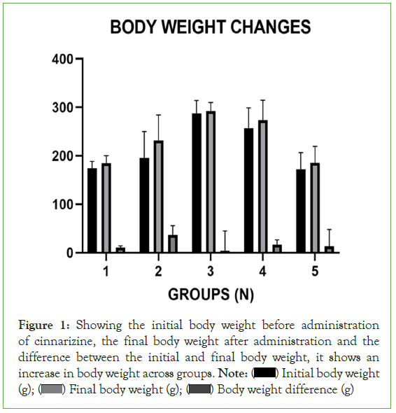
Figure 1: Showing the initial body weight before administration
of cinnarizine, the final body weight after administration and the
difference between the initial and final body weight, it shows an
increase in body weight across groups. Note: ( ) Initial body weight
(g); (
 ) Final body weight (g); (
 ) Body weight difference (g)
Oxidative stress makers
Superoxide Dismutase (SOD): Figure 2 shows the recorded mean of SOD level on the graph group two administered with 10 mg/ kg of cinnarizine for 21 days showed a very significant decrease in SOD level when compared to the control group on one-way ANOVA (*=significant at p-value˂0.05)., the control group shows an SOD level of 23.53 ± 5.94 which could be taken as normal, when compared to the second group that were administered with 10 mg/kg of cinnarizine it showed a decline with a significant value of 12.20 ± 3.16* which indicate the presence of lipid peroxidation, other groups that were administered with high dose of 20 mg/kg of cinnarizine showed no significant difference when compared to the control group on one-way ANOVA (*=significant at p-value˂0.05).
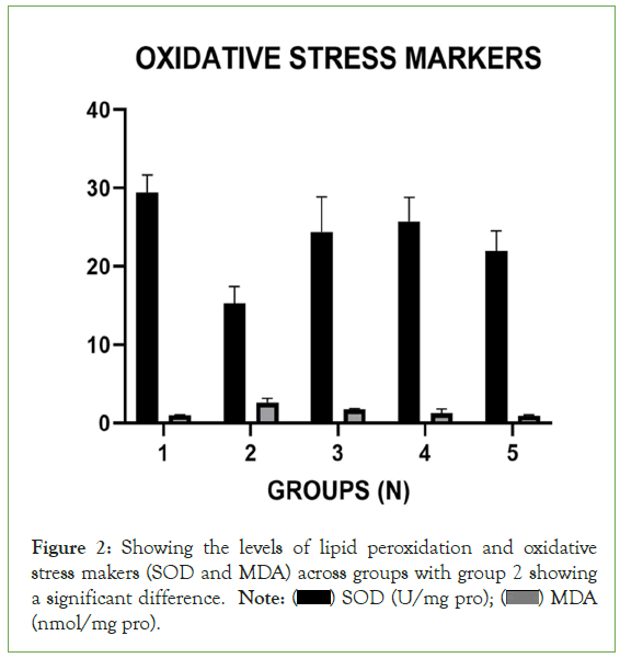
Figure 2: Showing the levels of lipid peroxidation and oxidative
stress makers (SOD and MDA) across groups with group 2 showing
a significant difference. Note: ( ) SOD (U/mg pro); (
 ) MDA
) MDA
(nmol/mg pro).
Malondialdehyde (MDA): Figure 2 graph shows difference in the level of MDA across the groups when compared to the control group on one-way ANOVA (*=significant at p-value˂0.05) the recorded mean of MDA of group one was 0.79 ± 0.20, this is compared to group two that were treated with low dose of 10 mg/ kg of cinnarizine recorded a high mean of MDA level of 2.08 ± 0.56* this is a significant increase in the level of MDA other groups treated with high dose of cinnarizine like the third, fourth and fifth group shows some increase when compared to the control group, the third group administered with 10 mg/kg for 14 day showed an increase of 1.38 ± 0.35 but this is not statistically significant as the second group when compared with the control. Using oneway ANOVA (*=significant at p-value˂0.05). the remaining groups, four and five which were treated with 20 mg/kg of cinnarizine for 14 and 21 days respectively showed no statistically significant difference when compared with group one (control) using one-way ANOVA (*=significant at p-value˂0.05).
Assay of inflammatory cytokine (TNF-α)
Tumor Necrosis Factor (TNF-α): Figure 3 shows the activity level of inflammatory cytokine; tumor necrosis factor (TNF-α) from the graph it shows that there is slight increase of 29.38 ± 7.35* at the second group administered with 10 mg/kg of cinnarizine for 21 days when compared with the control group which has a value of 20.68 ± 5.18 on one-way ANOVA (*=significant at p-value˂0.05). Other groups administered with 10 mg/kg for 14 days and those administered with 20 mg for 21 days and 14 days respectively show no significant difference when compared with the control group on one-way ANOVA (*=significant at p-value˂0.05).
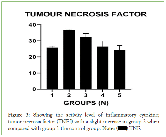
Figure 3: Showing the activity level of inflammatory cytokine;
tumor necrosis factor (TNF-α) with a slight increase in group 2 when
compared with group 1 the control group. Note: ( ) TNF.
Cognitive test
From Figure 4 the graph below the group 4 and 5 of rats administered with high dose of cinnarizine which is 20 mg/kg show lesser escape latency when compared to the control group and other groups administered with low dose of 10 mg/kg for a lesser period of time, this shows that cinnarizine improved their learning and memory ability when compared to other groups that were treated with lesser dose and the control group that was not given cinnarizine at all we can conclude that cinnarizine has the ability to improve learning and memory especially when administered in higher doses.
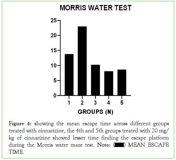
Figure 4: showing the mean escape time across different groups
treated with cinnarizine, the 4th and 5th groups treated with 20 mg/
kg of cinnarizine showed lesser time finding the escape platform
during the Morris water maze test. Note: ( ) MEAN ESCAPE
TIME.
Histological profile
The histoligical profile of all group were represented as Figures 5a-5e when observed they show the following characteristics as explained below; The cells were intact with well stained nuclei. The regions of the principal cell layer of the pyramidal layer were intact through CA1 to CA4 and well demonstrated in the control group.
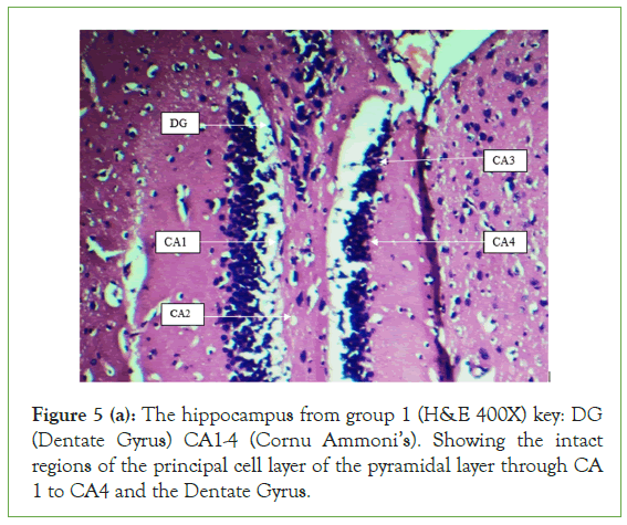
Figure 5 (a): The hippocampus from group 1 (H&E 400X) key: DG (Dentate Gyrus) CA1-4 (Cornu Ammoni’s). Showing the intact regions of the principal cell layer of the pyramidal layer through CA 1 to CA4 and the Dentate Gyrus.
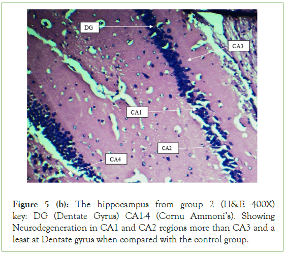
Figure 5 (b): The hippocampus from group 2 (H&E 400X) key: DG (Dentate Gyrus) CA1-4 (Cornu Ammoni’s). Showing Neurodegeneration in CA1 and CA2 regions more than CA3 and a least at Dentate gyrus when compared with the control group.
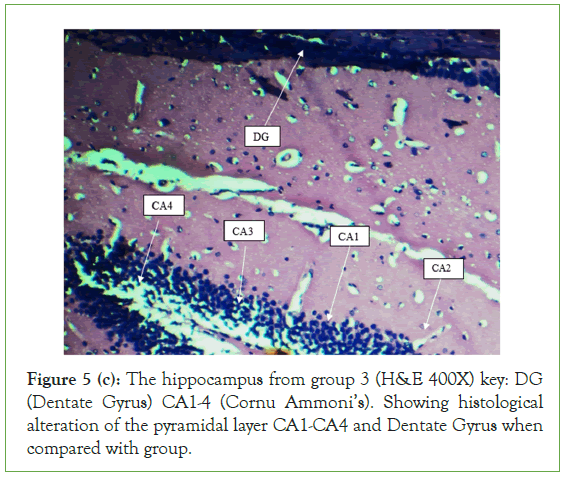
Figure 5 (c): The hippocampus from group 3 (H&E 400X) key: DG (Dentate Gyrus) CA1-4 (Cornu Ammoni’s). Showing histological alteration of the pyramidal layer CA1-CA4 and Dentate Gyrus when compared with group.
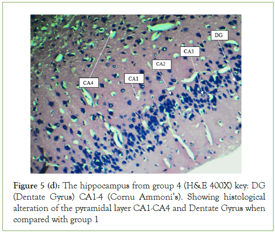
Figure 5 (d): The hippocampus from group 4 (H&E 400X) key: DG (Dentate Gyrus) CA1-4 (Cornu Ammoni’s). Showing histological alteration of the pyramidal layer CA1-CA4 and Dentate Gyrus when compared with group 1
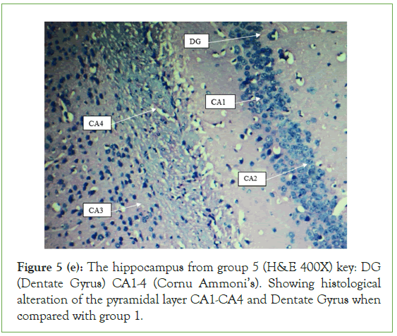
Figure 5 (e): The hippocampus from group 5 (H&E 400X) key: DG (Dentate Gyrus) CA1-4 (Cornu Ammoni’s). Showing histological alteration of the pyramidal layer CA1-CA4 and Dentate Gyrus when compared with group 1.
The Dentate Gyrus (DG) was intact and well stained. Its principal cell layer, the granular layer was well demonstrated. The layers of the hippocampus of the groups administered with 10 mg/kg for 14 and 21 days respectively and the group administered with 20 mg/kg for 14 days and 21 days respectively all showed alterations in few places. They showed extensive degeneration of the pyramidal layer as the cells appear vacuolated against when compared to the control group. The neurodegeneration was more noticeable in the CA1 and CA2 region than CA3. The cells of the CA4 region, the hilus of DG and granular cell layer of DG, were least affected. The stratum radiatum also shows extensive histological alterations when compared with the control. The hippocampus of group 3 and 4 administered with 10 mg/kg and 20 mg/kg respective showed more histological alteration when compared with those of the control group and group 2. The cells of the pyramidal layer CA1-CA4 were more packed when compared to those of group 5 administered with 20 mg/kg and in the cells of the principal cell layer of the hippocampus in group 5, the nuclei of some of the neurons were intact and well demonstrated. The stratum radiatum layer of group 3 and 4, showed less histological alteration when compared to those of group 5, especially in the CA2 region. Cells of the hilus of DG and CA4 regions were more demonstrated than those of group 5. When compared with group 5, the granular layer of the DG seems to have resisted neurodegeneration (Figures 5a-5e).
Discussion
The present study examined the effect of cinnarizine an antihistamine drug that has been approved for the treatment of motion sick on the body weight, oxidative stress makers, tumor necrosis factor and memory and learning ability in adult wistar rats from the results and data collected cinnarizine showed the following effects on the wistar rat.
There was a significant increase in the weight of the rats when compared to group one the following group two, four and five showed a very significant increase in body weight this could be attributed to the intake of cinnarizine as previous research by Abdul-Salam, reported that cinnarizine given to mice at 5 mg/ kg dose made the mice to suffer from oedema, which increased their body weight. In this study the rats in group two were treated with 10 mg/kg of cinnarizine which is close to what used in their study, it could be that the rats in group 2 and other groups also suffered from oedema which was responsible for the significant increase in weight. This increase in body weight can also be attributed to increase in appetite and feeding in treated rats which is in line with a research by Nilla-Orthen Ganbill, who reported that the administration of antihistamines which brings about the blockage of histamine stimulates appetite and brings about long lasting increase in food intake, cinnarizine is an antihistamine and could have caused an increase in appetite and feeding which led to the significant increase in weight [18]. In another study, reported that antihistamines which are active antipsychotic like cinnarizine excerpt sedative effect on patients which causes weight gain as a side effect of this antihistamines and cinnarizine falls under this category [19]. Also, Navarro-Badenes, also reported that cinnarizine caused weight gain in four female patients aged 50-57 and as such concluded that cinnarizine could be a cause of weight gain as observed with other drugs in the same class, and this present study showed clearly that cinnarizine is responsible for the increase in body weight [20].
Superoxide Dismutase (SODs) are a group of metalloenzymes that are found in all kingdoms of life. SODs form the front line of defense against Reactive Oxygen Species (ROS)-mediated injury [21]. From the result of this study it shows a significant reduction in the level of SOD in group when compared to the control group this could be attributed to increase in the level of Peroxidation which signifies damage to the hippocampal tissue, although across the groups especially group 3 and 5 and a bit of group 4 there seem to be an increase in the level of SOD towards the normal level, this is in line with studies conducted by Abdel-Fattah where cinnarizine used in treating induced Bronchial Asthma, showed a reversion back to normal of SOD level in rats treated with cinnarizine which brought them to the conclusion that cinnarizine possess some antiinflammatory effect which could be able to prevent cell death [22]. Also in another study by Abdel-Aal, where the neuroprotective effect of Cinnarizine was checked on rats with brain ischemia it showed an increase in the level of SOD after a reduction by ischemia, this can also be related to the significant reduction in SOD level of group two, that cinnarizine at a dose of 10 mg/kg could cause necrosis of hippocampal cells although other group showed a bit of increase which is in line with the study by Abdel- Aal, which showed increase in SOD level after treatment with cinnarizine we can deduce that cinnarizine could possess some neuroprotective effects.
Malondialdehyde (MDA) is known to be the final product of lipid peroxidation. Therefore, the serum or plasma levels of MDA can be used as a marker of lipid peroxidation. An increase in free oxygen radicals can cause excessive lipid peroxidation, causing oxygen damage in tissues [23]. Increased MDA levels in cerebrospinal fluid and plasma can cause oxidative damage to the brain [24]. From the results of this study it shows clearly an increase in MDA levels across groups but significantly at group two, increased level of MDA can cause oxidative damage to the brain and this can be mitigated by enzymatic antioxidants like SOD which this study analyzed and found out that at group two which shows a significant decrease in the level of SOD also showed a significant increase in the level of MDA which shows that there was oxidative stress which brought about the increase in the level of MDA, across all group there was an increase though more significant in group two, although previous studies by Omar stated that cinnarizine brought about a decrease in the level of MDA which was increased by Haloperidol induced oxidative stress [25]. Also, Atarbashe and Abu-Raghif, reported that prevented paraquat cinnarizine significantly reduced MDA level in ulcerated colitis male rats, and also Çeçen reported that cinnarizine induced increase in MDA level in mice [26]. In another study by Abdel-Aal reported that cinnarizine could not affect the increased level of MDA in rats that had induced ischemia. This study also showed an increase in level of MDA especially in group two and it seemed not affected by cinnarizine except for group five which show a non-significant reduction in MDA level, therefore from this study we could conclude that the increase in MDA level which is due to oxidative stress which could be due to the kind of diet the rats were fed with, according to Apel and Hirt, who reported that diets with high level of fats or carbohydrates have shown to increase oxidative stress, which could be the reason why there was an increase in MDA level across groups and also other factors could be responsible like environment, pollution, heavy metals in the diet or drinking water could be responsible for the increase in oxidative stress that brought about increase in MDA level [27]. Comparing the increased level of MDA to the reduced level of SOD we could say that there was oxidative stress that the enzymatic antioxidants (SOD) is trying to reduce to prevent, cell aging and death, we could come to the conclusion that cinnarizine possess the ability of causing damage as against other reports but also other factors like diet could strongly be the cause of the increased oxidative stress, while cinnarizine was acting as a therapeutic agent as reported by other study, and also considering that at other groups apart from group two there seem to be a bit of reversion in the level of SOD towards the normal level.
Tumor Necrosis Factor Alpha (TNF-α) is a cytokine that has pleiotropic effects on various cell types, it has been identified as a major regulator of inflammatory responses and is known to be involved in the pathogenesis of some inflammatory and autoimmune diseases [28]. From this study the results show a significant level of increase in the level of TNF-α in group two who were treated with 5 mg/kg of cinnarizine when compared to the control group, TNF-α as a signaling factor that brings about inflammation an Elevated levels of circulating TNF-α have been linked to a wide variety of diseases, including arthritis, diabetes, Crohn's disease, and cachexia associated with terminal cancer and AIDS [29]. Although previous studies conducted by Abdel- Fattah stated that administration of cinnarizine brought about the reduction of TNF-α to normal level, the study focused on the effect cinnarizine on induced asthma in rats while this study is focused on the effect of cinnarizine on the hippocampus and other oxidative makers, this could be the factor that led to the discrepancy in results, in another study by Attarbashee and Abu-Raghif, revealed that cinnarizine brought about the reduction in the level of TNF-α which brought that study to a conclusion that cinnarizine possess some therapeutic effects, although that study was focused on the effect of cinnarizine and on induced ulcerative colitis in male rats [22,30]. This study was focused on both male and female rats and the effect of cinnarizine on the brain specifically the hippocampus. Haloperidol an antipsychotic drug was reported to have elevated the level of TNF-α in the brain by Omar cinnarizine is also an antipsychotic drug which could be the reason for the increase in the level of TNF-α across the group with a significance in group two. It could be concluded that cinnarizine might thus be able to exacerbate inflammatory process in the brain that could lead to Neurodegeneration.
From the result demonstrated above this study is in agreement with previous study by Zhang who reported that neuron of pyramidal layers and dentate gyrus of rats treated with Cinnarizine were neatly arranged and dense without nuclear shrinkage after a pathological damage by microwaves [31]. Zhang also reported in another study were the hippocampus of rats were damaged by microwaves and treated with cinnarizine through nasal delivery the study showed that cinnarizine restored the alteration and damages caused by microwaves, this could be interpreted that cinnarizine possess neuroprotective abilities with regards to that study but according to results from this study there were alterations in some parts of the hippocampus especially in group 3, 4 and 5 when compared with the control group, other factors can be responsible for this damage one of which could be stress, which according to McLaughlin who reported that the use of chronic restraint stress, with wire mesh, for 6 h/21 d is a reliable and efficient method to produce psychological stress and to cause CA3 dendritic retraction and spatial memory deficits in male Sprague-Dawley rats [31,32]. This could have been the cause of the observed damages observed because the rats used for this study were kept in a wire mesh cage which could be resulted in psychological stress. The diet of the experimental animals could be the cause of the observed damages in the hippocampus as reported, that Alzheimer's disease and other hippocampal histological damages in rats can be linked with intake of a Western diet, characterized by high levels of saturated fats and simple carbohydrates [33]. We can conclude that these damages observed could be from other factors and it can be from the administration of cinnarizine as administration routes, doses, time and environment of administration of the reported cases of the neuroprotective abilities of cinnarizine are different from that of this study.
This study showed that the ability of the treated rats to locate the escape platform in Morris water maze test is less for the group of rats treated with higher dose of cinnarizine when compared with the control group. Previous research showed that rats impaired with haloperidol took longer time to locate the escape platform with Morris water maze test but this was greatly improved by treatment with cinnarizine which served as an ameliorator for that memory impairment, although this study is focused on the effect of cinnarizine on the memory and ability of rats and from results showed that high dose can enhance the ability of rats to learn and still have this memory, this could be attributed to the fact that Drugs with dopaminergic agonist activity have been shown to improve memory function [25]. Cinnarizine exhibits a dopamine D2 receptor blocking property and might therefore impair working memory. The drug, however, has been shown to exhibit a potential antipsychotic effect with an atypical profile [34].
This study is also in line with previous study by Zhang who reported that cinnarizine significantly improved the microwave-induced spatial memory through nasal administration of cinnarizine in another study he conducted he reported that cinnarizine improved the recovery of spatial memory and spontaneous explanatory behavior after been impaired with microwaves and treated with cinnarizine dissolving microneedles, In this study cinnarizine didn’t impair working memory in rats and also improved the memory and learning ability of rats in the groups that were given high doses of cinnarizine, this result could also be due to the level of TNF-α in the brain as studies by Golan who reported that TNF-α in the brain has a role to play in proper development of the hippocampus and also concluded that there is a possible involvement of TNF-α in the regulation of learning and memory under physiological and pathological conditions [31,35]. Although this study used mice but current study is in agreement with the claim because Golan also concluded that at basal levels of TNF-α it may be specifically involved in the regulation of hippocampus development and function, which could have been the cause of improved memory and learning ability as current study showed that rats in group four and five which had the least levels of TNF-α had the least escape time as compared to group two who had the highest level of TNF-α had the highest escape time, which brought us to the conclusion from this reports that high and low levels of TNF-α could affect memory and learning ability and the hippocampus and also in agreement with other research Cinnarizine possess the ability of improving impaired memory and learning abilities [36-40].
Conclusion
From the results and data collected and also from reports of previous study, cinnarizine possess some neuro protective abilities. Although there is still some alteration in the histological makeup of the hippocampus, but still didn’t affect the learning and memory ability of the treated rats. In this research which bring us to the conclusion that cinnarizine possess the ability of improving learning and memory in treated rats.
References
- Gilbert PE, Brushfield AM. The role of the CA3 hippocampal subregion in spatial memory: A process oriented behavioral assessment. Prog Neuropsychopharmacol Biol Psychiatry.2009;33(5):774-781.
[Crossref] [Google Scholar] [Pubmed]
- Last RJ. Philadelphia: Churchill-Livingstone. Hippocampal anatomy.1999;460.
- Dhikav V. Can phenytoin prevent Alzheimer’s disease? Med hypotheses.2006;67(4):725-728.
[Crossref] [Google Scholar] [Pubmed]
- Dhikav V, Anand K. Potential predictors of hippocampal atrophy in Alzheimer’s disease. Drugs Aging. 2011;28:1-1.
[Crossref] [Google Scholar] [Pubmed]
- Fumagalli M, Priori A. Functional and clinical neuroanatomy of morality.Brain.2012;135(7):2006-2021.
[Crossref] [Google Scholar] [Pubmed]
- Zhang LL, Liu HQ, Yu XH, Zhang Y, Tian JS, Song XR, et al. The combination of scopolamine and psychostimulants for the prevention of severe motion sickness. CNS Neurosci Ther.2016;22(8):715-722.
[Crossref] [Google Scholar] [Pubmed]
- Bertolini G, Straumann D. Moving in a moving world: A review on vestibular motion sickness. Front Neurol. 2016;7(14).
[Crossref] [Google Scholar] [Pubmed]
- Emanuel MB, Will JA. Cinnarizine in the treatment of peripheral vascular disease: Mechanisms related to its clinical action. Proc R Soc Med.1977;70(8):07-12.
[Crossref] [Google Scholar] [Pubmed]
- National formulary of India, fourth edition, 2011.
- Soto E, Vega R. Neuropharmacology of vestibular system disorders. Curr Neuropharmacol.2010;8(1):26-40.
[Crossref] [Google Scholar] [Pubmed]
- Drugs Controller General of India. List of approved drugs from 01.01.2010 to 31.12.2010.
- Haasler T, Homann G, Duong Dinh TA, Jungling E, Westhofen M, Luckhoff A. Pharmacological modulation of transmitter release by inhibition of pressure-dependent potassium currents in vestibular hair cells. Naunyn Schmiedebergs Arch Pharmacol.2009;380:531-538.
[Crossref] [Google Scholar] [Pubmed]
- Nicholson AN, Stone BM, Turner C, Mills SL. Central effects of cinnarizine: Restricted use in aircrew. Aviat Space Environ Med.2002;73(6):570-574.
[Google Scholar] [Pubmed]
- Nunez J. Morris water maze experiment. JoVE. 2008;(19):e897.
[Crossref] [Google Scholar] [Pubmed]
- Saalu LC, Osinubi AA, Jewo PI, Oyewopo AO, Ajayi GO. An evaluation of influence of Citrus paradisi seed extract on doxorubicin-induced testicular oxidative stress and impaired spermatogenesis. Asian J Sci Res. 2010;3(1):51-61.
- Winterbourn CC, Hawkins RE, Brian M, Carrell RW. The estimation of red cell superoxide dismutase activity. J Lab Clin Med.1975;85(2):337-341.
[Google Scholar] [Pubmed]
- Rukmini C, Sathiya Moorthy P, Viswanathan P, Krishnamoorthy P. Antioxidant potentials of flaxseed by In vivo model. J Agric Food Chem.2004;52(5):1391-1395
- Orthen N, Gambill M. Blockade of histamine receptors in the treatment of obesity. J Am Osteopath Assoc. 1988; 88(11):1429-1431.
- Ratliff JC, Barber JA, Palmese LB, Reutenauer EL, Tek C. Association of prescription H1 antihistamine use with obesity: Results from the National Health and Nutrition Examination Survey. Obesity. 2010;18(12):2398-2400.
[Crossref] [Google Scholar] [Pubmed]
- Navarro-Badenes J, Martínez-Mir I, Palop V, Rubio E, Morales-Olivas FJ. Weight gain associated with cinnarizine. Ann Pharmacother.1992;26(7-8):928-930.
[Crossref] [Google Scholar] [Pubmed]
- Kangralkar VA, Patil SD, Bandivdekar, AH, Rege NN. Antioxidant system in prostate cancer: A review. Cancer Letters.2010;297(2):129-135.
- Abdel-Fattah MM, Messiha BA, Salama AA. Assessment of the mechanistic role of cinnarizine in modulating experimentally-induced bronchial asthma in rats. Pharmacol.2015;96(3-4):167-174.
[Crossref] [Google Scholar] [Pubmed]
- Herken HU, Uz E, Ozyurt H, Sogut S, Virit O, Akyol O. Evidence that the activities of erythrocyte free radical scavenging enzymes and the products of lipid peroxidation are increased in different forms of schizophrenia. Mol Psychiatry.2001;6(1):66-73.
[Crossref] [Google Scholar] [Pubmed]
- Mahadik SP, Evans D, Lal H. Oxidative stress and role of antioxidant and ω-3 essential fatty acid supplementation in schizophrenia. Prog Neuropsychopharmacol Biol. Psychiatry.2001;25(3):463-493.
[Crossref] [Google Scholar] [Pubmed]
- Omar MH, Abdullah R, Hassan Z, Ibrahim N. Cinnarizine improves spatial working memory in rats: comparison with clozapine and haloperidol. Pharmacol Biochem Behav.2012;100(2):342-348.
- Attarbashee RK, Abu-Raghif A. Comparative treatment of induced ulcerative colitis in male rat model by using cinnarizine and sulfasalazine. J Vet Sci.2020;34(2):465-472.
- Apel K, Hirt H. Reactive oxygen species: Metabolism, oxidative stress, and signal transduction. Annu Rev Plant Biol.2004;55:373-399.
[Crossref] [Google Scholar] [Pubmed]
- Bradley J. TNF‐mediated inflammatory disease. J Pathol.2008;214(2):149-160.
[Crossref] [Google Scholar] [Pubmed]
- Omar Matthys P, Billiau A. Cytokine network in viral hepatitis. Cytokine Growth Factor Rev.1997;8(1):27-38.
- Attarbashee W, Abu-Raghif AR. Protective effects of cinnarizine against ulcerative colitis induced by acetic acid in male rats. J Biol Sci.2019;26(5):933-939.
- Zhang C, Shao J, Wei T, Dong Y, Xue Y, Xue X. Improved Microwave-Induced spatial memory with cinnarizine. ACS Chem Neurosci.2022;13(3):361-370.
- McLaughlin KJ, Gomez JL, Baran SE, Conrad CD. The effects of chronic stress on hippocampal morphology and function: An evaluation of chronic restraint paradigms. Brain Res.2007;1161:56-64.
[Crossref] [Google Scholar] [Pubmed]
- Kanoski SE, Davidson TL. Western diet consumption and cognitive impairment: Links to hippocampal dysfunction and obesity. Physiol Behav.2010;103(1):59-68.
[Crossref] [Google Scholar] [Pubmed]
- Dall'Igna OP, Porciúncula LO, Souza DO, Cunha RA. Cinnarizine improves spatial memory in rats by dopaminergic and histaminergic mechanisms. Psychopharmacology.2005 180(4), 612-620.
- Golan H, Levav T, Mendelsohn A, Huleihel M. Involvement of tumor necrosis factor alpha in hippocampal development and function. Cereb Cortex.2004;14(1):97-105.
[Crossref] [Google Scholar] [Pubmed]
- Abdel-Aal M, Zakaria MN, EL-Fayoumi MM, Oraby MA. Effect of some Neuroprotective drugs on certain biochemical parameters during experimental brain ischaemia. J Pharm Sci.2015;23(2):01-15.
- Söylemezoglu T. The effectiveness of n-acetylcysteine and cinnarizine on paraquat-induced neuro and hepato-toxicity in mice. J Fac Phann Gazi.2001;18(1):49-57.
- Diel P, Ruocco LA, Schröder J, Schreckenberger M. Imaging of stress and stress-related disorders with PET and SPECT. Curr Neurol Neurosci Rep.2016;16(9):88.
- Dkhil MA, Al-Quraishy S, Abdel Moneim AE. In vivo effect of cinnarizine on the liver of mice. J basic appl zool.2007 49(3), 107-117
- Pilar F, Dehaene S, Lambert J. Bridging levels of consciousness: Dynamical aspects of neural top-down control. Curr Opin Neurobiol.2018;49:218-230.
Citation: Akunna GG, Sule OO, Lucyann CA, Saalu LC (2023) Motion Sickness Medication Cinnarizine Could Impair Hippocampal Morphology, Memory and Learning in Wistar Rat Models. J Clin Med Sci.7:239.
Copyright: © 2023 Akunna GG, et al. This is an open access article distributed under the terms of the Creative Commons Attribution License, which permits unrestricted use, distribution, and reproduction in any medium, provided the original author and source are credited.
