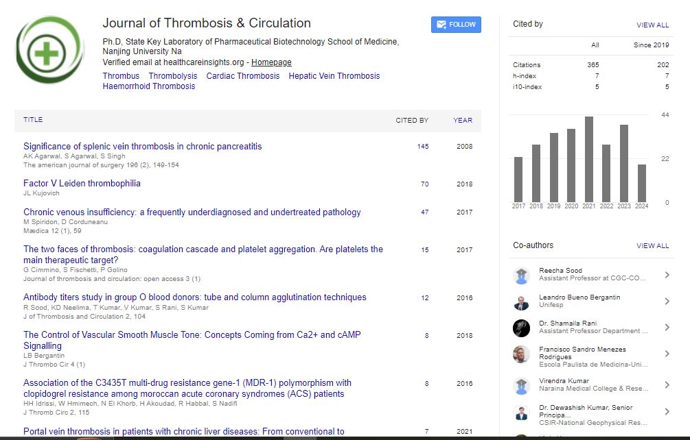

Indexed In
- RefSeek
- Hamdard University
- EBSCO A-Z
- Publons
- Google Scholar
Useful Links
Share This Page
Journal Flyer
Open Access Journals
- Agri and Aquaculture
- Biochemistry
- Bioinformatics & Systems Biology
- Business & Management
- Chemistry
- Clinical Sciences
- Engineering
- Food & Nutrition
- General Science
- Genetics & Molecular Biology
- Immunology & Microbiology
- Medical Sciences
- Neuroscience & Psychology
- Nursing & Health Care
- Pharmaceutical Sciences
Opinion - (2023) Volume 9, Issue 3
Imaging Techniques for Diagnosis: Identifying Deep Vein Thrombosis in the Presence of Inferior Venacava Agenesis
Dahl Turi*Received: 19-Apr-2023, Manuscript No. JTCOA-23-21493; Editor assigned: 21-Apr-2023, Pre QC No. JTCOA-23-21493 (PQ); Reviewed: 05-May-2023, QC No. JTCOA-23-21493; Revised: 12-May-2023, Manuscript No. JTCOA-23-21493 (R); Published: 22-May-2023, DOI: 10.35248/2572-9462.23.9.225
Description
Deep Vein Thrombosis (DVT) is a potentially life-threatening condition characterized by the formation of blood clots within the deep veins of the body. It commonly affects the lower extremities and can lead to severe complications such as pulmonary embolism. While DVT can occur due to various factors, one rare and challenging cause is Inferior Vena Cava (IVC) agenesis. This condition involves the absence or underdevelopment of the IVC, a major blood vessel responsible for returning deoxygenated blood from the lower body to the heart. The absence of the IVC disrupts normal blood flow, leading to an increased risk of clot formation in the deep veins.
Causes and pathophysiology
IVC agenesis is a congenital condition, meaning it is present at birth. The exact cause of IVC agenesis is unknown, but it is thought to result from improper development of the embryonic veins during early fetal development. As a consequence, the iliac veins, which drain blood from the lower limbs, connect to alternate pathways to return blood to the heart.
In individuals with IVC agenesis, the blood from the lower extremities is rerouted through collateral veins, such as the ascending lumbar veins, hemizygous and azygos systems, and pelvic venous plexus. This rerouting of blood creates a turbulent flow and stasis within the collateral veins, predisposing individuals to the formation of blood clots.
Symptoms and complications
The symptoms of DVT provoked by IVC agenesis are similar to those of DVT caused by other factors. Common symptoms include pain, swelling, and redness in the affected leg. However, due to the anatomical abnormality, the symptoms may be more severe and occur earlier in life compared to typical cases of DVT.
If left untreated, DVT can lead to serious complications such as pulmonary embolism, a condition where a clot travels to the lungs and obstructs blood flow. Symptoms of pulmonary embolism include shortness of breath, chest pain, and coughing up blood. Prompt diagnosis and treatment are crucial to prevent such life-threatening complications.
Diagnosis
Diagnosing DVT provoked by IVC agenesis can be challenging due to its rarity and the need for specialized imaging techniques. Several diagnostic modalities are used to identify the condition and assess the extent of clot formation. These include:
Doppler ultrasound: This non-invasive test uses sound waves to visualize blood flow and detect clots within the deep veins.
Magnetic Resonance Venography (MRV): MRV provides detailed images of the blood vessels using Magnetic Resonance Imaging (MRI) technology. It helps identify the absence or underdevelopment of the IVC and visualizes the collateral veins.
Computed Tomography Venography (CTV): CTV utilizes X-ray imaging and contrast dye to visualize blood vessels, allowing for the detection of clots and abnormal venous anatomy.
Treatment options
The management of DVT provoked by IVC agenesis aims to prevent clot progression, alleviate symptoms, and reduce the risk of complications. The treatment options commonly employed include:
Anticoagulant therapy: Medications such as heparin and warfarin are prescribed to thin the blood and prevent clotting. Anticoagulant therapy may be administered for a prolonged period, depending on the individual's condition.
Thrombolysis: In cases of extensive clot burden or severe symptoms, thrombolysis may be considered. This procedure involves the administration of clot-dissolving medications directly into the affected veins to break down the clots.
Inferior vena cava filter placement: In certain situations, when anticoagulant therapy is contraindicated or ineffective, an inferior vena cava filter may be inserted to prevent clot migration to the lungs. This filter catches the clots and prevents them from reaching critical organs.
Citation: Turi D (2023) Imaging Techniques for Diagnosis: Identifying Deep Vein Thrombosis in the Presence of Inferior Venacava Agenesis. J Thrombo Cir. 9:225.
Copyright: © 2023 Turi D. This is an open-access article distributed under the terms of the Creative Commons Attribution License, which permits unrestricted use, distribution, and reproduction in any medium, provided the original author and source are credited.