
Indexed In
- Open J Gate
- Genamics JournalSeek
- Academic Keys
- JournalTOCs
- ResearchBible
- China National Knowledge Infrastructure (CNKI)
- Scimago
- Ulrich's Periodicals Directory
- Electronic Journals Library
- RefSeek
- Hamdard University
- EBSCO A-Z
- OCLC- WorldCat
- SWB online catalog
- Virtual Library of Biology (vifabio)
- Publons
- MIAR
- Scientific Indexing Services (SIS)
- Euro Pub
- Google Scholar
Useful Links
Share This Page
Journal Flyer
Open Access Journals
- Agri and Aquaculture
- Biochemistry
- Bioinformatics & Systems Biology
- Business & Management
- Chemistry
- Clinical Sciences
- Engineering
- Food & Nutrition
- General Science
- Genetics & Molecular Biology
- Immunology & Microbiology
- Medical Sciences
- Neuroscience & Psychology
- Nursing & Health Care
- Pharmaceutical Sciences
Research Article - (2025) Volume 16, Issue 3
Green Synthesis of Chromium(III) Oxide Nanoparticles and their Evaluation against Skin Infections
Nadia Bukhari1, Muhammad Waris2, Muhammad Iqbal Khan1, Naila Gul1 and Farhana Anjum3*2Department of Environmental Science, Aror University of Art, Architecture, Design and Heritage, Sindh, Pakistan
3Department of Chemical and Life Sciences, Qurtuba University of Science and Technology, Khyber Pakhtunkhwa, Pakistan
Received: 07-Nov-2024, Manuscript No. JNMNT-24-27481; Editor assigned: 11-Nov-2024, Pre QC No. JNMNT-24-27481 (PQ); Reviewed: 25-Nov-2024, QC No. JNMNT-24-27481; Revised: 14-Oct-2025, Manuscript No. JNMNT-24-27481 (R); Published: 21-Oct-2025, DOI: 10.35248/2157-7439.25.16.790
Abstract
Nanotechnology is an important area of contemporary research since it addresses the size, structure, and composition of the products to be produced. Chromium(III) Oxide (Cr2O3) nanoparticles were prepared using fixed ratio of plant extract (Psidium guajava) which contain reducing sugars as reducing agent and Potassium Dichromate (K2Cr2O7) solution. The applications of Cr2O3 nanoparticles against different human pathogens were investigated. The synthesized nanoparticles were characterized with different characterization techniques which were UV-Vis, XRD, SEM and FTIR. UV-Vis analysis was used to confirm the initial spectra of Cr2O3 nanoparticles. Broad absorbance was observed in the range of 310 to 500 nm and high absorbance at 370 nm confirmed the synthesis of Cr2O3 nanoparticles. The nanoparticles were found to have the size of 33 nm according to XRD. All the desired peaks of plant and metal oxide were also present in FT-IR spectra. SEM showed particles were irregular in shape and poly dispersed. To know about the antifungal activities the plates were incubated at 28°C for 48 h, and after this period fungal growth inhibition zone were measured. It was found that Cr2O3 nanoparticles have shown good antifungal activities against different skin infection pathogens.
Keywords
Cr2O3; Green synthesis; Psidium guajava; Antifungal activity
Introduction
Metal oxide According to World Health Organization, poor diagnosis, inadequate attention and harmful antibiotic resistance are the major factors which has increased the ratio of fungal diseases and will also be increase in the near future. They having a great distinction in its functions and can be used as sensors, superconductors, Magnetic Resonance Imaging (MRI), adsorbents, catalyst, and sun based cells as well as its role in biological system have attracted much attention of researchers. Notable metal oxide nanoparticles with good antifungal activities are CuO, CaO, MgO and ZnO. Cr2O3 nanoparticles have received special consideration due to its multitudinal varied uses, including, layering of a particular or any substance that covers a surface, heterogenous catalyst, green pigments. Biomedical aspect of Cr2O3 has shown that it has significant efficiency against several bacteria. Cr2O3 nanoparticles can be synthesized with different methods, and it have a great contribution in the field of nanotechnology. The synthesis of Cr2O3 nanoparticles can be broadly divided into three main categories i.e., solution based synthesis, vapour based synthesis and biological synthesis which basically depends upon the type of the medium in which the oxidation reaction occurs [1]. Among all the above mentioned methods, the green synthesis method having plant extracts have gained much consideration as it is simple, feasible and one of the best substitute to the other methods, because such methods are injurious in one way or the other to the environment. The present fresh look is concerned with the achievability of metal oxide nanoscopic particle synthesis using plant extracts. Among the biological substitutes, plants and plant extracts suggest to be the best option. Because they are safe, clean, non-toxic and eco-friendly, in which microorganisms of plants like fungi and bacteria, are used [2].
Materials and Methods
Psidium guajava leaf extract was used as a capping and stabilizing agent for the Cr2O3 nanoparticles synthesis. Psidium guajava, (Family Myrtaceae) is known as guava is a small medium sized evergreen tree which is traditionally used for medicinal purposes and can be broadly planted all over the tropics. It can be cultivated by both methods either vegetatively or by seeds. The guava plant has become fostered whenever the climate in a particular area become suitable for its cultivation and it do not need much care. It also has the ability to tolerate scarcity conditions. Two known varieties of guava are the white (P. guajava var. pyrifera) and the red (P. guajava var. pomifera). In this study we used a wide diversity of plants are used medicinally for curing purposes, either a distinct part of it (leaves, root, seeds, fruit and flowers). The P. guajava leaves contains the quercetin-3 O-arabinoside, flavonol morin, morin-3-O-arabinoside, quercetin and morin-3-O-lyxoside. Literature survey have shown that the aqueous extracts of guava leaves were used, for the treatment of many type of gastrointestinal disturbances such as spasmolytic activity, vomiting, gastroenteritis, flatulence, diarrhea, dysentery, inhibition of the peristaltic reflex, gastric pain and abdominal distention. The antimicrobial and antifungal activities of guava leaf extracts (P. guajava L.) have also been proved, their inhibiting activity on the proliferation of yeast strains (C. tropicalis and C. albicans) (Figure 1) [3].
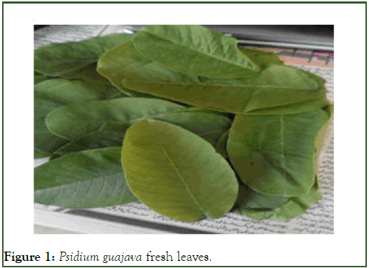
Figure 1: Psidium guajava fresh leaves.
Therefore, the primary goals of this work are to evaluate the potential of Psidium guajava and explore the prospective applications of its aqueous extract for the synthesis of Cr2O3 nanoparticles and the treatment of skin infections.
Experimental
Reagents and materials: For the synthesis of Cr2O3 NPs and for its biological activities, the following chemicals were used: Plant leaves, Potassium Dichromate (K2Cr2O7), Methanol (CH3OH), Dimethyl Sulphoxide (DMSO), distilled water, fluconazole, nutrient broth and fungal strains.
Synthesis of Chromium(III) Oxide nanoparticles: The Pisidium guajava leaves were collected from the Kohat city, Khyber Pakhtunkhawa, Pakistan, area. After being gathered in April, the leaves underwent three rounds of thorough washings in distilled water and fresh water to get rid of any contaminants. The popularity of this plant among the indigenous population and the unique uses it has in medicine were taken into consideration when choosing the leaves to use. About 25 g of fresh guava leaves were harvested, washed with distilled water, chopped into small pieces, and then placed in an Erlenmeyer flask with 200 ml of distilled water. The flask was heated to 70°C and rotated at 140 rpm for 30 minutes. After bringing the extract to room temperature, Whattman filter paper (no. 42) was used to finely filter it [4].
Distilled water was used to create a 0.5 M solution of solid Potassium Dichromate (K2Cr2O7). The nanoparticles were prepared by adding 14.70 g (0.5 M) of solid potassium dichromate to 100 ml of distilled water and stirring for 30 minutes at room temperature. Potassium dichromate solution in an orange solution was made. Subsequently, various ratios of potassium dichromate solutions and plant extract were combined, namely 1:1, 1:3, 1:5, 1:7, and 1:9, as well as 3:1, 5:1, 7:1, and 9:1, in order to obtain the best results. To ensure the reaction was completed, each of the various ratio mixtures was agitated for 30 minutes at 140 rpm while being maintained at room temperature [5].
From the reaction mixture nanoparticles were separated by centrifugation and was washed three times by using methanol. Then by centrifugation biosynthesized nanoparticles were collected, using centrifuge model. For purification, the nanoparticles suspension was redispersed in distilled water so as to remove the unbounded organics, and finally centrifuged. The brown solution will be kept in oven for drying at 196°C for 3 hrs, then calcined at 1000°C. At the end the brown colour of the sample changed to dark green.
Results and Discussion
Characterizations of Cr2O3 NPs
Characterization of Cr2O3 nanoparticles will be done by different techniques which are mentioned below:
UV-Vis spectra analysis of chromium (III) oxide NPs: The synthesized Chromium(III) Oxide (Cr2O3) nanoparticles were initially proved by UV-Vis analysis, prepared in different ratios and all had different colours in all ratios, having different absorption bands with difference in their peaks sharpness. Conventionally the marked region for the synthesized chromium(III) oxide nanoparticles is (310-500 nm). UV-Vis absorption analysis have shown that, 3:1 is the optimized ratio with maximium absorption peak at 365 nm (Figure 2) [6].
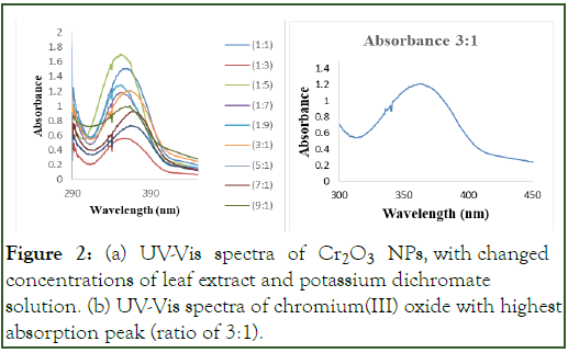
Figure 2: (a) UV-Vis spectra of Cr2O3 NPs, with changed concentrations of leaf extract and potassium dichromate solution. (b) UV-Vis spectra of chromium(III) oxide with highest absorption peak (ratio of 3:1).
Chromium(III) oxide nanoparticles formed at optimized ratio 3:1, absorbed at a wavelength of 365 nm, was employed for further characterization and for antifungal activities [7].
FT-IR spectroscopy: FTIR analysis is one of the fastest method, which is basically used to know about the main groups of compounds, which are the reason for the reduction and stabilization of chromium(III) oxide nanoparticles synthesized by the leaf extract of Pisidium guajava plant. Metal oxide Cr2O3 generally reveal absorption bands below 1000 cm-1 due to inter atomic vibrations. The peaks at 1301 cm-1 indicate the presence of (-COO-) carboxylate ions, responsible for stabilizing the Cr2O3 nanoparticles. The peak at 947 cm-1 and 547 cm-1 indicates Cr=O and Cr-O vibration of Cr2O3 nanoparticles [8].
The FT-IR analysis of our sample shows no –OH peak, because at 900°C temperature our sample was compleletly dehydrated and there was no any content of adsorbed water molecules. The strong narrowing of the Cr2O3 bands (from 750 to 500 cm-1) with increasing temperature is an indication of temperature induced ordering that could translate into the onset of a crystallization process in the layers. Ramesh, et al. synthesized chromium(III) oxide nanoparticles by the reduction of potassium dichromate solution with Arachis hypogaea leaf extract containing reducing sugars which act as reducing agent. The representative absorption peaks in FT-IR spectra of the synthesized nanoparticles located mainly at 3411 cm-1, 1311 cm-1, 947 cm-1 and 564 cm-1 in the region 400 cm-1-4000 cm-1 (Figure 3 and Table 1) [9].
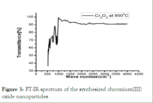
Figure 3: FT-IR spectrum of the synthesized chromium(III) oxide nanoparticles.
| Peak Position | Functional groups | Mode of vibration |
| 1301 cm-1 | -COO- | Stretching vibration |
| 947 cm-1 | Cr=O | Stretching vibration |
| 547 and 623 cm-1 | Cr-O | Stretching vibration |
Table 1: FTIR peaks of the prepared chromium(III) oxide nanoparticles with Pisidium guajava leaf extract.
X-Ray diffraction analysis: The synthesized chromium(III) oxide nanoparticles using plant extract (Psidium guajava) after drying in oven at 196°C, then annealed at 500°C, 600°C, 700°C, 800°C, and 900°C. The particles synthesized at 900°C were more crystalline in nature then the others. XRD analysis basically mentioned the crystallinity mode: The chromium(III) oxide nanoparticles. XRD spectra of the nanoparticles is mentioned below in the Figure 4. Similar results were reported by Anandhi, et al.; synthesized chromium(III) oxide nanoparticles by using Cr(NO3)3.9H2O. The materials were calcinated in electronic oven at 200°C, for 1 hour followed by annelation at 500°C, 700°C and 900°C. The crystallinity of the sample annealed at 900°C was found to be much better than that annealed at 500°C or 700°C.
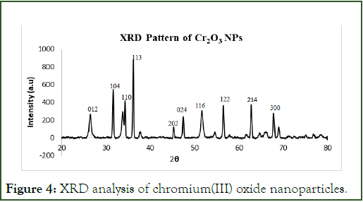
Figure 4: XRD analysis of chromium(III) oxide nanoparticles.
The peak width at half maximum was used to evaluate the crystallite size (D) by using the following Debby Scherer formula:
D=Kλ/(β cos θ)
Where, K=0.9 is the Scherer constant, λ: is the wavelength of the X-ray radiation (=1.54056 Å), β: is the peak full width at half maximum in radians and (θ): is the Bragg diffraction angle. Using this formula, the average crystallite size of as-grown nanostructures was found to be equal to (29-33) nm. An average particle size of the synthesized nanoparticles was found i.e., 33 nm, calculated by using the Debye-Scherrer equation. It having hexagonal structure.
SEM analysis: Esam J. Al-Kalifawi, et al. in 2017 synthesized chromium(III) oxide nanoparticles having spherical or semispherical nanoparticles formation, aggregated in the form of clusters. He also reported that the shape and size of the nanoparticles have significant effect in the antibacterial activity. For these reasons, nanoparticles must be examined with a scanning electron microscope to determine the shape of the nanoparticles. SEM analysis was performed to study the morphological studies of the chromium(III) oxide nanoparticles as shown in the given Figure 5 below.
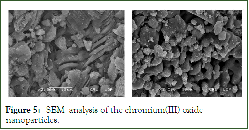
Figure 5: SEM analysis of the chromium(III) oxide nanoparticles.
The results have shown that the chromium(III) oxide nanoparticles are spherical plus irregular in shape with rough surfaces and agglomerated, which is probably due to the dispersing ability of the solvent.
Antifungal activities of Cr2O3 NPs
Different concentrations of Cr2O3 NPs were used to evaluate activity against Candida albicans and C. tropicalis. It was observed that 1 mg/mL concentration of Cr2O3 NPs were more effective against Candida species and have shown high zone of inhibition Figures 6 and 7. The Cr2O3 NPs have shown highest zone of inhibition at 1 mg/mL concentration followed by 150 ug/mL, 100 μg/mL and 50 μg/mL. The zone of inhibition of different concentration of Cr2O3 NPs have been (Table 2).
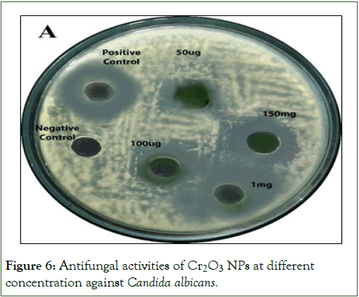
Figure 6: Antifungal activities of CCr2O3 NPs at different concentration against Candida albicans.
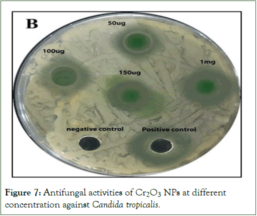
Figure 7: Antifungal activities of Cr2O3 NPs at different concentration against Candida tropicalis.
| Species | Nanoparticles Name concentration (μg/well) | Control | ||||
| 1000 | 150 | 100 | 50 | Positive | Negative | |
| C. albicans | 19 | 14 | 11 | 9 | 22 | NI |
| C. tropicalis | 17 | 12 | 11 | 8 | 19 | NI |
| Note: NI: No Inhibition, Fluconazole was used as a positive control and DMSO was used as a negative control | ||||||
Table 2: Evaluation of Cr2O3 NPs' antifungal activity at different concentrations in agar well diffusion against Candida albicans and Candida tropicalis.
Conclusion
This work successfully synthesised Cr2O3 nanoparticles in a green manner by employing Psidium guajava leaf extract as a capping and reducing agent. The morphological analysis showed that although the chromium oxide nanoparticles had irregular and spherical shapes, the chromium nanoparticles themselves were similar in size (below 400 nm). The nanoparticles were found to have the size of 33 nm according to XRD. The EDX spectra shows peaks for oxygen and carbon in addition to chromium, indicating that the chromium was reduced from the precursor slat. The green synthesis of chromium oxide nanoparticles was further validated by the XRD and UV-VIS investigations. When tested against both C. albicans and C. tropicalis bacterial strains, the green synthesised Cr2O3 nanoparticles demonstrated excellent antibacterial performance and were on par with commercially available drugs. Psidium guajava leaf extracts can be used to create a wide range of additional biocompatible nanomaterials for biological uses.
References
- Yang M, He J, Hu X, Yan C, Cheng Z, Zhao Y, et al. Copper oxide nanoparticle sensors for hydrogen cyanide detection: Unprecedented selectivity and sensitivity. Sens Actuators B Chem. 2011;155(2):692–698.
- Segev-Bar M, Haick H. Flexible sensors based on nanoparticles. ACS Nano. 2013;7(10):8366–8378.
[Crossref] [Google Scholar] [PubMed]
- O’Regan B, Grätzel M. A low-cost, high-efficiency solar cell based on dye-sensitized colloidal TiO2 films. Nature. 1991;353(6346):737–740.
- Pillai V, Kumar P, Hou MJ, Ayyub P, Shah DO. Preparation of nanoparticles of silver halides, superconductors and magnetic materials using water-in-oil microemulsions as nano-reactors. Adv Colloid Interface Sci. 1995;55:241–269.
- Zhou J, Song H, Chen X, Zhi L, Yang S, Huo J, et al. Carbon-encapsulated metal oxide hollow nanoparticles and metal oxide hollow nanoparticles: A general synthesis strategy and its application to lithium-ion batteries. Chem Mater. 2009;21(13):2935–2940.
- Kim DK, Zhang Y, Kehr J, Klason T, Bjelke B, Muhammed M. Characterization and MRI study of surfactant-coated superparamagnetic nanoparticles administered into the rat brain. J Magn Magn Mater. 2001;225(1–2):256–261.
- Sawai J, Yoshikawa T. Quantitative evaluation of antifungal activity of metallic oxide powders (MgO, CaO and ZnO) by an indirect conductimetric assay. J Appl Microbiol. 2004;96(4):803–809.
[Crossref] [Google Scholar] [PubMed]
- Rotter H, Landau MV, Carrera M, Goldfarb D, Herskowitz M. High surface area chromia aerogel efficient catalyst and catalyst support for ethylacetate combustion. Appl Catal B. 2004;47(2):111–126.
- Rakesh SA, Gowda NMM. Synthesis of Chromium (III) Oxide nanoparticles by electrochemical method and Mukia maderaspatanaplant extract, characterization, KMnOâ?? decomposition and antibacterial study. Imp J Interdiscip Res. 2016;2016(37).
Citation: Bukhari N, Waris M, Khan MI, Gul N, Anjum F (2025) Green Synthesis of Chromium(III) Oxide Nanoparticles and their Evaluation against Skin Infections. J Nanomed Nanotech. 16:790.
Copyright: © 2025 Bukhari N, et al. This is an open access article distributed under the terms of the Creative Commons Attribution License, which permits unrestricted use, distribution, and reproduction in any medium, provided the original author and source are credited.