
Indexed In
- Open J Gate
- The Global Impact Factor (GIF)
- Open Archive Initiative
- VieSearch
- International Society of Universal Research in Sciences
- China National Knowledge Infrastructure (CNKI)
- CiteFactor
- Scimago
- Ulrich's Periodicals Directory
- Electronic Journals Library
- RefSeek
- Directory of Research Journal Indexing (DRJI)
- Hamdard University
- EBSCO A-Z
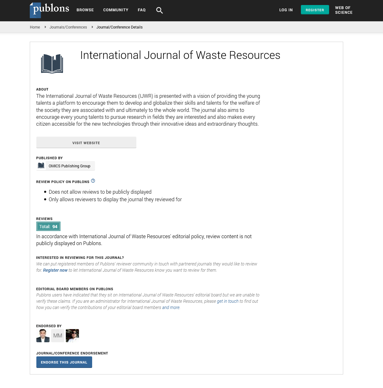
- Publons
- Google Scholar
Useful Links
Share This Page
Journal Flyer
Open Access Journals
- Agri and Aquaculture
- Biochemistry
- Bioinformatics & Systems Biology
- Business & Management
- Chemistry
- Clinical Sciences
- Engineering
- Food & Nutrition
- General Science
- Genetics & Molecular Biology
- Immunology & Microbiology
- Medical Sciences
- Neuroscience & Psychology
- Nursing & Health Care
- Pharmaceutical Sciences
Opinion - (2023) Volume 13, Issue 4
E-Waste and its Impact towards Clinical Diseases
Lynda Wibowo*Received: 15-Jun-2023, Manuscript No. IJWR-23-22090; Editor assigned: 19-Jun-2023, Pre QC No. IJWR-23-22090(PQ); Reviewed: 04-Jul-2023, QC No. IJWR-23-22090; Revised: 11-Jul-2023, Manuscript No. IJWR-23-22090(R); Published: 18-Jul-2023, DOI: 10.35248/2252-5211.23.13.539
Description
According to the UN, electronic waste is any object that has been thrown that has a battery or plug and contains harmful and poisonous materials like mercury, which can seriously endanger both human and environmental health. In addition to being known as Virchow-Robin spaces, perivascular spaces are the regions around perforating cerebral arteries. The majority of the early study on perivascular spaces was devoted to anatomy and disease; however, with the advancement of Magnetic Resonance Imaging (MRI) technology, they have attracted more and more attention, and a number of clinical investigations on perivascular spaces have been conducted. Perivascular spaces connected blood vessels and nerves and served as a conduit for the movement of substances from the brain to the Cerebrospinal Fluid (CSF), suggesting that they may be relevant for disorders of the cerebrum, the nervous system, and even the immune system. It is thought that vascular stiffness and aging, brain atrophy, and the deposition of metabolic chemicals may all have a role in the onset and progression of EPVS in older persons, even if the precise mechanisms underlying this link are still not entirely known. As a result, we describe and explain the relationships between these aspects in this review. We also examine the state of our understanding of anatomy, physiology, and key imaging features, as well as the risk factors affecting the perivascular gaps and the development of clinical research on age-related disorders. Overall, this work offers fundamental knowledge for next clinical research.
A significant part of the circadian system that controls the sleep cycle is the hormone melatonin, which is so reliably conserved phylogenetically that it is present in all known aerobic phyla. The typical circadian system-influencing chemical (also known as a chronobiotic) is melatonin. The circadian pattern of melatonin production in the pineal gland and its release as a hormone into the bloodstream is closely tied to the rhythm of sleep in both healthy individuals and blind patients. It has been demonstrated that the onset of evening fatigue coincides with the onset of nighttime melatonin secretion, which starts about 2 hours before a person's typical bedtime. The physiological regulation of circadian mechanisms that determine sleep propensity is linked to plasma melatonin levels, according to several studies. The most visible source of circulating melatonin in humans is the pineal gland, and a decline in plasma melatonin is one of the signs of aging. In addition to its function as the major coordinator of circadian rhythmicity, the hormone melatonin exhibits strong cytoprotective properties and has significant antioxidant, antiinflammatory, and immunoregulatory activity. Melatonin therapy may therefore be a useful way to control the low-grade inflammation associated with aging. The benefits and drawbacks of using melatonin to treat sleep disorders in the elderly are the main topics of this review.
One of the key factors contributing to permanent blindness worldwide is glaucoma. Primary Open-Angle Glaucoma (POAG), the most prevalent type, is an optic neuropathy marked by a progressive loss of Retinal Ganglion Cells (RGCs) and their axons, which causes structural alterations in the optic nerve head and related visual field deficits. The most significant modifiable risk factor for POAG continues to be increased Intraocular Pressure (IOP). The equilibrium of Aqueous Humor (AH) production and drainage controls IOP. The ciliary body produces AH, which is removed from the eye by the trabecular meshwork and non-conventional uveoscleral outflow channels. More recent research indicates that AH draining occurs and that unique lymphatic channels exist in the human ciliary body.
Conclusion
We offer a hypothesis that suggests that decreased perivascular waste clearance and reduced glymphatic transport in the optic nerve may result from vascular and CSF variables, at least in part. Numerous vascular-related disorders, such as systemic hypertension, peripheral vascular disease, diabetes mellitus, Raynaud syndrome, and migraine headache, may limit arterial pulsatility, which in turn causes decreased glymphatic activity in the optic nerve and, ultimately, GON. It is possible that systemic hypotension, another known risk factor for NTG, could lessen CSF production, which will adversely affect glymphatic transport in the optic nerve. By lowering glymphatic flow in the optic nerve, compromised CSF dynamics in the SAS of the optic nerve may also play a role in the pathophysiology of NTG. Additionally, we hypothesize that some cases of NTG might be a reflection of glymphatic dysfunction in normal brain aging and CNS diseases like AD. Clearly, further research is required to understand the relative contributions of these factors and situations to decreased glymphatic transport in the optic nerve.
Citation: Wibowo L (2023) E-Waste and its Impact towards Clinical Diseases. Int J Waste Resour. 13:539.
Copyright: © 2023 Wibowo L. This is an open-access article distributed under the terms of the Creative Commons Attribution License, which permits unrestricted use, distribution, and reproduction in any medium, provided the original author and source are credited.