Indexed In
- Open J Gate
- Academic Keys
- ResearchBible
- China National Knowledge Infrastructure (CNKI)
- Centre for Agriculture and Biosciences International (CABI)
- RefSeek
- Hamdard University
- EBSCO A-Z
- OCLC- WorldCat
- CABI full text
- Publons
- Geneva Foundation for Medical Education and Research
- Google Scholar
Useful Links
Share This Page
Journal Flyer
Open Access Journals
- Agri and Aquaculture
- Biochemistry
- Bioinformatics & Systems Biology
- Business & Management
- Chemistry
- Clinical Sciences
- Engineering
- Food & Nutrition
- General Science
- Genetics & Molecular Biology
- Immunology & Microbiology
- Medical Sciences
- Neuroscience & Psychology
- Nursing & Health Care
- Pharmaceutical Sciences
Case Report - (2023) Volume 11, Issue 2
A Case Report on Nasal Entomophthoromycosis: An Uncommon Infection in a Twenty-Four-Year Young Male
Upasana Das1*, Rupa Das1 and Smruti Swain22Department of ENT, University of Pandit Raghunath Murmu, Baripada, India
Received: 17-Mar-2023, Manuscript No. JTD-23-20184 ; Editor assigned: 23-Mar-2023, Pre QC No. JTD-23-20184 (PQ); Reviewed: 07-Apr-2023, QC No. JTD-23-20184 ; Revised: 17-Apr-2023, Manuscript No. JTD-23-20184 (R); Published: 25-Apr-2023, DOI: 10.35241/2329-891X.23.11.376
Abstract
Entomopthoromycosis is a fungal infection of the nose and Para nasal sinuses that is rather uncommon. Conidiobolus and Basidiobolus are the pathogenic organisms of medical significance. Basidiobolus, affects the limbs, buttocks, and back, while Conidiobolus spp, and affects the face and nose. Rhino facial infection invades the surrounding structures and causes facial deformity. The gold standard for diagnosing Entomopthoromycosis is histological proof of fungus and culture isolation. A twenty-four-year-old male with a nasal and forehead tumour is presented here. Broad, acetate hyphae surrounded by Splendore- Hoepple material and intense inflammatory infiltration was seen in the histological investigation. The fungus was visible using special stains like PAS and GMS. After surgical debridement, the patient was given Itraconazole. The edema was reduced after a month of constant antifungal treatment. Because this illness is unusual, clinicians should suspect it right away and refer the patient for a biopsy to avoid unnecessary delays in therapy.
Keywords
Rhinoentemopthoromycosis; Conidiobolomycosis; Facial disfigurement
Introduction
Entomopthoromycosis is a fungal infection caused by a pathogenic fungus belonging to the Entomopthoromycotina subphylum of which Basidiobolus and Conidiobolus are the only genera that cause infections in humans [1,2]. These fungi are saprophytic and they can be found all over the world. They thrive in dirt, rotting plants, animal dung, and parasites in insects. Conidiobolus, which is both saprophytic and invasive in humans, thrives in a warm and humid environment. India, as a tropical country, has a large number of instances reported in the literature. The illness is spread mostly through inhalation and takes up residence in the nose and Para nasal sinuses. It may penetrate deeper organs, causing fibrosis and scarring, which leads to facial deformity, regardless of immune condition. Dissemination of fungal infection in immunocompromised persons can lead to catastrophic cardiopulmonary complications. A case of nasal entomophthoromycosis in a young man is presented here.
Case Report
The patient is a 24-year-old immunocompetent male who had gradual nasal swelling over the course of four months. Initially, he complained of a right nostril blockage and persistent coryza.
As the modest, painless swelling became larger, it entirely blocked the right-side nasal cavity. Externally, the right nasolabial fold was missing, and the nose was disfigured due to symmetry loss (Figure 1). Another painless swelling on the forehead was discovered. There was no antecedent trauma or history of epistaxis. The right inferior turbinate was found to be diffusely enlarged on anterior rhinos copy. CT scan showed a mass in the right nasal cavity involving the maxillary sinus (Figure 2). Fine needle aspiration cytology was performed on nose and forehead swellings. The presence of lymphocytes and large cells in cytology smears suggested a nonspecific inflammatory reaction. Under local anaesthetic, a biopsy of the nasal tumour was conducted. Greyish-black pieces of tissue were received. Classic clear, wide, septate hyphae surrounded by a dense eosinophilic cellular substance described as Splendore- Hoepple material were seen in an H&E slide. Granuloma encircled the fungal ingredient. On Periodic Acid–Schiff (PAS) and Grocott Methenamine Silver (GMS) stains, the fungal hyphae are clearly visible as lilac and black, respectively (Figures 3 and 4). The patient was given itraconazole and followed up for 2 months. The size of nasal edema was dramatically reduced after one month of antifungal medication.
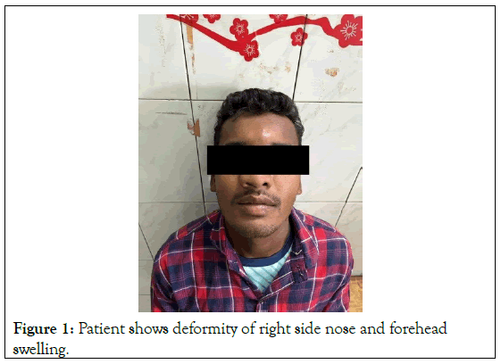
Figure 1: Patient shows deformity of right side nose and forehead swelling.
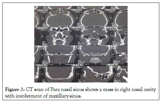
Figure 2: CT scan of Para nasal sinus shows a mass in right nasal cavity with involvement of maxillary sinus.
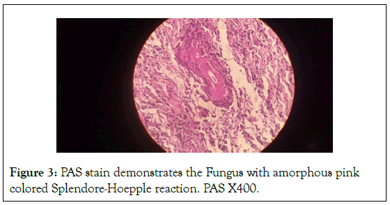
Figure 3: PAS stain demonstrates the Fungus with amorphous pink colored Splendore-Hoepple reaction. PAS X400.
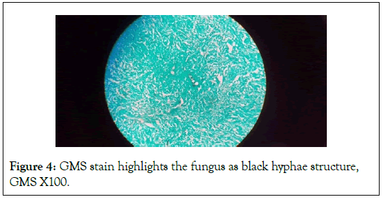
Figure 4: GMS stain highlights the fungus as black hyphae structure, GMS X100.
Discussion
Entomophthoro is a name used to describe fungi that are infectious to insects [2]. Entomophthoromycosis in humans has been observed in tropical nations around the world, as the warm and humid climate encourages the growth of this fungus. Zygomycetes were previously classified as Entomophthorales. Basidiobolus and Conidiobolus, the pathogenic fungi of interest, have recently formed a novel subphylum Entomopthoromycotina. These fungi have the ability to infect the skin, subcutaneous tissue, and even deeper organs. Immunocompromised individuals may require immediate treatment due to the risk of infection spreading to the mediastinum, kidney failure, and catastrophic cardiorespiratory consequences.
Insect bites, minor trauma, and iatrogenic causes are also possible sources of infection. In an autopsy performed on infected patients, the tracheobronchial tree has been identified as the pathogen's entrance point. The host responds by forming granulomas made up of lymphocytes, multinucleated giant cells, and epithelioid cells [3]. Despite the great number of instances in tropical locations around the world, literature report is scarce, which explains the fungi's low pathogenicity in humans. Conidiobolus produces enzymes like elastase, lipase, collagenase, and esterase in vitro, which may explain the pathogenicity of fungus in human hosts. These enzymes help fungus to enter the body and invade deeper viscera [4].
Agricultural field workers are particularly susceptible to entomophthorales because they are more common among insects. Our patient is a young agricultural field worker, which helps to explain how the condition developed. After a common cold infection, repeated exposure, or during a partial immunosuppressive phase, low pathogenic members of this phylum may impact humans [5]. Conidiobolus attacks the nose and sinuses. Blumenthrath, et al. identified atypical mucocutaneous conidiobolomycosis due to diffuse face involvement [6]. Basidiobolus, on the other hand, infects the buttocks, perineum, thigh, back, and thorax [4]. In Basidiobolus infection, deeper invasion and visceral involvement are less common. Rhinomycosis affects the nasal mucosa at first, and then develops into a nodule that grows in size and obstructs the airway. It could be ulcerated, causing pus to develop. Longterm untreated instances can infiltrate and destroy deeper organs such as the Para nasal sinuses and orbital floor, resulting in proptosis, visual impairment, periorbital edema, and reduced ocular reflexes [7]. Meningitis and elevated intracranial tension have been documented in immunocompromised patients as the fungi infiltrate the cerebral cavity.
The gold standard tests for diagnosing Entomophthoromycosis are histological identification and culture. The classic Splendor- Hoeppli phenomenon can be seen, albeit it is not pathognomonic, in various fungal illnesses such as sporotrichosis and nocardiosis. In a routine H&E stain, the broad, aseptate fungal hyphae may not be clearly visible. Entomophthorales stain poorly, despite the fact that Periodic acid–Schiff (PAS) emphasizes the hyphae of most fungus. To highlight the fungi, Grocott Methenamine Silver (GMS) is a better choice. Biopsy of the tissue rather than pus released from the ulcerated nodule is recommended because fungus is more invasive and concentrated in the tissue [8]. The Entomophthorales are distinguished from other species by their large, sparsely septate, and right-angled hyphae [9]. Polymerase Chain Reaction (PCR) and Immunohistochemistry (IHC) are required for definitive species identification. In Sabouraud Dextrose agar and potato agar, typical beige to brown flat colonies with aerial hyphae can be detected. The treatment of choice is surgical debridement followed by longterm antifungal medication. Our patient responded to itraconazole within 2 months of therapy. A similar situation was recently reported by Thomas, et.al [10]. Plastic surgery can be used to correct facial deformity. Culture was not performed in our case because the diagnosis was established by biopsy. Due to the lack of these infrastructures in our set up, PCR and IHC were not performed.
Conclusion
Entomophthoromycosis is a rare rhinomycosis seen in ordinary clinical practice. Disease is restricted to the nose and paranasal sinuses in immunocompetent patients, but immunocompromised patients may develop deadly consequences as a result of excessive treatment delays. Clinical awareness is required for early suspicion of the fungus and fast diagnosis in biopsy. The investigation method of choice is histological identification and culture. In these circumstances, both surgical debridement and medicinal therapy are essential.
References
- Kwon-Chung KJ. Taxonomy of fungi causing mucormycosis and entomophthoramycosis (zygomycosis) and nomenclature of the disease: molecular mycologic perspectives. Clin Infect Dis. 2012;54(suppl_1):S8-15.
[Crossref] [Google Scholar] [PubMed]
- Hibbett DS, Binder M, Bischoff JF, Blackwell M, Cannon PF, Eriksson OE, et al. A higher-level phylogenetic classification of the Fungi. Mycol. Res. 2007;111(5):509-547.
[Crossref] [Google Scholar] [PubMed]
- Kimura M, Yaguchi T, Sutton DA, Fothergill AW, Thompson EH, Wickes BL. Disseminated human conidiobolomycosis due to Conidiobolus lamprauges. J Clin Microbiol. 2011;49(2):752-756.
[Crossref] [Google Scholar] [PubMed]
- Shaikh N, Hussain KA, Petraitiene R, Schuetz AN, Walsh TJ. Entomophthoramycosis: a neglected tropical mycosis. Clin Microbiol Infect. 2016;22(8):688-694.
[Crossref] [Google Scholar] [PubMed]
- Vikram HR, Smilack JD, Leighton JA, Crowell MD, De Petris G. Emergence of gastrointestinal basidiobolomycosis in the United States, with a review of worldwide cases. Clin Infect Dis. 2012;54(12):1685-1691.
[Crossref] [Google Scholar] [PubMed]
- Blumentrath CG, Grobusch MP, Matsiégui PB, Pahlke F, Zoleko-Manego R, Nzenze-Aféne S, et al. Classification of rhinoentomophthoromycosis into atypical, early, intermediate, and late disease: a proposal. PLoS Negl Trop Dis. 2015;9(10):e0003984.
[Crossref] [Google Scholar] [PubMed]
- Wüppenhorst N, Lee MK, Rappold E, Kayser G, Beckervordersandforth J, de With K, Serr A. Rhino-orbitocerebral zygomycosis caused by Conidiobolus incongruus in an immunocompromised patient in Germany. J Clin Microbiol. 2010; 48(11):4322-4325.
[Crossref] [Google Scholar] [PubMed]
- Chowdhary A, Randhawa HS, Khan ZU, Ahmad S, Khanna G, Gupta R, et al. Rhinoentomophthoromycosis due to Conidiobolus coronatus. A case report and an overview of the disease in India. Sabouraudia. 2010;48(6):870-879.
[Crossref] [Google Scholar] [PubMed]
- El‐Shabrawi MH, Arnaout H, Madkour L, Kamal NM. Entomophthoromycosis: A challenging emerging disease. Mycoses. 2014;57:132-137.
[Crossref] [Google Scholar] [PubMed]
- Thomas G, Thekkethil JS, Koshy JM, Thomas SM. Rhinoentomophthoromycosis: An uncommon but not rare fungal infection of the nose. J Curr Res Sci Med. 2019;5(2):114.
Citation: Das U (2023) A Case Study on Nasal Entomophthoromycosis: An Uncommon Infection in a Twenty-Four-Year Young Male. J Trop Dis. 11:376.
Copyright: © 2023 Das U. This is an open-access article distributed under the terms of the Creative Commons Attribution License, which permits unrestricted use, distribution, and reproduction in any medium, provided the original author and source are credited.

