Indexed In
- The Global Impact Factor (GIF)
- CiteFactor
- Electronic Journals Library
- RefSeek
- Hamdard University
- EBSCO A-Z
- Virtual Library of Biology (vifabio)
- International committee of medical journals editors (ICMJE)
- Google Scholar
Useful Links
Share This Page
Journal Flyer

Open Access Journals
- Agri and Aquaculture
- Biochemistry
- Bioinformatics & Systems Biology
- Business & Management
- Chemistry
- Clinical Sciences
- Engineering
- Food & Nutrition
- General Science
- Genetics & Molecular Biology
- Immunology & Microbiology
- Medical Sciences
- Neuroscience & Psychology
- Nursing & Health Care
- Pharmaceutical Sciences
Case Report - (2024) Volume 23, Issue 3
A Case Report on Disproportionate Dwarfism with Differential Diagnosis
B. Karthika*, M. Pavani, T. Sridhar Reddy and S. KarthikaReceived: 27-Apr-2023, Manuscript No. OHDM-23-21139; Editor assigned: 01-May-2023, Pre QC No. OHDM-23-21139 (PQ); Reviewed: 15-May-2023, QC No. OHDM-23-21139; Revised: 13-Sep-2024, Manuscript No. OHDM-23-21139 (R); Published: 30-Sep-2024, DOI: 10.35248/2247-2452.24.23.1120
Abstract
Dwarfism is a condition wherein an individual has a short stature when compared to normal individuals. It is of two types, proportionate and disproportionate dwarfism. Achondroplasia is considered to be most common form of dwarfism. It is a genetic disorder that occurs due to mutation of Fibroblast Growth Factor Receptor (FGFR3). Following is a case report of a patient with dwarfism.
Keywords
Radiology; Dwarfism; Medicine diagnosis; Achondroplasia; Turner syndrome
Introduction
Dwarfism is a condition where an individual is exceptionally small. It is sometimes defined as an adult height of less than 147 centimeters. Regardless of gender, the average adult height with dwarfism is 122 centimeters. Dwarfism can be proportionate or disproportionate. Individuals with short limbs or a short torso are considered to be of disproportionate dwarfism. In cases of proportionate dwarfism, both limbs and torso are usually small. Intelligence is usually normal, and most of them have a nearly normal life expectancy [1]. The most common and recognizable form of dwarfism in humans is achondroplasia [2]. Achondroplasia is a form of disproportionate dwarfism. It is a genetic disorder that is due to mutation of Fibroblast Growth Factor Receptor (FGFR3) gene and it results in dwarfism, usually an autosomal dominant trait. The classical clinical features seen in disproportionate dwarfism are rhizomelic shortening of proximal limbs, short fingers and toes with trident hands, large head, frontal bossing, small mid-face, flattened nasal bridge, kyphosis or lordosis, virus (bow leg), and valgus (knock knee) deformities [3]. Oral manifestations include macroglossia, tongue thrust swallowing pattern, posterior crossbite, anterior open bite, anterior reverse overjet etc. Growth hormone deficiency is responsible for most of the other cases of dwarfism. Short stature with proportional body parts has a hormonal cause, such as growth-hormone deficiency, which is once called pituitary dwarfism.
Case Presentation
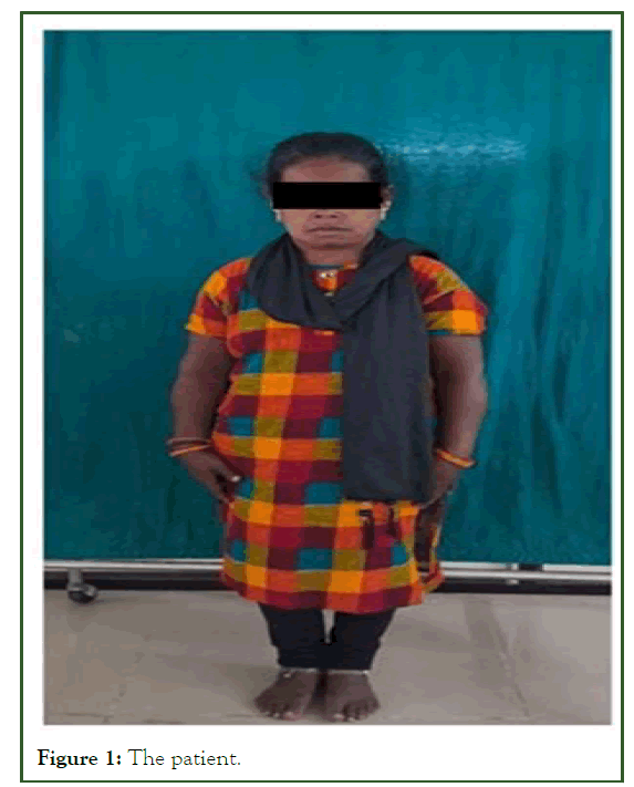
A 40 years old female reported to the department of oral medicine and radiology with a chief complaint of pain in her lower right back tooth region for past 5 days. History reveals that the pain is dull continuous, which radiates to the ears and head, aggravates upon eating and relieves upon rest. Patient gives a history of hyperthyroidism for past 10 years and is under medication. Past dental history reveals that the patient has undergone uneventful extraction before 1 month. Patient’s family history reveals that the patient’s parents had consanguineous marriage, patient’s elder sister is slightly taller than her and had a good intellectual ability when compared to the patient. Upon general examination patient is calm and cooperative but signs of low intellectual ability seen and had dull face and a short stature. The patient’s height was measured to be around 137 centimeters. On extraoral examination, the following signs are appreciated.
Face
• Frontal bossing (Figure 1).
• Flattening of malar prominence.
• Flattening of nasal bridge.
• Puffy face.
• Bimaxillary protrusion.
• Jaw deviation towards left side while opening.
• Clicking sound in right TMJ.

Figure 1: The patient.
Extremities
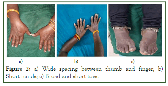
• Short extremities (Figures 2a and 2b).
• Broad and short fingers.
• Short toes (Figure 2c).
• Wide spacing between thumb and index finger.

Figure 2: a) Wide spacing between thumb and finger; b) Short hands; c) Broad and short toes.
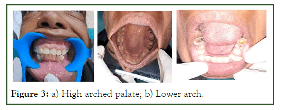
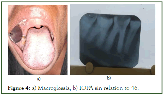
On intraoral examination, multiple teeth were decayed in relation to 17,18,26,35,38,46 and 48, root stumps in relation to 16, missing teeth in relation to 25 and 37, and a retained deciduous tooth in relation to 73 were seen. Tongue appeared bulbous, suggestive of macroglossia (Figure 3a). Palate is Ushaped, deep, and high arched (Figure 3b). In accordance with the history and clinical features, the case was provisionally diagnosed as dental caries with apical periodontitis in 46 .

Figure 3: a) High arched palate; b) Lower arch.
Intraoral periapical radiograph in relation to 46 showed radiolucency involving enamel, dentin and pulp with widening of periodontal ligament space (Figure 4a). Upon correlating with the history, clinical features and radiological findings this case was diagnosed as dental caries with apical periodontitis in 46. The patient had differential diagnosis for Dwarfism. The conditions in which dwarfism can be seen include, pituitary dwarfism due to growth hormone deficiency, achondroplasia, downs syndrome, turner syndrome, Spondyloepiphyseal Dysplasias (SED), noonan syndrome (Figure 4b).

Figure 4: a) Macroglossia; b) IOPA sin relation to 46.
Results and Discussion
Short stature seen in dwarfism could be due to an underlying disorder or a standard growth variation.
Growth hormone deficiency in children, also known as pituitary dwarfism occurs when the pituitary gland fails to produce adequate supply of growth hormone. Deficiency of growth hormone is a relatively common cause of proportionate dwarfism. The hypothalamic-pituitary axis maintains the levels of growth hormones in the body, which directly or indirectly through Insulin-like Growth Factor-1 (IGF-1), stimulates growth of soft tissue and cartilage and elongation of bones. Lower levels of IGF-1 is linked with short stature [4].
Achondroplasia is a type of short-limbed dwarfism. The prevalence of Achondroplasia is about 1 in 20,000 livebirths. Patients with achondroplasia have a gain-of-function mutation in the Fibroblast Growth Factor Receptor 3 gene (FGFR3 gene) [5]. In Achondroplasia there is abnormal endochondral ossification (Figure 5).
Downs syndrome also known as trisomy 21 is the most common chromosomal abnormality occurring in human population leading to mental retardation and retarded growth [6]. It is considered to be a genetic disorder with defect in all or part of a third copy of chromosome 21. Down’s syndrome is also associated with a number of other phenotypes including learning disability, heart problems, and leukemia in childhood as well as alzheimer’s disease [7]. Its incidence is around 1:660 live births [8]. Abnormalities in sexual development are also noted to be significant with delayed puberty in both genders.
Turner Syndrome (TS), also called as congenital ovarian hypoplasia syndrome is a genetic disorder where X chromosome is partially or completely missing in females. Clinical manifestations include growth disorders, abnormalities of reproductive system, cardiovascular abnormalities, and autoimmune diseases. This condition occurs in approximately 1 in every 2000 female births [9]. It is highly prevalent in China [10]. Short stature is one of the main causes of diagnosis in patients with turner syndrome [11]. Short stature and gonadal dysgenesis are the cardinal features of Turner syndrome. Affected girls may also encounter a wide range of problems from conductive deafness to schooling difficulties [12].
Spondyloepiphysial dysplasia congenita is a rare and inherited genetic disorder which results in short stature (dwarfism) and skeletal anomalies that primarily affects the spine and long bones of arms and legs. It is autosomal dominant. In SED growth deficiency usually occurs before birth and continues throughout life. Disproportionate short stature. There is a primary defect in type II collagen synthesis. Other manifestations include the flat face, cleft palate, barrel-shaped chest, muscle hypotonia, clubfoot, myopia and sensorineural deafness [13].
Noonan syndrome is an autosomal dominant genetic disorder with mildly unusual facial features such as short stature, congenital heart disease, bleeding problems, and skeletal malformations. Facial features include widely spaced eyes, lightcolored eyes, low-set ears, a short neck, and a small lower jaw. It has been discovered that mutations in the PTPN11 gene are the primary cause of Noonan syndrome. NS is characterized by distinct features including hypertelorism, down-slanting palpebral fissures, a high arched palate, and low set posteriorly rotated ears, malar hypoplasia, ptosis and a short or webbed neck. The general body appearance is similar to that of turner syndrome, and hence NS is also known as “pseudoturner syndrome” in females and “male turner syndrome” in males.
Conclusion
Thus, dwarfism can occur in various clinical conditions. Usually in most of the conditions short stature is one clinical manifestation among several others. A complete clinical and family history is important in establishing the exact cause of dwarfism.
References
- Pauli RM, Adam MP, Ardinger HH, Pagon RA, Wallace SE, Bean LJH, et al. Achondroplasia. Gene Rev. 2012.
- Cevik B, Colakoglu S. Anesthetic management of achondroplastic dwarf undergoing cesarean section-a case report. Middle East J Anaesthesiol. 2010;20(6):907-910.
[Google Scholar] [PubMed]
- Swathi KV, Maragathavalli G. Achondroplasia: A form of disproportionate dwarfism-A case report. Indian J Dent Res. 2020;31(5):794-798.
[Crossref] [Google Scholar] [PubMed]
- Ornitz DM, Legeai-Mallet L. Achondroplasia: Development, pathogenesis, and therapy. Dev Dyn. 2017;246(4):291-309.
[Crossref] [Google Scholar] [PubMed]
- Leonard H, Wen X. Epidemiology of mental retardation. Hum Mol Genet. 2009;18:75-83.
- Bertelli EC, Biselli JM, Bonfim D, Goloni-Bertollo EM. Clinical profile of children with Down syndrome treated in a genetics outpatient service in the southeast of Brazil. Rev Assoc Med Bras. 2009;55(5):547-552.
[Crossref] [Google Scholar] [PubMed]
- Nielsen J, Wohlert M. Chromosome abnormalities found among 34,910 newborn children: Results from a 13-year incidence study in Arhus, Denmark. Hum Genet. 1991;87(1):81-83.
[Crossref] [Google Scholar] [PubMed]
- Cui X, Cui Y, Shi L, Luan J, Zhou X, Han J. A basic understanding of Turner syndrome: Incidence, complications, diagnosis, and treatment. Intractable Rare Dis Res. 2018;7(4):223-238.
[Crossref] [Google Scholar] [PubMed]
- Orbananos IR, Desojo AV, Martinez-Indart L, Bolado GG, Estevez AR, Echevarria IR. Turner syndrome: From birth to adulthood. Endocrinol Nutr. 2015;62(10):499-506.
[Crossref] [Google Scholar] [PubMed]
- Donaldson MD, Gault EJ, Tan KW, Dunger DB. Optimising management in turner syndrome: From infancy to adult transfer. Arch Dis Child. 2006;91(6):513-520.
[Crossref] [Google Scholar] [PubMed]
- Saleem S, Anwar A, Iftikhar PM, Anjum Z, Tariq Z. Spondyloepiphyseal dysplasia congenita: A rare cause of respiratory distress. Cureus. 2019;11(7):e5101.
[Crossref] [Google Scholar] [PubMed]
- Noonan JA. Noonan syndrome and related disorders: Alterations in growth and puberty. Rev Endocr Metab Disord. 2006;7(4):251-255.
[Crossref] [Google Scholar] [PubMed]
- Lee SM, Cooper JC. Noonan syndrome with giant cell lesions. Int J Paediatr Dent. 2005;15(2):140-145.
[Crossref] [Google Scholar] [PubMed]
Citation: Karthika B, et al (2024) A Case Report on Disproportionate Dwarfism with Differential Diagnosis. Oral Health Dent Manag. 2024;23(3):1120
Copyright: © 2024 Karthika B, et al. This is an open access article distributed under the terms of the Creative Commons Attribution License, which permits unrestricted use, distribution, and reproduction in any medium, provided the original author and source are credited.
