Citations : 2345
Dentistry received 2345 citations as per Google Scholar report
Indexed In
- Genamics JournalSeek
- JournalTOCs
- CiteFactor
- Ulrich's Periodicals Directory
- RefSeek
- Hamdard University
- EBSCO A-Z
- Directory of Abstract Indexing for Journals
- OCLC- WorldCat
- Publons
- Geneva Foundation for Medical Education and Research
- Euro Pub
- Google Scholar
Useful Links
Share This Page
Journal Flyer

Open Access Journals
- Agri and Aquaculture
- Biochemistry
- Bioinformatics & Systems Biology
- Business & Management
- Chemistry
- Clinical Sciences
- Engineering
- Food & Nutrition
- General Science
- Genetics & Molecular Biology
- Immunology & Microbiology
- Medical Sciences
- Neuroscience & Psychology
- Nursing & Health Care
- Pharmaceutical Sciences
Research Article - (2020) Volume 10, Issue 6
Three-Dimensional Reconstruction and Measurement for Endodontic Assessment of Complex Root Morphologies with an Application Framework for Cone Beam Computed Tomography Images
Inbaraj Anand Sherwood1*, James L. Gutmann2 and Manu Unnikrishnan32Department of Endodontics, Nova Southeastern University College of Dental Medicine, Florida, USA
3Department of Conservative Dentistry and Endodontics, Chettinad Dental College, Kanchipuram, Tamil Nadu, India
Received: 03-Aug-2020 Published: 26-Aug-2020, DOI: 10.35248/2161-1122.20.10.564
Abstract
The purpose of this paper is to present a customizable application framework by using MeVisLab image processing and visualization platform for three-dimensional reconstruction and evaluation of tooth with complex root canal morphology using Cone Beam Computed Tomography (CBCT) images. Eleven different types of CBCT scans from teeth with complex root anatomy were reconstructed with a customizable application framework based on MeVisLab. This application framework provided an economical platform for three-dimensional assessments of complex root anatomies.
Keywords
Cone beam computed tomography; Complex root morphology; MeVisLab; Three-dimensional reconstruction
Introduction
One of the critical themes in endodontic research is the study of pulp and root canal morphology. Three-Dimensional (3D) reconstruction from high resolution data is a popular and valuable tool [1]. Micro-Computed Tomography (micro-CT) is non-destructive and allows for rapid examination of morphologic characteristics of a tooth in a detailed and accurate manner. While micro-CT is a costly 3D imaging tool that cannot be used clinically, however, Cone Beam Computed Tomography (CBCT) has been a tremendous clinical adjunct dentistry [2-5]. Gao et al. [1] in 2009, described about a freely available software package MeVisLab for endodontic 3D analysis using micro-CT scan images. MeVisLab (MeVis Research, Bremen, Germany) is a framework system for medical image processing and visualization platform for Windows (Microsoft Corp, Redmond, WA); it provides an easy to learn modular visual programming interface with a comprehensive suite of image processing and visualization tools [1]. Later the same software was applied and validated for volumetric analysis of regenerative endodontic procedures [5,6]. Maret et al. [7] highlighted the accuracy of CBCT images usage for 3D reconstruction compared with micro-CT.
Apart from volumetric analysis MeVisLab has other features uses, such as 3D reconstruction of tooth and root canal, geometric measurement of tooth and canal, evaluation of root canal preparation and risk analysis during virtual removal of broken instrument [1]. The purpose of this article is to create a modified customized application framework by using MeVisLab software for 3D reconstruction and assessing of complex pulp and root canal morphologies using patient CBCT images.
Materials and Methods
The MeVislab software package is freely available from www. mevislab.de/index. php=4 provides a visual data-flow program environment on its Graphic User Interface (GUI) (Figure 1). The boxes on the GUI are called modules and the wires connecting them are termed data pipes. Each module is assigned a specific function; it has a parameter panel providing a control to its functions the data pipes carry input and output data between them. As a whole the module and data pipes comprise a data-flow framework. Complex image processing networks can be established with different image modules combinations.
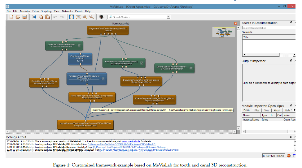
Figure 1: Customized framework example based on MeVisLab for tooth and canal 3D reconstruction.
CBCT image acquisition and export
Preoperative CBCT scans were acquired (Kodak CS 8100 3D,Carestream Dental, Atlanta, USA) with a FOV to a maximum of 8 teeth with an exposure of 80 KV, 5 mA, 19.96 s and a voxel size of 90 μm. This case series included 11 different permanent teeth from patients who presented to the Endodontic department for root canal treatment in the years between 2016 and 2020 (Figures 2-5). The CBCT images were acquired for these patients to better assess their variation in root canal morphology to enhance the treatment delivery. All the data of CBCT images were exported using the Digital Imaging and Communication in Medicine (DICOM) file format with voxel size of 90 μm.
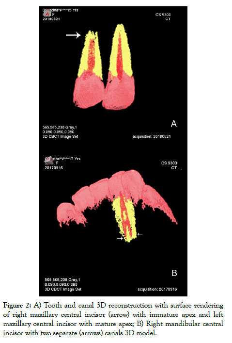
Figure 2: A) Tooth and canal 3D reconstruction with surface rendering of right maxillary central incisor (arrow) with immature apex and left maxillary central incisor with mature apex; B) Right mandibular central incisor with two separate (arrows) canals 3D model.
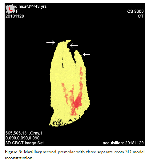
Figure 3: Maxillary second premolar with three separate roots 3D model reconstruction.
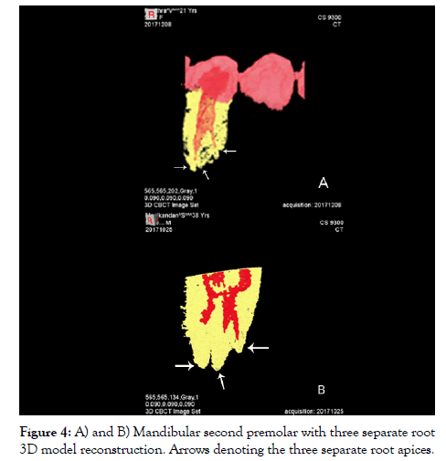
Figure 4: A) and B) Mandibular second premolar with three separate root 3D model reconstruction. Arrows denoting the three separate root apices.
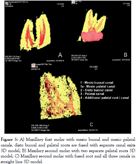
Figure 5: A) Maxillary first molar with mesio buccal and mesio palatal canals, disto buccal and palatal roots are fused with separate canal exits 3D model; B) Maxilary second molar with two separate palatal roots 3D model; C) Maxillary second molar with fused root and all three canals in straight line 3D model.
3D Reconstruction of tooth and canal
Only the DICOM files that depicted the crown and root portion of the concerned tooth structure were used for segmentation of the root canal system, dentin and enamel to generate the 3D models. This was done to reduce the computation time [5]. Individual steps in the workflow are depicted in Figure 1. The major steps included: (1) import the concerned tooth scanned image and create a 3D data set; (2) segment the root canal, dentin and enamel with region growing macro module; and, (4) create rendering views of canal, dentin and enamel with View 3D module. 3D model rendered can be saved as TIF (.tif) image file or as video file (.mp4).
Three segmentation steps were used to extract dentin from surrounding bone and to separate enamel from dentin and root canal space from dentin. Following segmentation, the 3D reconstruction of hard tissue and root canal was done without smoothening to preserve its raw volume measurement. Pulp space volume could be assessed using WEMinfo module.
Results
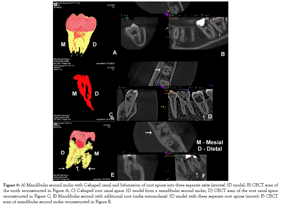
CBCT images of 11 different tooth types with varied canal anatomy were used for 3D reconstruction. Two incisors, premolars; three maxillary and mandibular molars included for the image acquisition. Figure 2 depicts a maxillary central incisor with immature apex along with mature apex contralateral tooth (Figure 2A) and mandibular incisor with two canals (Figure 2B). Figures 3 and 4 show maxillary and mandibular premolars with three roots, respectively. Figure 5 depict maxillary molars with two mesio-buccal canals with fused disto-buccal and palatal roots, two palatal roots, and three canals in a straight line in a fused root. Figure 6 shows a mandibular second molar with a C-shaped canal and additional roots; along with pulp canal 3D reconstruction of a C-shaped root. Figure 6 also depicts the concerned CBCT scan images of patients from which 3D reconstruction was obtained.
Figure 6: A) Mandibular second molar with C-shaped canal and bifurcation of root apices into three separate exits (arrows) 3D model; B) CBCT scan of the tooth reconstructed in Figure A; C) C-shaped root canal space 3D model from a mandibular second molar; D) CBCT scan of the root canal space reconstructed in Figure C; E) Mandibular second with additional root (radix entomolaris) 3D model with three separate root apices (arrow); F) CBCT scan of mandibular second molar reconstructed in Figure E.
Discussion
This study used a modified application framework described by Gao et al. [1] to reconstruct 3D tooth model with CBCT images of teeth with complex root anatomy. Modification from earlier framework was necessary as the present observation was with CBCT scan rather than micro-CT [1]. The teeth selected for this reconstruction had varied and complex root anatomy that extended the limits of MeVisLab software package. Reconstructed tooth/canal model adequately represented the actual morphology of tooth/canal. Previous research with MeVisLab in endodontic 3D reconstruction incorporated teeth subjected to regenerative endodontic procedures, mostly being single-rooted teeth [5,6]. In the current study teeth were selected, which had high degree of root/canal complexities along with maxillary incisor presenting immature apex to understand the full capabilities of this software. The models presented by the software showed that 3D reconstruction was acceptable for all the teeth in this study. The package used in this proof-of-principle, MeVisLab, consists of large set of powerful functions and allows the operator to customize with easy to use interface. In this study, an application framework is presented; thus, no statistically supported conclusions were presented. The present experimentation observed the performance of framework and usefulness for dental imaging analysis.
3D models constructed could be used for education of tooth and root morphology along with for some advanced applications. Root canal dimensions reconstruction along with metric information can be used for understanding and researching the changes in canal space. Computer-aided dental visualization is not only sufficient to display tooth data, but allows for performing simulations on the basis of 3D data for treatment planning and evaluation [1]. Gao et al. [1] recommended the CBCT because of its lower level radiation compared to micro-CT together with the framework based on MeVisLab software will enable endodontists to effectively evaluate root canal morphology. It was also predicted that CBCT images with MeVisLab software will provide better tool for assessing root and alveolar fractures and pre-surgical planning in root-end surgeries [1].
Conclusion
In conclusion, application framework based on MeVisLab with CBCT image enables a standardized method of 3D reconstruction and metrics of canal even in tooth with complex root and canal anatomy. This powerful tool could be widely used for contemporary 3D endodontic research in teeth with complex anatomy along with CBCT scan.
Conflict of Interest
None
REFERENCES
- Gao Y, Peters OA, Wu H, Zhou X. An application framework of three-dimensional reconstruction and measurement for endodontic research. J Endod. 2009;35:269-274.
- Esposito SA, Huybrechts B, Slagmolen P, Cotti E, Coucke W, Pauwels R, et al. A novel method to estimate the volume of bone defects using cone-beam computed tomography: an in vitro study. J Endod. 2013;39:1111-1115.
- Shahbazian M, Jacobs R, Wyatt J, Denys D, Lambrichts I, Vinckier F, et al. Validation of the cone beam computed tomography-based stereolithographic surgical guide aiding auto transplantation of teeth: clinical case-control study. Oral Surg Oral Med Oral Pathol Oral Radiol. 2013;115:667-675.
- Yang F, Jacobs R, Willems G. Dental age estimation through volume matching of teeth imaged by cone-beam CT. Forensic Sci Int. 2006;159:S78-S83.
- EzEldeen M, Van Gorp G, Van Dessel J, Vandermeulen D, Jacobs R. 3-dimensional analysis of regenerative endodontic treatment outcome. J Endod. 2015;41:317-324.
- Chrepa V, Joon R, Austah O, Diogenes A, Hargreaves KM, Ezeldeen M, et al. Clinical outcomes of immature teeth treated with rgenerative endodontic procedures-a san antonio study. J Endod. 2020;46:1074-1084.
- Maret D, Molinier F, Braga J, Peters OA, Telmon N, Treil J, et al. Accuracy of 3D reconstructions based on cone beam computed tomography. J Dent Res. 2010;89:1465-1469.
Citation: Sherwood IA, Gutmann JL, Unnikrishnan M (2020). Three-Dimensional Reconstruction and Measurement for Endodontic Assessment of Complex Root Morphologies with an Application Framework for Cone Beam Computed Tomography Images. Dentistry 10: 564. doi: 10.35248/2161- 1122.20.10.564
Copyright: © 2020 Sherwood IA, et al. This is an open-access article distributed under the terms of the Creative Commons Attribution License, which permits unrestricted use, distribution, and reproduction in any medium, provided the original author and source are credited.
Sources of funding : Nil
