Citations : 2345
Dentistry received 2345 citations as per Google Scholar report
Indexed In
- Genamics JournalSeek
- JournalTOCs
- CiteFactor
- Ulrich's Periodicals Directory
- RefSeek
- Hamdard University
- EBSCO A-Z
- Directory of Abstract Indexing for Journals
- OCLC- WorldCat
- Publons
- Geneva Foundation for Medical Education and Research
- Euro Pub
- Google Scholar
Useful Links
Share This Page
Journal Flyer

Open Access Journals
- Agri and Aquaculture
- Biochemistry
- Bioinformatics & Systems Biology
- Business & Management
- Chemistry
- Clinical Sciences
- Engineering
- Food & Nutrition
- General Science
- Genetics & Molecular Biology
- Immunology & Microbiology
- Medical Sciences
- Neuroscience & Psychology
- Nursing & Health Care
- Pharmaceutical Sciences
Research Article - (2025) Volume 15, Issue 3
Orthopantomographic Evaluation of the Oral and Dental Condition of the Population at the Biamba Marie Mutombo Hospital in Kinshasa
Nsonde Nsiku Guillain1*, Mukaya Tshibola Jean1,2, Molua Aundu Antoine1, Lelo Tshikwela Michel1, Luyeye Mvila Gertrude1, Yanda Tongo Stephane1, Malenga Mpaka serge1, Moyo Kimfuidi Nancy1, Mbongo Tanzia Angèle1, Nsiala suamunu Erba3, Tshienda Batuambila Marie3 and Mbungu Mabiala Richard3Department of Radiology and Medical Imaging, Kinshasa General Reference Hospital, Kinshasa, Democratic Republic of Congo
2Department of Radiology and Medical Imaging, Biamba Marie Mutombo Hospital, Kinshasa, Democratic Republic of Congo
Received: 09-Jan-2024, Manuscript No. DCR-24-24566; Editor assigned: 12-Jan-2024, Pre QC No. DCR-24-24566 (PQ); Reviewed: 26-Jan-2024, QC No. DCR-24-24566; Revised: 11-Mar-2025, Manuscript No. DCR-24-24566 (R); Published: 18-Mar-2025
Abstract
Introduction: Oral health is an important parameter for assessing the oral health of a population in order to adopt an adapted health policy with preventive purposes but also to assess the needs of this population with regard to oral care-dental.
Materials and methods: This is a single-center documentary study of data from the medical files of patients who performed an orthopantomogram at the Biamba Marie Mutombo hospital. Over a period of 24 months, going from January 2020 to January 2022, which included 688 patients of all ages without discrimination of sex.
Results: With a total of 688 panoramics of patients without gender predominance with a sex ratio of 1:1. Agemean was 40.16 ± 20.29 years with the extremes of 6 years to 89 years. The age group of 20 to 39 was the most represented. The teeth of the incisor-canine sector were the most present. Wisdom teeth were the most absent, whatever the age group, with a female predominance, whether mandibular or maxillary; followed by the 2nd premolars and the 1st maxillary molars. The premolars were the most dilapidated; and the teeth in the posterior sector are those, which have benefited the most from composite treatment.
Keywords
Oral condition; Orthopantomogram; Oral diseases
Introduction
The objective of assessing the oral health of a given population is to adopt an adapted health policy with preventive purposes, but also to assess the needs of this population with regard to oral care. Oral diseases currently constitute, taking into account their prevalence in the world, a real public health problem. All over the world, in industrialized countries as well as, increasingly, in developing countries, especially in the poorest communities [1,2].
This article reports the results of a survey carried out among 688 patients residing in Kinshasa and having attended the radiology and medical imaging department of the Biamba Marie Mutombo hospital in Kinshasa in the Democratic Republic of Congo. The aim is to establish an orthopantomographic profile of the oral and dental condition of the said population.
Materials and Methods
This was a single-center documentary study of data from the medical files of patients who underwent OPT at the Biamba Marie Mutombo Hospital (HBMM). Over a period of 24 months, from January 2020 to January 2022; it involved 688 patients without discrimination of age or sex. The sample was divided into four groups based on age as follows: 0 to 19 years old; from 20 to 39 years old; from 40 to 59 years old and from 60 years old and over.
The assessment of the oral condition of the population was carried out according to four parameters: Intact teeth, missing teeth, teeth with composites and dilapidated teeth.
Variations depending on age and sex of these parameters were taken into account.
Results
Oral condition of the study population
The following graph (Figure 1) represents the distribution of the different parameters of the study. We immediately notice that the teeth in the incisor-canine sector are the most present in the mouth and that they are the least affected. We also note a high proportion of missing wisdom teeth, whether mandibular or maxillary.
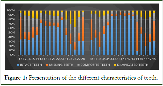
Figure 1: Presentation of the different characteristics of teeth.
Missing teeth
The graph below (Figure 2) represents the distribution of missing teeth in the oral cavity and we see a high proportion of missing wisdom teeth, whether mandibular or maxillary. Except for the 3rd molars, it is the 2nd premolars and the 1st maxillary molars which are most often absent. The incisor-canine sector is the most preserved sector since the average percentage of absence does not exceed 10%. The most common teeth are the canines and more particularly the mandibular canines.
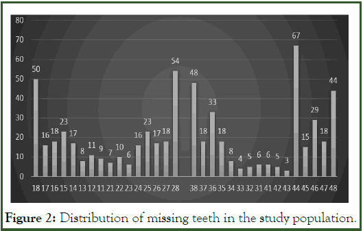
Figure 2: Distribution of missing teeth in the study population.
We note whatever the age, it is always the third molars which are the most absent (Table I). We also note a slight increase in the percentage of absence of canines and more particularly in the mandible. Statistically, we see that the p value is less than 0.01. This means that age influences the number of missing teeth.
| 18 | 17 | 16 | 15 | 14 | 13 | 12 | 11 | 21 | 22 | 23 | 24 | 25 | 26 | 27 | 28 | |
|---|---|---|---|---|---|---|---|---|---|---|---|---|---|---|---|---|
| 0 to 19 years old | 22.3 | 1.1 | 2.1 | 8.3 | 5.4 | 0 | 2.4 | 1.6 | 0.4 | 1 | 0 | 4.8 | 7.4 | 1.1 | 1.1 | 21 |
| 20 to 39 years old | 57.1 | 13.6 | 14.3 | 18.8 | 12.3 | 2.6 | 6.5 | 4.5 | 5.2 | 7.1 | 1.9 | 11 | 20.1 | 14.3 | 14.9 | 52.6 |
| 40 to 59 years old | 71.6 | 26.4 | 37.1 | 40.7 | 35 | 14.3 | 22.1 | 20 | 17.1 | 20 | 14.3 | 28.6 | 37.9 | 32.1 | 32.1 | 75.7 |
| ≥ 60 years old | 79 | 42 | 47 | 46 | 42 | 21 | 23 | 23.1 | 24 | 23.2 | 24 | 20.3 | 39 | 47 | 45.1 | 84.2 |
| 48 | 47 | 46 | 45 | 44 | 43 | 42 | 41 | 31 | 32 | 33 | 34 | 35 | 36 | 37 | 38 | |
| 0 to 19 years old | 20.5 | 2.2 | 3.3 | 5.4 | 2.7 | 0 | 0.5 | 0 | 0 | 0.5 | 0 | 3.4 | 6.3 | 2.9 | 1.5 | 19.5 |
| 20 to 39 years old | 40.4 | 14.6 | 38.8 | 23.9 | 11.9 | 4.5 | 3.7 | 5.2 | 4.5 | 3.7 | 3.7 | 11.2 | 15.7 | 44 | 25.4 | 52.2 |
| 40 to 59 years old | 58.2 | 23.9 | 38.8 | 23.9 | 11.9 | 4.5 | 3.7 | 5.2 | 4.5 | 3.7 | 5 | 13.6 | 29.3 | 55.7 | 35 | 61.4 |
| ≥ 60 years old | 68 | 50 | 52 | 33 | 15 | 9 | 13 | 18 | 21 | 13 | 6 | 18 | 30 | 52 | 41 | 65 |
Table I: Distribution of missing teeth according to age and location in the mouth.
Figure 3 indicates that there is on average a slightly higher percentage of missing teeth in women, with the main difference being in wisdom teeth (p<0.001). For the other teeth, the difference is not significant.

Figure 3: Distribution of missing teeth according to sex.
Dilapidated teeth
The following graph (Figure 4) represents the distribution of dilapidated teeth and we note that the most dilapidated teeth are those of the posterior mandibular or maxillary sector, in particular the premolars and two first molars.
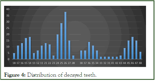
Figure 4: Distribution of decayed teeth.
Composite teeth
The following figure (Figure 5) represents the teeth having benefited from treatment with composites.
We note that the teeth in the posterior, mandibular or maxillary sector were those which benefited the most from composite treatment; in revenge, the incisor-canine block, particularly the mandibular block, was particularly not treated with composite.
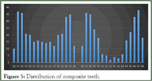
Figure 5: Distribution of composite teeth.
Discussion
In our study, we note that the teeth of the incisor-canine sector are the most present in the mouth and that they are the least affected. We also note a high proportion of missing wisdom teeth, whether mandibular or maxillary.
Indeed a very high proportion of missing wisdom teeth evolve in increasing age groups with already from 0 to 19 years an average percentage of 21.7% at the maxillary level and 20% at the mandible; for the age group of 60 years and over the average is 81.5% at the maxillary level and 66.5% at the mandible. We can therefore see that its position in the oral environment which makes it difficult to clean and its high prevalence of inclusion lead to a high proportion of pathology and therefore to its extraction. So, for Akadri OA, et al. [3] and Olasoji HO, et al. [4] the main reason for extraction is pericoronitis and for Adeyemo WL, et al. [5], the main cause of extractions is caries (and its consequences) for 62.3%.
The first mandibular molars and the maxillary premolars also represent a high percentage of absence, most probably related to caries and its consequences, periodontal problems and orthodontics as highlighted by several authors [6,7].
Composite teeth with restoration have a higher prevalence in the first two molars, whether maxillary or mandibular, consistent with the studies of Adeyemo WL [5] and Bourgeois D, et al. [8].
Concerning dilapidated teeth, they are mainly found in the posterior sectors. These results corroborate those of the entire literature [9,10].
Conclusion
In conclusion, the orthopantomographic evaluation of the oral and dental condition of the population at Biamba Marie Mutombo Hospital in Kinshasa reveals significant insights into common dental issues prevalent in the area. The study highlights a high prevalence of dental caries, periodontal diseases, and other oral health complications. It underscores the need for increased preventive measures, oral hygiene education, and accessible dental care. The findings call for strengthened public health initiatives to address the growing burden of dental conditions and improve overall oral health outcomes for the population in Kinshasa.
References
- Petersen PE. Rapport sur la sante bucco-dentaire dans le monde. 2003:48.
- Pr JM. Sante bucco-dentaire du jeune enfant: Connaissances et pratiques des professionnels de sante de perinatalite.
- Akadiri OA, Okoje VN, Fasola AO, Olusanya AA, Aladelusi TO. Indications for the removal of impacted mandible third molars at Ibadan--any compliance with established guidelines?. Afr J Med Med Sci. 2007;36(4):359-363.
[Google Scholar] [PubMed]
- Olasoji HO, Odusanya SA, Ojo MA. Indications for the extraction of impacted third molars in a semi-urban Nigerian Teaching Hospital. Niger Postgrad Med J. 2001;8(3):136-139.
[Google Scholar] [PubMed]
- Adeyemo WL, James O, Ogunlewe MO, Ladeinde AL, Taiwo OA, Olojede AC. Indications for extraction of third molars: a review of 1763 cases. Niger Postgrad Med J. 2008;15(1):42-46.
[Google Scholar] [PubMed]
- Al Najam Y, Tahmaseb A, Wiryasaputra D, Wolvius E, Dhamo B. Outcomes of dental implants in young patients with congenital versus non-congenital missing teeth. Int J Implant Dent. 2021;7:92.
[Crossref] [Google Scholar] [PubMed]
- Albrektsson T, Zarb G, Worthington P, Eriksson AR. The long-term efficacy of currently used dental implants: A review and proposed criteria of successs. Int J Oral Maxillofac Implants. 1986;1(1):11-25.
[Google Scholar] [PubMed]
- Bourgeois D, Berger P, Hescot P, Leclercq MH, Doury J. Oral health status in 65-74 years old adults in France, 1995. Rev Epidemiol Sante Publique. 1999;47(1):55-59.
[Google Scholar] [PubMed]
- Kirkevang LL, Orstavik D, Horsted-Bindslev P, Wenzel A. Periapical status and quality of root fillings and coronal restorations in a Danish population. Int End J. 2000;33(6):509-515.
[Crossref] [Google Scholar] [PubMed]
- Kronstrom M, Palmqvist S, Soderfeldt B. Changes in dental conditions during a decade in a middle-aged and older Swedish population. Acta Odontol Scandin. 2001;59(6):386-389.
[Crossref] [Google Scholar] [PubMed]
Citation: Guillain NN, Jean MT, Antoine MA, Michel LT, Gertrude LM, Stephane YT, et al. (2025) Orthopantomographic Evaluation of the Oral and Dental Condition of the Population at the Biamba Marie Mutombo Hospital in Kinshasa. J Dentistry. 15:725.
Copyright: © 2025 Guillain NN, et al. This is an open access article distributed under the terms of the Creative Commons Attribution License, which permits unrestricted use, distribution, and reproduction in any medium, provided the original author and source are credited.
