

Indexed In
- Open J Gate
- Academic Keys
- ResearchBible
- China National Knowledge Infrastructure (CNKI)
- Centre for Agriculture and Biosciences International (CABI)
- RefSeek
- Hamdard University
- EBSCO A-Z
- OCLC- WorldCat
- CABI full text
- Publons
- Geneva Foundation for Medical Education and Research
- Google Scholar
Useful Links
Share This Page
Journal Flyer
Open Access Journals
- Agri and Aquaculture
- Biochemistry
- Bioinformatics & Systems Biology
- Business & Management
- Chemistry
- Clinical Sciences
- Engineering
- Food & Nutrition
- General Science
- Genetics & Molecular Biology
- Immunology & Microbiology
- Medical Sciences
- Neuroscience & Psychology
- Nursing & Health Care
- Pharmaceutical Sciences
Case Report - (2026) Volume 15, Issue 2
Isolated Primary Hydatid Cyst of the Eyelid: A Rare Case Report
Nidhi Pandey, S.L. Adile, Richa Nayak* and Suprio DattaReceived: 13-Nov-2024, Manuscript No. JTD-24-27500; Editor assigned: 15-Nov-2024, Pre QC No. JTD-24-27500 (PQ); Reviewed: 29-Nov-2024, QC No. JTD-24-27500; Revised: 15-May-2026, Manuscript No. JTD-24-27500 (R); Published: 22-May-2026, DOI: 10.35241/2329-891X.26.15.511
Abstract
Eccinococcus granulosus otherwise known as Eccinococcus alveolaris tapeworm causes parasitic disease known as hydatid cyst. It affects mostly the liver, lungs and CNS, and rarely bones and orbit. Orbital hydatid cyst comprises <1% of all hydatid cases. Our case showed hydatid cyst in the eyelid which is very rare. Treatment of choice is surgery without rupturing the cyst, adjuvant medical therapy with oral albendazole decreases the chance of relapse and size of cyst.
Keywords
Hydatid cyst; Eyelid; Orbit; Eccinococcus granulosus
Introduction
Hydatid disease is a parasitic infection caused by the larval stage of Ecchinococcus granulosus. Found to be endemic in rural population of the Mediterranean regions, South Africa, South America, Australia and Middle East. In India the disease is still endemic, more common in Southern States (predominantly in Andhra Pradesh and Tamil Nadu). Dog is definitive host of the disease, cow and sheep intermediate host and humans are accidental intermediate hosts in the life cycle of Eccchinococcus granulosus. Common sites of involvement being liver (60-70%), lungs (30%), brain, rarely orbit. Orbital hydatid cysts are extremely rare and accounts for <1% of all Hydatid eye diseases. Complete surgical removals without rupture of the cyst remains gold standard.
Case Presentation
A 6 years old male child presented to the ophthalmology OPD with swelling in the upper eyelid of the left eye since past 3 months, gradually progressive in size with on and off pain. On examination swelling in upper eyelid of left eye measuring 2 × 1.5 cm close to the lateral canthus but away from lid margin, cystic in consistency with well-defined borders, mild tenderness was present and skin over it was free. Swelling adherent to the deeper structures. Moderate mechanical ptosis of left upper eyelid, conjunctival congestion underneath the swelling. No extension of mass into the orbit on manual examination. BCVA 6/6 both eyes. Rest anterior and posterior segment examination was within normal limits of both eyes. History of contact with cattles present. There was no history of trauma to the eye. No history of any other chronic illness. Hordeolum Internum was suspected and a course of antibiotics was given to the patient. On review the swelling did not subside and patient was posted for surgery under GA [1].
Patient underwent routine blood investigations which came out to be within normal limits. Chest X-ray and USG abdomen revealed normal findings. X-ray of orbit was normal. Patient underwent excision of mass under general anesthesia after pre-anesthetic clearance [2].
Incision was made on the eyelid skin (8 mm from eyelid margin), subcutaneous layer including orbicularis fibers dissected and a well encapsulated mass measuring approx 2 × 1 × 1 cm delivered in toto and was sent for histopathological examination. Eyelid was repaired using 7-0 vicryl suture. Histopathology showed wall of cyst with lamellated wall with germinal epithelial linning consistent with hydatid cyst with granulomatous response [3].
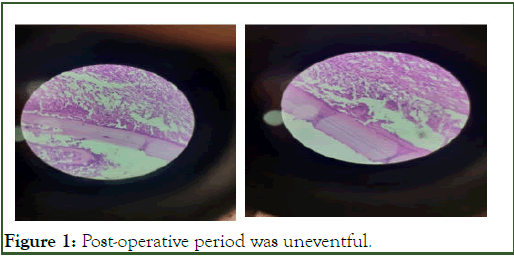
Figure 1: Post-operative period was uneventful.
On-post Op day 1 mild ptosis with lid edema was present. Post operatively LPS function was normal and EOM were full and free in all directions. The patient was evaluated for other foci of the disease in the body and was consulted with department of infectious diseases. No other organ was found to be involved which was rare occurrence. Patient was discharged on oral albendazole 15 mg/kg for 28 days and topical antibiotic. Follow up was done twice and wound was healthy [4]. No recurrences till date.
Hydatid disease, caused by the Echinococcosis species, is a parasitic infection typically affecting organs such as the liver and lungs. It is contracted through the ingestion of eggs from infected canines. While the condition is most commonly seen in the abdominal organs, ocular and orbital involvement remains exceedingly rare. This report presents a unique case of an isolated primary hydatid cyst found in the eyelid, a highly unusual location for this disease.
Hydatid cysts generally form as a result of the parasitic larvae being deposited in a host's tissues. The cysts grow over time and can cause damage to surrounding organs. In most cases, hydatid cysts are secondary, meaning they arise from distant metastasis of the primary cyst in organs like the liver or lungs. However, primary hydatid cysts can occasionally be found in unusual locations, including the eyelid, where they present significant diagnostic challenges [5].
A 45-year-old male patient presented with a gradually enlarging, painless swelling on his left upper eyelid. The swelling had been present for several months, without associated pain, redness, or discharge. On clinical examination, a firm, cystic mass was palpable, but there were no signs of acute infection or inflammation. Initially, the differential diagnosis included benign lesions such as a sebaceous cyst or chalazion, which are more common causes of eyelid masses [6].
Further imaging studies were performed, revealing a wellcircumscribed cystic lesion within the eyelid. Ultrasonography showed an anechoic mass, suggestive of a cystic structure, while a contrast-enhanced CT scan of the orbit confirmed its location and size without involvement of adjacent structures. The patient had no systemic symptoms or other health issues, and there was no history of trauma to the eyelid. Interestingly, upon further inquiry, the patient disclosed past travel to a region known for endemic Echinococcus infections, raising suspicion for a hydatid cyst.
Surgical excision of the cyst was carried out, and the specimen was sent for histopathological examination. The pathology report confirmed the diagnosis of an isolated primary hydatid cyst, revealing the laminated membrane and protoscolices typical of Echinococcus granulosus. The patient recovered well after the surgery, with no recurrence or complications noted during follow-up [7].
This rare case underscores the importance of considering hydatid disease in the differential diagnosis of eyelid masses, particularly in individuals with a history of exposure to regions endemic to Echinococcus. While primary hydatid cysts of the eyelid are extremely uncommon, awareness of this potential diagnosis is crucial for timely and effective management [8].
Results and Discussion
Orbital hydatid cyst is a rare occurrence and is most commonly observed in younger people as compared to older, it can be primary or secondary depending on the organs of involvement. Primary hydatid cyst of eyelid is extremely rare, as presented in our case [9].
The cyst is a two walled structure, containing antigenic fluid. Outer layer is laminated ectocyst which acts as a wall and avoids tissue invasion; inner germinal layer contains daughter cyst which produces scolioses giving rise to tapeworms. Hence, cyst removal without damaging the wall is very important so as to prevent recurrences.
The CASONI’S Intradermal test shows immediate hypersensitivity reaction with sensitivity and low specificity. USG, CT, MRI are valuable diagnostic aid. In USG a characteristic ‘double wall sign’ is often diagnostic. However histopathological examination remains the definitive diagnostic modality. Serological tests are used for diagnosis of hydatid cyst, are usually negative in case of primary orbital involvement. In India, only one case of Eyelid hydatid cyst has been reported in 1973, in West Bengal. No new cases were reported after that till now in eyelid.
Treatment for ocular hydatid illness consists of surgically excising the cyst after a 2–4 week course of anthelmintic medication, preferably albendazole. In order to avoid parasite seeding and allergic reactions in the event that the cyst bursts during manipulation during surgery, preoperative anthelmintic therapy is essential. If a cyst ruptures during surgery, further spread would be prevented by irrigation with hypertonic saline and hydrogen peroxide to kill the daughter cyst.
Conclusion
For the great majority of these individuals, timely surgical therapy following an early diagnosis of orbital hydatidosis produces excellent outcomes. This emphasizes how vital it is to get an accurate diagnosis. In the differential diagnostic list for orbital mass, the physician should always include the diagnosis of hydatid cyst, particularly in paediatric patients. The rise in global tourism and mobility should prompt clinicians to consider this extremely uncommon diagnosis, especially in endemic as well as non-endemic regions.
Results
- Baruah A, Sarma K, Barman B, Phukan P, Nath C, Boruah P, et al. Clinical and laboratory presentation of hydatid disease: A study from Northeast India. Cureus. 2020;12:e10260.
[Crossref] [Google Scholar] [PubMed]
- Almulhim AM, John S. Echinococcus granulosus. 2023.
- Hosaini SF, Qader AQ, Neyazi M, Razaqi N, Satapathy P, Afzali H, et al. Orbital hydatid cyst and its successful treatment: A case report from Afghanistan. Am J Ophthalmol Case Rep. 2024;36:102140.
[Crossref] [Google Scholar] [PubMed]
- Debela AS, Abore KW, Worke AB, Wendimagegn ST. Primary intra-orbital hydatid cyst: A case report of a rare cause of exophthalmos. Int Med Case Rep J. 2024;17:89-92.
[Crossref] [Google Scholar] [PubMed]
- Thambidurai L, Santhosham R, Dev B. Hydatid cyst: Anywhere, everywhere. Radiol Case Rep. 2015;6:486.
[Crossref] [Google Scholar] [PubMed]
- Aggarwal S, Bansal A. Laparoscopic management of renal hydatid cyst. JSLS. 2014;18:361-366.
[Crossref] [Google Scholar] [PubMed]
- Betharia SM, Sharma V, Pushker N. Ultrasound findings in orbital hydatid cysts. Am J Ophthalmol. 2003;135:568-570.
[Crossref] [Google Scholar] [PubMed]
- Sen SC, Mukherji AK. Hydatid cyst in eyelid. Indian J Ophthalmol. 1973;21:143-145.
[Google Scholar] [PubMed]
- Sayek I, Tirnaksiz MB, Dogan R. Cystic hydatid disease: Current trends in diagnosis and management. Surgery Today. 2004;34:987-996.
[Crossref] [Google Scholar] [PubMed]
Citation: Pandey N, Adile SL, Nayak R, Datta S (2026) Isolated Primary Hydatid Cyst of the Eyelid: A Rare Case Report. J Trop Dis. 15:511.
Copyright: © 2026 Pandey N, et al. This is an open access article distributed under the terms of the Creative Commons Attribution License, which permits unrestricted use, distribution, and reproduction in any medium, provided the original author and source are credited.