


Indexed In
- Open J Gate
- Genamics JournalSeek
- Academic Keys
- JournalTOCs
- CiteFactor
- Ulrich's Periodicals Directory
- Access to Global Online Research in Agriculture (AGORA)
- Electronic Journals Library
- Centre for Agriculture and Biosciences International (CABI)
- RefSeek
- Directory of Research Journal Indexing (DRJI)
- Hamdard University
- EBSCO A-Z
- OCLC- WorldCat
- Scholarsteer
- SWB online catalog
- Virtual Library of Biology (vifabio)
- Publons
- Geneva Foundation for Medical Education and Research
- Euro Pub
- Google Scholar
Useful Links
Share This Page
Journal Flyer

Open Access Journals
- Agri and Aquaculture
- Biochemistry
- Bioinformatics & Systems Biology
- Business & Management
- Chemistry
- Clinical Sciences
- Engineering
- Food & Nutrition
- General Science
- Genetics & Molecular Biology
- Immunology & Microbiology
- Medical Sciences
- Neuroscience & Psychology
- Nursing & Health Care
- Pharmaceutical Sciences
Research Article - (2023) Volume 14, Issue 4
Incidence of Foliage Fungal Diseases on Seedlings of Social Forest Nursery in Mysore, Karnataka, India
Naguvanahalli Somashekar Bhavana* and Gottravalli Ramanayaka JanardhanaReceived: 31-May-2023, Manuscript No. JPPM-23-21575; Editor assigned: 05-Jun-2023, Pre QC No. JPPM-23-21575 (PQ); Reviewed: 19-Jun-2023, QC No. JPPM-23-21575; Revised: 26-Jun-2023, Manuscript No. JPPM-23-21575 (R); Published: 03-Jul-2023, DOI: 10.35248/2157-7471.23.14.682
Abstract
Social forest nursery in Mysore district maintained by State Forest Department of Karnataka (India) was surveyed for the incidence of fungal diseases. Incidence, severity and spread of the disease were recorded and fungal pathogens were isolated and identified. Forestry species raised in nursery bags includes Azadirachta indica, Bauhinia variegata, Cinnamomum verum, Dalbergia latifolia, Dendrocalamus strictus, Mangifera indica, Phyllanthus emblica, Pterocarpus santalinus, Pterospermum acerifolium and Tamarindus indica were found infected with foliage diseases. Some of the important pathogens include species of fungi belonging to genera Alternaria, Cercospora, Cladosporium, Colletotrichum, Helminthosporium, Oidium, Ravenelia etc. Seedling congestion and overcrowding often resulted in spread of foliar fungal pathogens. The study helped us to understand the incidence of fungal pathogens on seedlings in social forest nursery of Mysore. Since seedlings grown in forest nurseries are the primary sources of planting stock, it is necessary to investigate the associated pathogens. There is a strong need for better understanding of these pathogens for developing effective management strategies.
Keywords
Diseases, Forest nursery, Fungal pathogens, Fungal disease
Introduction
Forestry in India is more than just about wood and fuel, which creates habitats for wildlife, serve in water and soil conservation, stabilize slopes, protect against winds and support human enjoyment and recreation [1]. Forestry as a segment plays an important role not only in maintaining ecological stability, but also in the socio- economic and rural development in a developing country like India. The majority of planting stock used in forestry comes from forest nurseries that specialize in its production. A forest nursery is a place where plants of tree species were propagated and grown to plantable size for regeneration of forests and social forestry. Healthy plants are goal of every nursery. This is not only restricted to forest nurseries but applies to nurseries of all sizes and levels of sophistication. Maintenance of seedlings in forest nurseries ranges from 3 to 18 months or more depending upon the tree species, climatic conditions as well as planting technique followed. Raising and maintaining disease free healthy planting stock for large scale planting program is often risky and failures of nurseries due to the outbreak of diseases are not uncommon. Large scale mortality in nurseries due to diseases could seriously affect the plantation programs by reducing stock of seedlings. The seedlings in nurseries are often infected by many pathogens like viruses, bacteria, fungi, nematodes and mycoplasma-like organisms. They cause heavy damage to seedlings and hence reduce both quality and quantity of planting stock. There is a relationship between the presence of the pathogen, host and suitable conditions for the pathogen, but in general there is always risk of plant diseases in forest nurseries [2]. Fungal pathogens are the most prevalent and the diseases caused by them are a serious problem in forest regeneration [3]. Abiotic stress caused by environmental conditions or injury can also predispose a large number of rapidly growing seedlings to fungal attack [2]. The conditions like high densities, irrigation, fertilization and herbicides that make nursery production of seedlings successful may also favor pathogenic fungi [4]. However, most diseases known to cause problems in forestry species today are fungal and gain access to the host plant during periods of physiological stress or through localized sites of damaged or diseased tissue [5]. The seedlings are susceptible to several diseases because of their tender tissues and they find difficulty in establishing themselves. In addition poor seedlings are likely to have slower growth and more liable to damage by insects and pests. When such diseased, substandard seedlings are used as planting stock, they further spread the diseases to plantations and to forests causing heavy damage [3]. Therefore raising disease free, healthy tree seedlings is not only important for maintaining a good nursery stock but also essential in establishing a healthy stand in field for better productivity. There is a strong need for better understanding of these pathogens in order to develop effective management strategies. Hence it was envisaged to investigate and to identify the fungal pathogens of plant seedlings in social forest nursery in Mysore district.
Materials and Methods
Sample collection
Mysore is selected as the study area located in Karnataka state, India, as it is consisting of many social forest nurseries. The forest nursery chosen for the investigation was the ‘Karnataka state forest urban greening nursery’, in Paduvaralli area of Mysore district in the month January to June 2022 (Figure 1). In the present investigation the tree species were identified, data of their economic importance were collected and diseased leaves from the infected seedlings were collected in separate sterilized polythene covers from the chosen forest nursery. The Botanical name of the infected seedlings in the nursery was identified with the help of a Taxonomist. The extent of damage caused and disease symptomology were photographed and recorded in the nursery. Symptoms on seedlings were carefully observed in forest nursery.

Figure 1: Map showing the location of social forest nursery selected for the study in Mysore district.
Morphological examination of plants
The collected plant parts were examined morphologically for their color, texture and appearance of infection on leaves were carefully observed with the help of magnifying glass. The nature of disease and symptoms of infection were recorded.
Identification of fungal pathogens
The diseased leaf samples were collected in transparent air tight polythene bags from nurseries and brought to the laboratory. Samples were washed gently under running tap water to remove debris and observed under the stereomicroscope for the identification of surface microorganisms. Next they were washed in distilled water. The samples were surface sterilized with 1% NaOCl (Sodium hypochlorite) solution for 2 to 3 minutes followed by rinsing in sterile distilled water for three to four times. The diseased parts of the leaves were cut into 1 cm2 and placed equidistantly on moistened sterilized blotter discs using Standard Blotter Method which were incubated at room temperature (28ºC) for 2-3 days for better growth of fungal flora [6]. After the period of incubation plates having fungal growth were seen under stereo binocular microscope to observe the colony morphology of the isolated fungus and further observed under compound microscope to study the hyphae morphology and spores. Cotton blue stain with lactophenol was used as mounting media for the preparation of slides. The observations were commenced under 10X, 40X and 100X oil immersion objectives. The fungal species were identified based on their habit, colony morphology and reproductive structures and confirmed by referring standard manuals [7-10].
Data analysis
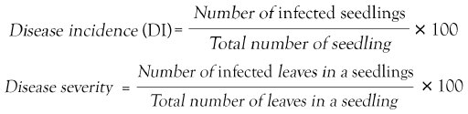
The disease incidence and severity was calculated using the formula [11].
Results
Incidence and severity of fungal diseases
Survey conducted in the government social forest nursery revealed, most of the plant species raised in root trainers suffered from one or the other foliage diseases. The incidence and severity of the diseases varied depending on the nursery management practices and prevailing environmental conditions. All the species were not grown in same number and same agronomic practices. Disease symptoms restricted mainly to the aerial parts of the plant body. The percentage of disease incidence and disease severity on seedlings of forest nursery in Mysore district is presented in Table 1. The highest disease incidence was recorded in Phyllanthus emblica, followed by Pterospermum acerifolium and lowest disease incidence was recorded in Pterocarpus santalinus. The highest disease severity was found in Tamarindus indica and lowest was found in Pterocarpus santalinus.
| Host seedlings | Percentage of disease incidence | Percentage of disease severity |
|---|---|---|
| Annona reticulata | 83.33% | 47.36% |
| Azadirachta indica | 94% | 64.28% |
| Bauhinia variegata | 28.57% | 43.47% |
| Cinnamomum verum | 71.42% | 86.95% |
| Dalbergia latifolia | 92% | 87.50% |
| Dendrocalamus strictus | 62.50% | 58.33% |
| Madhuca indica | 19.20% | 50% |
| Mangifera indica | 73.91% | 88.23% |
| Muntingia calabura | 30% | 83.33% |
| Phyllanthus acidus | 97.14% | 60% |
| Phyllanthus emblica | 100% | 93.75% |
| Pterocarpus marsupium | 78% | 73.07% |
| Pterocarpus santalinus | 16% | 24% |
| Pterospermum acerifolium | 100% | 75% |
| Sapindus saponaria | 42.85% | 41.93% |
| Tamarindus indica | 98% | 96.83% |
Table 1: Percentage of disease incidence and disease severity on social forest nursery seedlings.
Fungal diseases associated with host seedlings
Leaf spot symptom was the common disease observed in most of the forest seedlings. 10 different types of symptoms were on 16 seedlings of forest tree species. The fungal pathogens associated with seedlings includes Alternaria alternata causing leaf spot on Muntingia calabura, species of A. zinniae on Pterospermum acerifolium, Cercospora apiicola on Azadirachta indica, species of Cladosporium on Bauhinia variegata, Colletotrichum gloeosporioides causing anthracnose on Cinnamomum verum, C. graminicola on Pterocarpus santalinus, Drechslera longirostrata causing leaf blight on Sapindus saponaria, Helminthosporium maydis on Dendrocalamus strictus. Oidium mangiferae and O. tamarindii caused powdery mildew disease in Mangifera indica and Tamarindus indica respectively. Species of Pestalotia caused leaf spot on Madhuca indica, species of Phoma caused leaf spot on Annona reticulata and on Pterocarpus marsupium (Figure 2), Species of Phyllachora caused tar spot on Dalbergia latifolia, species of Ravenelia caused rust disease on Phyllanthus acidus and P. emblica. Symptoms on host seedlings and description of observed pathogens are represented in Table 2.
| Host seedlings | Disease | symptoms | Fungal species | Pathogen description |
|---|---|---|---|---|
| Annona reticulataLinn. | Leaf spot | Symptoms begin as small circular black spots on the surface of leaves. Spots enlarge in size surrounded by light and tanned borders. As the disease proceeds, browning and falling of leaves occurs. | Phomaspp. | Pycnidia are almost spherical, dark brown, thin-walled with one conspicuous protruding pore opening depressed in the leaf tissues. Pycnidiospores are hyaline, aseptate, unicellular and ellipsoidal measured about 5.08 µm in length and 2.02 µm width. Spores escape through ostiole in clusters. |
| Azadirachta indica A. Juss | Leaf spot | Leaves showed circular small spots with reddish brown borders and light tan centers on the upper surface of the leaf. There may be black speckles within the dead tissue of the leaf. The affected part of the leaves show chlorosis finally turning brown. Infected leaves gradually wilt and die. | Cercospora apiicola M. Groenewald, Crous and U. Braun | Conidiophores were arising in clusters and bursting out of leaf tissue bearing very long conidia. Conidia were hyaline, pale with conidial scar, 3-7 celled measuring about 19.50 µm long and 5.41 µm wide. |
| Bauhinia variegate (L.) Benth. | Leaf spot | Leaves showed circular, irregular lesions with light brown borders and greyish centers. Spot occurs along the veins of leaf which appears on both the surfaces of leaf. Infected leaves gradually turn yellow and wither | Cladosporium sp. | Spores are dark pigmented, one celled formed in simple chains and youngest conidia found at the top. Conidia are globose, 3-4 µm diameter, mid to dark olivaceous brown. Some are lemon shaped. This fungus commonly found on living and decaying plant material which spread by wind and may grow on surface when moisture is present. |
| Cinnamomum verum J. S. Persl. | Anthracnose | The leaves showed circular spots having light tan centers surrounded by reddish brown borders with water soaked tissues. With disease progression spots develop into patches resulting in dying of tissues finally leaves become yellow in appearance. | Colletotrichum gloeosporioides (Penz.) Penz. and Sacc. | Waxy acervuli produced in infected tissue which is subepidermal in position with setae. Setae dark, long and more looks like stiff bristles. Conidiophores simple, short and erect bearing hyaline, one-celled, ovoid to oblong dumbbell shaped conidia which measures about 12.46 µm in length and 3.60 µm in width. |
| Dalbergia latifolia Roxb. | Tar spot | Majority of younger seedlings showed round to irregular black, tar-like spots on the upper surface of infected leaves. Small black fruiting bodies may be visible on the upper surface of spots. As the fungus grows within the leaf, the infected areas develop slightly raised, shiny tar like black spots on the leaves. In case of severe infection defoliation can occur. | Phyllachorasp. | The fungus produces sexual spores called Ascospores in Asci. Asci found surrounded by Paraphyses which are colorless, filiform an abundant. Ascospores are aseptate, smooth and hyaline and piled on one other inside the Asci. |
| Dendrocalamus strictus (Roxb). Nees | Leaf blight | Small, greyish brown, spindle shaped lesions appear on young leaves. Around these lesions, pale yellow to yellowish orange haloes develop. The lesions coalesce to form large, spreading irregular lesions with dark brown borders, covering the entire leaf lamina. Such leaves become necrotic and blighted. Severe infection causes yellowing of the foliage, followed by leaf blight and withering. | Helminthosporium maydis Y. Nisik. and C. Miyake, | Conidiophores mid to dark brown, medium to long, slender, slightly curved and bear conidia at wide intervals. Conidia are distinctly curved, broad in the middle, simply tapering towards the ends, pale to mid-dark golden brown, smooth, 5-11 septate and measures about 64.44 µm long and 19.50 µm wide with dark point of attachment. Conidia dispersed through win or rain splashes invade the tissue, sporulates and cause necrosis under high humidity. |
| Madhuca indica J. F. Gmel. | Leaf spot | Symptoms begin as very small yellow brown spots on the youngest leaves. The spot with light tan centers become surrounded by dark brown borders. With the growth of seedlings disease development occur leading to the enlargement and spread of the spots. Further spot appeared as patches with irregular outline throughout the foliar surface. Infected leaves gradually wither from seedlings. | Pestalotia sp. | The fungus produces sooty, black, long curly, thick, thread like structures on leaf surface with white mycelium at base. These threads like structures consist of conidia which are ellipsoidal, broadest in the middle, narrower at both ends. Middle cells are dark brown where end cells are hyaline. Conidia re measured about 23.80 µm in length and 7.01 µm in width. A diagnostic feature is two whisker-like appendages were arising from the end cell. |
| Mangifera indica L. | Powdery mildew | The disease appeared as small scattered water soaked lesions on the under surface of young leaves as they are highly susceptible. Later directly under water spots, white powdery growth forming necrotic lesions. These irregular necrotic lesions enlarge and coalesce forming large dead tissue on the leaf. Infected leaves gradually curl and get destroyed. | Oidium mangiferae Berthet. | The conidiophores are simple, short, erect and hyaline. The conidia were aseptate, hyaline, smooth walled, elliptical to barrel shaped producing singly. Conidia measured about 35.12 µm in length and 15.63 µm in width. Conidial germination does not depend on rain and germinate in humidity. Germ tubes are of variable length depending upon humidity. |
| Muntingia calabura L. | Leaf spot | Leaf spot begins as small circular dark reddish brown spots with darker outline on the dorsal surface of foliage. As the disease progress the spots coalesce and make darker, irregular patches in appearance surrounded by dark yellow brown borders | Alternaria alternata (Fr.) Keissl. | Hyphae are dark brown, thick, septate and branched. Conidiophores simple dark pigmented and clustered producing dark pigmented conidia in an acropetal succession of simple or branched chains. Conidia have transverse and oblique septa, measures 42.5 µm in width. Conidia obovoid, obclavate, ellipsoidal, in an elongated terminal cell. Smooth walled conidia are pale to mid-golden brown. |
| Phyllanthus acidus (L.) Skeels | Rust | Symptoms begin as small pale yellow spots that further enlarge in size in circular manner. The spots usually turn dark brown with even darker outline. The ventral side of the spotted surface was tan in appearance indicating the invasion by rust fungus. | Raveneliasp. | The fungus produces colored masses of subepidermal Teliospores and Urediniospores on P. acidus in its life cycle. Teliospores are hemispherical, brown, the whole surface being covered with large brown verrucae. Echinulate urediniospores are circular to globose, hyaline to light brown with small spines on its wall surface. The diameter of Urediniospores measured about 18.72 µm. |
| Phyllanthus emblica L. | Rust | Rust symptoms appeared as circular to semicircular, reddish spots of 2-5 mm in diameters on dorsal surface which appeared as small circular in outline. As the disease spreads brown pustules appear throughout the seedlings gradually reducing the growth and yield. | Raveneliasp. | The fungus produces colored masses of Urediniospores on P.emblica in its uredinial phase of life cycle. Only pycnidial, uredial and telial stages occur in its life cycle. Microscopic examination of Raveneliasp. showed smooth clavateparaphyses with clavate, echinulate urediniospores having equatorial germ pores. Spores measured about 30.99 µm in length and 17.84 µm in width. |
| Pterocarpus marsupium Roxb. | Leaf spot | Symptoms begin as small circular dark brown spots with reddish brown borders followed by dark yellow borders on the upper surface of the leaves. Small pores coalesce to form larger irregular lesions. Infected leaves gradually turn yellow leading to chlorosis finally resulting in withering of leaf at higher rate. | Phomaspp. | Pycnidia are black and depressed in the leaf tissues. Pycnidiospores are hyaline, unicellular; ovoid to ellipsoidal measured about 5.48 µm in length and 2.03 µm width. |
| Pterocarpus santalinus L. f. | Anthracnose | Leaf spot begin as small water soaked dull whitish spots with dark green borders on the upper surface of the leaf. As the disease develops the spots cover the entire leaf lamina as pinkish irregular patches with brown margin around centers. | Colletotrichum graminicola (Ces.) G.W. Wilson | Acervuli are rounded, separate, and superficial with conspicuous darkly pigmented setae consisting of a gelatinous conidial mass. Conidiophores are hyaline, single-celled, falcate, fusiform, spindle shaped, with acute apices and measured 27.82 µm in length and 2.45 µm in width. Setae are brown with dark swollen base and a pale rounded tip. |
| Pterospermum acerifolium L. Wild. | Leaf blight | Symptoms initially appeared as circular, small dark brown spots on the leaf surface. The spots enlarge in size with grayish center and purple margined lesions on the surface and also along the edges of leaf lamina giving a burnt appearance to the seedlings. | Alternaria zinniae | Mycelium either sparse or abundant usually olive green to brown. Hyphae dark brown, thick, septate and branched. Conidiophores are simple, erect and often clustered. Conidia solitary, obclavate, pale to dark brown smooth and minutely verruculose with 5-9 transverse and several longitudinal septa. Conidia measures 39.7 µm in length and 12 µm wide. Surface wall pale to mid golden brown. |
| Sapindus saponaria L. | Leaf blight | Individual leaf spots are less than 1mm in diameter further expands to form irregular spots. The larger spots are bleached in appearance with surrounded light brown borders that girdle the leaf blade. These spots coalesce to form irregular patches. Severely infected leaves eventually wither and dry to light tan color. | Drechslera longirostrata (Subram.) comb. nov. | Growth of the fungus consists of short conidiophores bearing conidia. Conidia are brown, shiny, ellipsoid, longirostrata, smooth walled 3-12 septate. Conidia measured about 50.89 µm in length and 3.34 µm in width with well-developed protuberant hilum. |
| Tamarindus indica L. | Powdery mildew | Powdery white patches appear on the upper surface of leaves. As the infection proceeds, upper foliar surface becomes densely covered with white mycelium and hyphal net. Severe infection reduces effective photosynthesis areas of leaves. | Oidium tamarindi (J.M. Yen) U. Braun. | The conidia of Oidium spread by wind without depending on rain or dew from host to host. White mycelium appears external on host. Hyphae are hyaline with smooth cell wall producing short and erect conidiophores. Conidia re hyaline, unicellular, aseptate and cylindrical to globose. |
Table 2: Occurrence of pathogenic fungi, the type of disease, symptoms on host seedlings and description of pathogen.

Figure 2: Fungal pathogens associated with diseased seedlings of social forest nursery.
Note: A: Leaf spot of Annona reticulata caused by Phoma spp., a- Conidia of Phoma spp. (100X); B: Leaf spot of Azadirachta indica A. Juss caused by Cercospora apiicola; b- conidia of C. apiicola (100X); C: Leaf spot of Bauhinia variegate L. infected by Cladospriumsp. c- conidia of Cladosporium sp. (100X); D: Anthracnose of Cinnamonum verum J. Presl. caused by Colletotrichum goleosporioides Penz; d- conidia of C. gloeosporioides Penz. (40X); E: Tar spot of Dalbergia latifolia Roxb. caused by Phyllachora sp.; e- Ascospores of Phyllachora produced inside Asci (40X);F: Leaf blight of Dendrocalamus strictus Roxb. Caused by Helminthosporium maydis Nisikado and Miyake; f- Conidia of H. maydis (40X); G: Leaf spot of Madhuca indica Gmel. caused by Pestalotia sp.; g- Conidia of Pestalotia sp. (100X); H: Powdery mildew of Mangifera indica L. caused by Oidium mangiferae Berthet; h- Conidia of O. mangiferae (40X); I: Leaf spot of Muntingia calabura Plum. caused by Alternaria alternata (Fr.) Keissler; i- conidia of A. alternata (40X); J: Rust of Phyllanthus acidus L. caused by Ravenelia sp.; j- Urediniospores (100X); K: Rust of Phyllanthus emblica L. caused by Ravenelia sp.; k- Urediniospores of Ravenelia sp. (100X); L: Leaf spot of Pterocarpus marsupium Roxb. caused by Phoma spp. l- Conidia of Phoma spp. (100X); M: Anthracnose of Pterocarpus santalinus L.f. caused by Colletotrichum graminicola; m- Conidia of C. graminicola with setae (100X); N: Leaf blight of Pterospermum acerfolium L. caused by Alternaria zinniae; n- A. zinniae (100X); O: Leaf blight of Sapindus saponaria L. caused by Drechslera longirostrata (Subram.) Subram; o- Conidia of D. longirostrata (100X); P: Powdery mildew of Tamarindus indica L. caused by Oidium tamarindi; p- Germination of Oidium tamarindi conidium (100X).
Discussion
Fungal diseases are a serious problem in forest regeneration causing heavy mortality in forest nurseries. The present investigation revealed the high incidence of fungal diseases in social forest nursery ‘Karnataka state forest urban greening nursery’, located in Paduvaralli, Mysore. Many leaf spots, blights, anthracnose, nuts, powdery mildews diseases were spreading at high rate (90%-100%) of disease incidence. Hence there is need for management.
Seedlings of Annona reticulata and Pterocarpus marsupium revealed leaf spot symptoms caused by Phoma spp. Ojha et al., reported Phoma spp. causing fruit rot diseases in Leaf spot of Annona reticulata caused by Phoma spp., a- Conidia of Phoma spp. (100X) [12]. Pinto et al., reported same pathogen causing leaf blight in Annona sp [13]. Mohanan et al. reported pathogenic nature of Sclerotium rolfsii in P. marsupium in root trainer nurseries [14]. In spite of versatile medicinal properties neem is not free from fungal diseases. The leaf symptoms on seedlings of Azadirachta indica showed the association of Cercospora apiicola. Shivanna, reported Cercospora sp. causing leaf spot disease in the same host in Shimoga forest nurseries [15].
Species of Cladosporium was diagnosed as the causal agent for leaf spot disease in Bauhinia variegata in our nursery survey. The pathogen has been reported to cause diseases in B variegata fruits by Kumar et al., [16]. Colletotrichum gloeosporioides showed similar symptoms in Cinnamomum verum during the survey. The same species known to cause anthracnose in Cinnamomum sp. as reported by Rozwalka et al., [17].
Tar spot disease was most prevalent on seedlings of Dalbergia latifolia caused by species of Phyllachora in forest nursery. Workers like Shivanna and Naik reported Phyllachora sp. in the same host [18]. The pathogen that caused characteristic spindle shaped blight disease over the entire leaf blade of Dendrocalamus strictus was identified as Helminthosporium maydis. Rai and Mamatha, reported occurrence of leaf spot and blight diseases in seedlings of D. strictus by Myrothecium roridum and Cercospora apii in forest nursery [3].
Seedlings of Madhuca indica found infected with Pestalotia sp. causing leaf spot disease. Dube and Bilgrami, Shivanna, reported genus Pestalotia in Madhuca indica tree species [19]. In the present study powdery mildew disease was observed among the young seedlings of Mangifera indica. Mycological analysis revealed Oidium mangiferae as the causal agent. This fungus has been reported to cause powdery mildew as reported by other workers on the same host [20-22]. The present study revealed that occurrence of Alternaria alternata as causal agent of leaf spot and blight diseases in Muntingia calabura. Earlier workers reported Phellinus noxious causing brown root rot by Ann et al., Botrytis cinerea causing gray mold diseases in fruits of Muntingia calabura by Chen and Hsieh, [23,24]. There is no report of Alternaria alternata on this host. This is the first report on occurrence of Alternaria alternata on leaves of Muntingia calabura.
The seedlings observation of Phyllanthus acidus and P. emblica showed rust symptoms caused by species of Ravenelia. This fungal genus has been reported to cause similar symptoms in P. emblica as reported by Thaung and Shivanna [15,25]. Anthracnose disease caused by Colletotrichum graminicola in Pterocarpus santalinus in forest nursery. Earlier workers had also reported the pathogenic nature of Colletotrichum in forest nurseries by Mohanan and Shivanna [15,26].
Alternaria zinniae was diagnosed as the causal agent of leaf blight disease in Pterospermum acerifolium. The pathogenic nature of Alternaria sp. as leaf pathogen was reported by Mohanan et al., on Acacia auriculiformis [14]. A. zinniae causing leaf blight disease in this host is the first report. Drechslera longirostrata was observed to cause leaf blight disease in Sapindus saponaria. This is also found to be the first report. Powdery mildew disease was also observed on seedlings of Tamarindus indica caused by Oidium tamarindi. Shivanna reported the same pathogen on seedlings of T. indica in forest nurseries [15]. Pawar and Patil also reported this fungus in the same host [22].
16 tree species were surveyed for the incidence of different fungal diseases in social forest nursery out of which 10 fungal genera were isolated causing different types of disease symptoms. Seven species known to cause leaf spot disease were Alternaria spp., Cercosporaapiicola, Cladosporium sp., Pestalotia sp., Phoma spp. and Phyllachora sp. Two fungal species known to cause Anthracnose disease ( .) and other two seedlings species affected by powdery mildews caused by Oidium species. Ravenelia sp. was responsible for rust disease on two Phyllanthaceae members. Leaf blight affected seedlings showed the occurrence of Alternaria zinniae, Drechslera longirostrata and Helminthosporium maydis. The study revealed the occurrence of different fungal pathogens belonging to Deuteromycetes, Ascomycetes and Basidiomycetes respectively. The incidence of some of these diseases readily severe and hence proper forest nursery management practices need to be adopted for reducing the incidence of such diseases.
Conclusion
In the current study survey was carried out to know the occurrence of fungal diseases, their incidence and severity on seedlings of forest nursery in Mysore district, Karnataka. Fungal pathogens are one of the most frequent and challenging type of pathogens. Anthracnose, rust, powdery mildew, tar spot and leaf blight were commonly found and some fungal pathogens Phoma spp., C. apiicola, Cladosporium sp., Petalotia sp. and A. alternata showed leaf spots on infected plant seedlings. Therefore, a study of fungal pathogens and finding solutions to exterminate or prevent their infection is important. The present survey may help coordinators of forest nurseries to understand about successful production of pathogen free seedlings in nurseries with hygienic practices, sterilizing containers and surface area together with proper fertilization, ventilation and growing densities which aid in preventing entry or survival of fungal pathogens.
References
- Mustafa A. Seed mycoflora of Shisham (Dalbergia sissoo Roxb.) and their integrated management. 2009.
- Landis TD, Tinus RW, McDonald SF, Barnett JP. (1989). Diseases in container tree nurseries. U.S. Department of Agriculture Handbook, 674(5): 1-99.
- Rai VR, Mamatha T. Seedling diseases of some important forest tree species and their management. Working Pap. Finnish Forest Research Institute. 2005; 11:51-64.
- Lilja A, Lilja S, Kurkela T. Nursery practices and management of fungal diseases in forest nurseries in Finland. A review. 1997.
- Lilja A, Poteri M, Petäistö RL, Rikala R, Kurkela T, Kasanen R. Fungal diseases in forest nurseries in Finland.
- De Tempe J. The blotter method for seed health testing. Proc Int Seed Test Assoc. 1953; 21:133-151.
- Domsch KH, Gams W, Anderson TH. Compendium of soil fungi. Volume 1. Academic Press (London) Ltd.; 1980.
- Moss MO, K. Singh, JC Frisvad, U. Thrane and SB Mathur. An Illustrated Manual on Identification of some Seed-borne Aspergilli, Penicillia and their Mycotoxins. The Danish Government Institute of Seed Pathology for Developing Countries, Ryvangs Alle Denmark. ISBN 87-7026-317-5:1993;317-5.
- Barnett HL, Hunter BB. Illustrated genera of imperfect fungi 4th edition. APS, St. Paul, Minnesota. 1998.
- Mathur SB, Kongsdal O. Common laboratory seed health testing methods for detecting fungi. International Seed Testing Association. 2003.
- Sikdar AK, Krishnaswami S. Assessment of leaf yield loss of the two mulberry varieties due to leafspot disease. Indian J. Seric. 1980.
- Ojha S, Chakraborty M, Chatterjee NC. Antagonistic potentials of Trichoderma Spp. against fruit rot of custard apple caused by Phoma ligam. J Biosci. 2011; 19:15-21.
- Pinto PM, Alonso JA, Fernández VP, Casero JJ. Fungi isolated from diseased nursery seedlings in Spain. New Forests. 2006; 31:41-56.
- Mohanan C, Ratheesh N, Nair LP, Rajesh Kumar KC. Disease problems in root trainer forest nurseries in Kerala State and their management. In5th Meeting of IUFRO Working Party S 2005 (Vol. 7, pp. 7-12).
- Shivanna MB. Fungal diseases in forest nurseries in Shimoga district, Karnataka, India. Diseases and Insects in Forest Nurseries. 2014:27.
- Arvind K, Anuj K, Kharwar RN. Two new phytoparasitic hyphomycetes from Varanasi, India. Indian Phytopathol. 2006; 59(1):85-90.
- Rozwalka LC, Alves E, do Amaral DC. Ultrastructural study of conidiaof Colletotrichum gloeosporioides and Colletotrichum musae treated with essential oils. Interciencia. 2010; 35(12):912-915.
- Imkongsunep L, Naik ST. Survey and epidemiology of powdery mildew of Acacia auriculiformis (A. Cunn.). J Agric Sci. 2010; 23(2):400-401.
- Dube HC, Bilgrami KS. Morphology of fruiting body pustules in the genus Pestalotia. Mycopathologia. 1996;28(4):305-311.
- Akhtar KP, Alam SS. Powdery Mildew of Mango: A Review. Pak J Biol Sci. 2000.
- Galli JA, Silveira LC, Michelotto MD, Martins AL. Powdery mildew (Oidium mangiferae Bert.) infection in mango varieties. Bioscience. 2008 Jun 4; 24(2):43-46.
- Pawar VP, Patil VA. Occurrence of powdery mildew on some wild plants from Khandesh region of Maharashtra state. Recent res. sci. technol. 2011; 3(5):94-95.
- Ann PJ, Chang TT, Ko WH. Phellinus noxius brown root rot of fruit and ornamental trees in Taiwan. Plant Dis. 2002; 86(8):820-826.
[Crossref] [Google Scholar] [PubMed]
- Chen CH, Hsieh TF. First report of Botrytis cinerea causing gray mold of Jamaica cherry in Taiwan. 2009.
- Thaung MM. Rusts, smuts and their allies in Burma. Australasian Mycologist. 2005; 24(2):29-46.
- Mohanan C. Status of forest nursery diseases in India and emerging trends in seedling disease management. 2000.
Citation: Bhavana NS, Janardhana GR (2023) Incidence of Foliage Fungal Diseases on Seedlings of Social Forest Nursery in Mysore, Karnataka, India. J Plant Pathol Microbiol. 13:682.
Copyright: © 2023 Bhavana NS, et al. This is an open-access article distributed under the terms of the Creative Commons Attribution License, which permits unrestricted use, distribution, and reproduction in any medium, provided the original author and source are credited.