
Indexed In
- Academic Journals Database
- Open J Gate
- Genamics JournalSeek
- Academic Keys
- JournalTOCs
- China National Knowledge Infrastructure (CNKI)
- Ulrich's Periodicals Directory
- Electronic Journals Library
- RefSeek
- Hamdard University
- EBSCO A-Z
- OCLC- WorldCat
- SWB online catalog
- Virtual Library of Biology (vifabio)
- Publons
- Geneva Foundation for Medical Education and Research
- Euro Pub
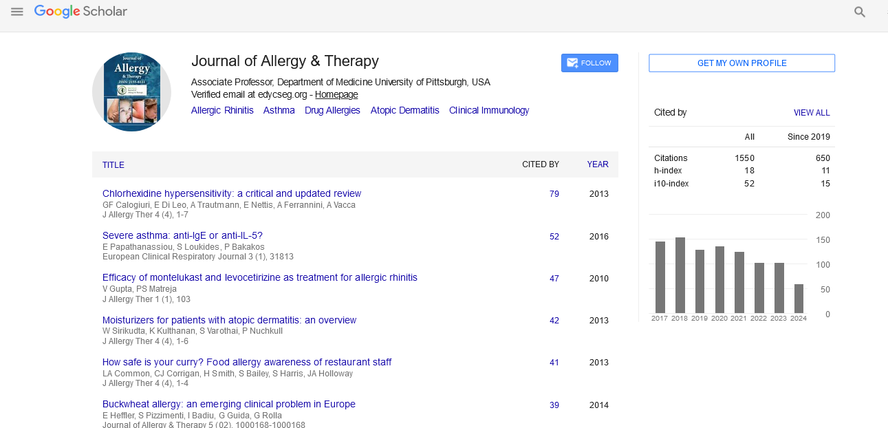
- Google Scholar
Useful Links
Share This Page
Journal Flyer
Open Access Journals
- Agri and Aquaculture
- Biochemistry
- Bioinformatics & Systems Biology
- Business & Management
- Chemistry
- Clinical Sciences
- Engineering
- Food & Nutrition
- General Science
- Genetics & Molecular Biology
- Immunology & Microbiology
- Medical Sciences
- Neuroscience & Psychology
- Nursing & Health Care
- Pharmaceutical Sciences
Short Communication - (2023) Volume 14, Issue 3
Emerging Findings Reveal Link between Allergy and Pterygium
Yotam Eyni1*, Elli Rosenberg2 and Erez Tsumi12Department of Clinical Immunology and Allergy, Soroka University Medical Center, Ben-Gurion University of the Negev, Beer Sheva, Israel
Received: 19-Jun-2023, Manuscript No. JAT-23-21900 ; Editor assigned: 22-Jun-2023, Pre QC No. JAT-23-21900 (PQ); Reviewed: 06-Jul-2023, QC No. JAT-23-21900 ; Revised: 13-Jul-2023, Manuscript No. JAT-23-21900 (R); Published: 21-Jul-2023, DOI: 10.35248/2156-6121.14.343
Description
Pterygium is a prevalent worldwide condition characterized by the benign growth of fibrovascular tissue on the bulbar conjunctiva of the eye. It affects a substantial proportion of the population, ranging from 3% to 19.5% [1]. The incidence is particularly higher in tropical and subtropical regions. Pterygium is associated with aesthetic concerns, irregular astigmatism, and potentially severe vision impairment [2]. The exact underlying mechanisms leading to pterygium formation remain unclear, although environmental, immunological, infectious, and genetic factors are believed to contribute. Notably, Ultraviolet-B (UV-B) radiation is implicated in damaging corneal limbus cells, which subsequently triggers cell proliferation, inflammation, and angiogenesis. These processes involve the activation of fibroblasts and mediated by cytokines, and growth factors [3-6]. Several known risk factors for pterygium include living in rural areas, dust exposure, low income, and older age [7-10]. Surgical removal is typically recommended. However, excision often presents challenges as recurrence rates are high, with recurrent pterygia being more problematic than their initial counterparts [11]. A better understanding of the pathogenesis of pterygium may potentially contribute to finding novel efficient treatments.
Pterygium and Allergy
Previous research has provided evidence suggesting a potential association between pterygium and allergy-mediated mechanisms. Studies have found elevated mast cell counts and increased levels of Immunoglobulin E (IgE) in pterygium specimens compared to normal conjunctiva [12,13]. In vitro experiments have shown that tranilast, an allergy medication, can inhibit the growth of pterygium-derived cells [14]. However, the precise role of mast cells and IgE antibodies in pterygium pathophysiology remains unclear, necessitating further investigation to fully comprehend their involvement. It is noteworthy that mast cells and IgE antibodies play essential roles in type-1 hypersensitivity conditions.
Additionally, studies have highlighted the significant involvement of fibroblasts in the proliferation of elastin and collagen observed in pterygium [3]. These fibroblasts are stimulated by various growth factors, including Transforming Growth Factor (TGF)-b1 and Matrix Metalloproteinases (MMPs). Notably, these growth factors also participate in type 1 allergic conditions. For instance, TGF-b1 and MMPs are implicated in fibroblast invasiveness during airway remodeling in asthma [15]. Furthermore, the TH2 cytokine IL-13 directs multiple aspects of airway remodeling through TGF-b1 and MMPs.
Recently, the term "entopy" has been introduced to describe locally restricted allergic reactions in specific mucosal regions, distinct from systemic atopy. Similar conditions involving elevated local IgE antibody levels within tissues have been observed in non-atopic individuals with allergic conditions like nasal polyps and allergic conjunctivitis [16]. These findings suggest the presence of a localized type 1 hypersensitivity mechanism. Although more research is needed, this mechanism could potentially apply to pterygium as well.
New Evidence for Clinical Association Between Allergy and Pterygium
A recent population-based case-control study conducted in the southern district of Israel examined 13,944 patients with pterygium and 41,832 matched controls [17]. The study identified several significant associations between pterygium and allergic periocular and ocular surface diseases, including Vernal Keratoconjunctivitis (VCK) and chronic allergic conjunctivitis. VCK is an allergic disease mediated by IgE antibodies and is often observed in individuals with a family history of other allergic conditions such as asthma and atopic dermatitis. The study has also found a higher prevalence of unspecified systemic allergies as a risk factor for pterygium. These findings strongly support the connection between pterygium and allergy.
Conclusion
In conclusion, pterygium is a prevalent fibrovascular growth affecting the conjunctiva of the eye. While the precise etiology remains unclear, environmental, immunological, and genetic factors are believed to contribute to its development. Studies have provided indications of a potential link between pterygium and allergic mechanisms, supported by elevated mast cells and IgE levels observed in pterygium specimens. Recent research has established significant associations between pterygium and allergic eye conditions. Further investigation is necessary to fully understand these connections and potentially develop effective treatment approaches.
References
- Rezvan F, Khabazkhoob M, Hooshmand E, Yekta A, Saatchi M, Hashemi H. Prevalence and risk factors of pterygium: a systematic review and meta-analysis. Surv Ophthalmol. 2018;63(5):719-735.
[Crossref] [Google Scholar] [PubMed]
- Errais K, Bouden J, Mili-Boussen I, Anane R, Beltaif O, Meddeb Ouertani A. Effect of pterygium surgery on corneal topography. Eur J Ophthalmol. 2008;18(2):177-181.
[Crossref] [Google Scholar] [PubMed]
- Di Girolamo N, Chui J, Coroneo MT, Wakefield D. Pathogenesis of pterygia: role of cytokines, growth factors, and matrix metalloproteinases. Prog Retin Eye Res. 2004;23(2):195-228.
[Crossref] [Google Scholar] [PubMed]
- Moran DJ, Hollows FC. Pterygium and ultraviolet radiation: a positive correlation. Br J Ophthalmol. 1984;68(5):343-346.
[Crossref] [Google Scholar] [PubMed]
- Coroneo MT. Pterygium as an early indicator of ultraviolet insolation: a hypothesis. Br J Ophthalmol. 1993;77(11):734.
[Crossref] [Google Scholar] [PubMed]
- Liu T, Liu Y, Xie L, He X, Bai J. Progress in the pathogenesis of pterygium. Curr Eye Res. 2013;38(12):1191-1197.
[Crossref] [Google Scholar] [PubMed]
- Saw SM, Tan D. Pterygium: prevalence, demography and risk factors. Ophthalmic Epidemiol. 1999;6(3):219-228.
[Crossref] [Google Scholar] [PubMed]
- Gazzard G, Saw SM, Farook M, Koh D, Widjaja D, Chia SE, et al. Pterygium in Indonesia: prevalence, severity and risk factors. Br J Ophthalmol. 2002;86(12):1341-1346.
[Crossref] [Google Scholar] [PubMed]
- Luthra R, Nemesure BB, Wu SY, Xie SH, Leske MC. Frequency and risk factors for pterygium in the Barbados Eye Study. Arch Ophthalmol. 2001;119(12):1827-1832.
[Crossref] [Google Scholar] [PubMed]
- Liu L, Wu J, Geng J, Yuan Z, Huang D. Geographical prevalence and risk factors for pterygium: a systematic review and meta-analysis. BMJ open. 2013;3(11):e003787.
[Crossref] [Google Scholar] [PubMed]
- Coster D. Pterygium--an ophthalmic enigma. Br J Ophthalmol. 1995;79(4):304.
[Crossref] [Google Scholar] [PubMed]
- Deveci H, Yücetürk A, Yardım BG, Aydın S. Primer ve Nüks Pterjium Patogenezinde Mast Hucrelerinin Rolu. Turk J Ophthalmol. 2012;42(3).
- Pinkerton OD, Hokama Y, Shigemura LA. Immunologic basis for the pathogenesis of pterygium. Am J Ophthalmol. 1984;98(2):225-228.
[Crossref] [Google Scholar] [PubMed]
- Isaji M, Kikuchi S, Miyata H, Ajisawa Y, Araki-Inazawa K, Tsukamoto Y, et al. Inhibitory effects of tranilast on the proliferation and functions of human pterygium-derived fibroblasts. Cornea. 2000;19(3):364-368.
[Crossref] [Google Scholar] [PubMed]
- Ingram JL, Huggins MJ, Church TD, Li Y, Francisco DC, Degan S, et al. Airway fibroblasts in asthma manifest an invasive phenotype. Am J Respir Crit Care Med. 2011;183(12):1625-32.
[Crossref] [Google Scholar] [PubMed]
- Powe DG, Bonnin AJ, Jones NS. â??Entopyâ??: local allergy paradigm. Clin Exp Allergy. 2010;40(7):987-997.
[Crossref] [Google Scholar] [PubMed]
- Eyni Y, Kerman T, Hazan I, Rosenberg E, Lev Ari O, Knyazer B, et al. Are Periocular and Systemic Allergy Conditions Risk Factors for Pterygium?. Semin Ophthalmol.2023;1-5.
[Crossref] [Google Scholar] [PubMed]
Citation: Eyni Y, Rosenberg E, Tsumi E (2023) Emerging Findings Reveal Link between Allergy and Pterygium. J Allergy Ther. 14:343.
Copyright: © 2023 Eyni Y, et al. This is an open-access article distributed under the terms of the Creative Commons Attribution License, which permits unrestricted use, distribution, and reproduction in any medium, provided the original author and source are credited.