
Indexed In
- Open J Gate
- Genamics JournalSeek
- Academic Keys
- JournalTOCs
- ResearchBible
- Ulrich's Periodicals Directory
- Access to Global Online Research in Agriculture (AGORA)
- Electronic Journals Library
- RefSeek
- Hamdard University
- EBSCO A-Z
- OCLC- WorldCat
- SWB online catalog
- Virtual Library of Biology (vifabio)
- Publons
- MIAR
- Geneva Foundation for Medical Education and Research
- Euro Pub
- Google Scholar
Useful Links
Share This Page
Journal Flyer
Open Access Journals
- Agri and Aquaculture
- Biochemistry
- Bioinformatics & Systems Biology
- Business & Management
- Chemistry
- Clinical Sciences
- Engineering
- Food & Nutrition
- General Science
- Genetics & Molecular Biology
- Immunology & Microbiology
- Medical Sciences
- Neuroscience & Psychology
- Nursing & Health Care
- Pharmaceutical Sciences
Case Report - (2026) Volume 17, Issue 4
Echinococcal cyst of the lung at the Parasitology Laboratory of Fann Hospital: A case report
Souleye Lelo 1,2 * , Babacar Faye 1 , Khadime Sylla 1,2 , Roger Tine 1,2 , Aissatou Gaye 1,2 , Cheikh S Kane 2 , Amadou Dioulde Diallo 3 , Moussa Seck Diop 3 , Moussa Maréme Samba 3 and Souleymane Diatta 32Parasitology and Mycology Laboratory, Fann University Hospital, Dakar, Senegal
3Department of Cardiovascular Surgery, Fann University Hospital, Dakar, Senegal
Received: 20-Jul-2026 , Manuscript No. JBP-26-32828; Editor assigned: 22-Jul-2026 , Pre QC No. JBP-26-32828 (PQ); Reviewed: 05-Aug-2026 , QC No. JBP-26-32828; Revised: 12-Aug-2026 , Manuscript No. JBP-26-32828 (R); Published: 19-Aug-2026 , DOI: 10.35248/2155-9597.26.17.616
Abstract
Hydatid cysts of the lung are a rare condition in sub-Saharan Africa. They are the second most common site after the liver. Treatment is primarily surgical, although medical treatment has been shown to be effective. We report the first case managed by the thoracic surgery department at the fann university hospital in Dakar. This is a 49-year-old female patient, originally from Mauritania, with no significant medical history, who was referred to us for management of a pulmonary cyst discovered incidentally on a chest Computed Tomography (CT) scan during a routine check-up, with a negative serological test for Echinococcus granulosus. The clinical examination revealed no abnormalities. laboratory results showed white blood cells: 8,620/mm³, haemoglobin: 9.4 g/dl, haematocrit: 30.8 per cent, platelets: 517,000/ mm³. The chest CT scan revealed well-defined, homogeneous, hypodense lesions without vegetations, of fluid like density, located in the right anterobasal region and the left polylobar region (Fowler’s position). Surgery was performed to remove the cyst. Direct examination of the specimen between a slide and a coverslip under a 40 × objective revealed hooks of Echinococcus granulosus. Pulmonary hydatid cysts are rare. Treatment is surgical, whether radical or conservative, and yields good results.
Keywords
Pulmonary hydatid cyst; Dakar; Senegal.
Introduction
Hydatid disease also known as hydatid echinococcosis or hydatid cyst is a zoonosis caused by the development in humans of the hydatid larva of the tapeworm Echinococcus granulosus [1]. It is transmitted to humans through the ingestion of eggs (embryophores) excreted into the environment by infected dogs, as well as through the consumption of contaminated meat [1,2]. It is a parasitic disease that affects more than one million people and accounts for more than 3 billion dollars in expenditure each year [3]. Any organ can be affected by cystic echinococcosis, but liver and lung involvement are more commonly found in patients [4]. However, some cases involving bone involvement have also been reported [5]. The rarity of this zoonosis, and the difficulty in diagnosing and treating it, prompted this case study conducted at fann university hospital.
Case Presentation
Patient details
This is a 49-year-old female patient. Her symptoms are thought to have begun 5 months ago, characterised by bilateral lower chest pain, predominantly on the right, described as a tingling sensation, which is relieved by standard painkillers, accompanied by a cough with yellowish sputum containing no traces of blood and stage III dyspnoea according to the Sadoul classification. All of this occurred against a background of a high fever accompanied by a deterioration in her general condition (non-selective anorexia, physical asthenia, unquantified weight loss). This led to a consultation at the nouakchott national hospital (Mauritania), where a physical examination was requested and carried out. Following an analysis of the results, she was referred to the department of cardiovascular and thoracic surgery at fann university hospital (Senegal) for further management.
Clinical findings
The patient was in good general health. The cardiovascular and gastrointestinal examinations were normal.
The pleuropulmonary examination revealed normal breathing and a thorax of normal morphology.
The X-ray revealed a rounded opacity in the lower third of the right lung field, with a watery appearance and regular margins, and a bilobed opacity in the middle third of the left lung field, also with a watery appearance (Figure 1).
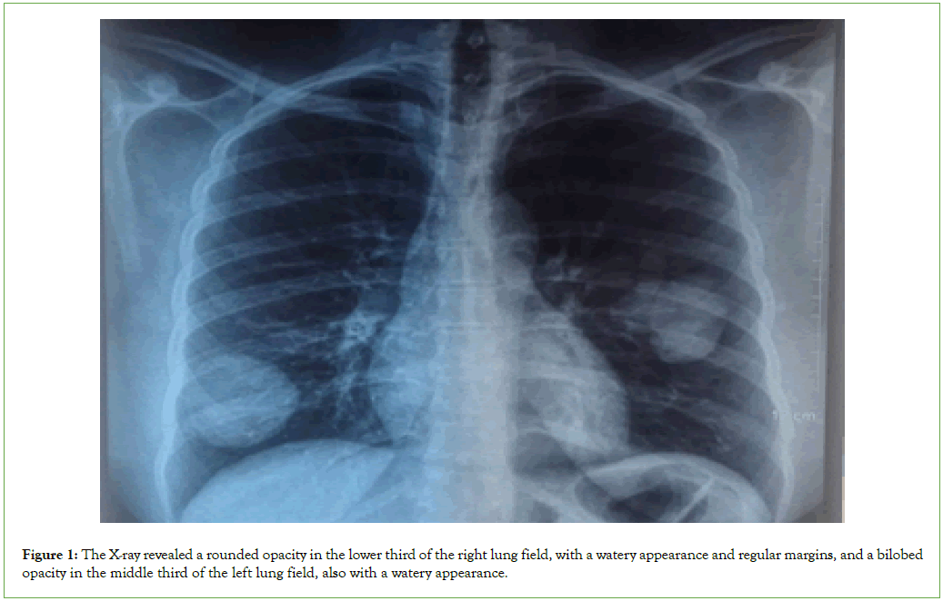
Figure 1: The X-ray revealed a rounded opacity in the lower third of the right lung field, with a watery appearance and regular margins, and a bilobed opacity in the middle third of the left lung field, also with a watery appearance.
CT scan revealed, in the thoracic region, the presence of welldefined, homogeneous, hypodense lesions without vegetations, of fluid-like density, located in the right anterobasal region and the left polylobar region (Fowler’s position). No bronchial dilatation. No pleuropericardial effusion or thickening. No abnormality in cardiac volume. No thoracic adenomegaly. No abnormality of the great vessels (Figure 2).
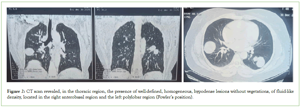
Figure 2: CT scan revealed, in the thoracic region, the presence of well-defined, homogeneous, hypodense lesions without vegetations, of fluid-like density, located in the right anterobasal region and the left polylobar region (Fowler’s position).
Diagnostic approach
The haematological results were as follows: white blood cells: 8,620/mm³, haemoglobin: 9.4 g/dl, haematocrit: 30.8 per cent, platelets: 517,000/mm³, blood group: A, Rhesus negative, prothrombin time: 90 per cent. The biochemical results were as follows: Urea: 0.17 g/l, Creatinine: 5.2 mg/l, Na: 135 mmol/l, K: 4.4 mmol/l, Cl: 103 mmol/l.
An intraoperative biopsy-type sample was received at the parasitology and mycology laboratory of fann university hospital for parasitological examination. Direct examination of the sample on a slide under a 40 × objective revealed hooks of Echinococcus granulosus (Figure 3,4).
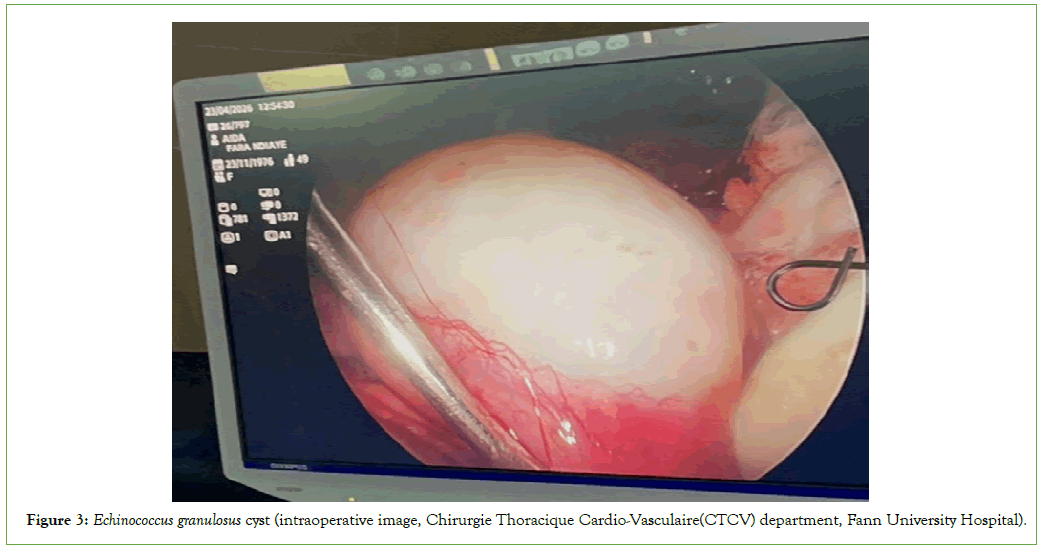
Figure 3: Echinococcus granulosus cyst (intraoperative image, Chirurgie Thoracique Cardio-Vasculaire(CTCV) department, Fann University Hospital).
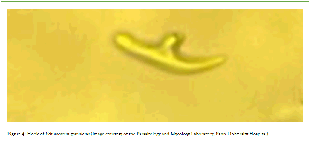
Figure 4: Hook of Echinococcus granulosus (image courtesy of the Parasitology and Mycology Laboratory, Fann University Hospital).
The hydatid serology test was negative.
Treatment
The patient was prescribed the anti-parasitic drug albendazole at a dose of 400 mg twice daily, and the antibacterial drug cefazolin, to be taken three times a day at 1 g per dose. Other adjunctive treatments were also used: Troynoxa, perfalgan, morphine, omeprazole, vitafer forte, glucose solution and NaCl. Surgery was performed to remove the cyst, and a post-operative X-ray was taken for follow-up.
Discussion
Echinococcosis is a cosmopolitan parasitic disease. The hydatid cyst results from the development within tissue of the larva, or hydatid, of the echinococcus tapeworm, which in its adult form parasitises the small intestine of canids. It is a cosmopolitan anthropozoonosis, prevalent in livestock-rearing areas (sheep, cattle, goats, pigs, camelids, equines, etc.) [6]. Pulmonary involvement is more common, as illustrated by our case, followed by hepatic and then splenic involvement [7]. A similar case of pulmonary hydatid disease was reported by Sarr, et al at the Le Dantec University Hospital [8]. Ultrasound, CT and Magnetic Resonance Imaging (MRI) provide essential radiological assessment prior to any surgical intervention, accurately determine the location, size and number of cysts, and form part of post-treatment monitoring [6].
X-ray findings were positive in all cases of pulmonary hydatid disease, according to Lakhdar, et al [9]. The diagnosis must be confirmed by laboratory analysis of hydatid fluid obtained via intraoperative ultrasound-guided aspiration, or of hydatid vomitus or surgical specimens, as it is dangerous to puncture a suspected cyst [4]. The presence of hooks in fresh specimens enables a histological diagnosis of hydatid disease to be made [6]. The hydatid serology test had come back negative; Souki, et al. had found negative hydatid serology results in 46 per cent of cases of hydatid disease, whilst Zaghba, et al. had found negative results in 47.97 per cent [10,11].
Conclusion
Echinococcosis is a rare but still prevalent condition, transmitted mainly indirectly through the consumption of contaminated meat. Medical imaging is crucial for diagnosis but must be combined with parasitological examination to confirm the diagnosis. Surgical treatment is the treatment of choice for removing the parasite, in combination with antiparasitic and antibacterial medication.
Acknowledgements
We would like to acknowlegdge the fann teaching hospital, parasitology and mycology laboratory, and cardiovascular surgery department, fann.
Ethical Approval and Consent to Participate
This study was conducted according to the declaration of Helsinki and existing national legal and regulatory requirements.
This study was not funding.
Availability of Data and Material
All data generated or analysed during this study are included in this manuscript and are available from the corresponding author on reasonable request.
Competing Interests
The authors declare that they have no competing interest.
Author Contributions
Souleye Lelo, Khadime Sylla, Roger Tine conceived and designed the study. Souleye Lelo, Cheikh S Kane, Amadou Dioulde Diallo supervised the data collection. Souleye Lelo wrote the first draft of the manuscript. All authors read and approved the final manuscript.
References
- African society of parasitology (SoAP)
- Bourée P, Bisaro F. Hydatidosis: Epidemiological and diagnostic aspects. Antibiotics. 2007;9(4):237-245.
- Higuita ANI, Brunetti E, Mccloskey C. Cystic echinococcosis. J Clin Microbiol. 2016;54(3):518-523.
[Crossref] [Google Scholar] [Pubmed]
- Hydatidosis: Epidemiological and diagnostic aspects.
- Loudiye H, Aktaou S, Hassikou H, Bardouni A, Manouar M, Fizazi M, et al. Bone hydatidosis: Study of 11 cases. Rev Rhum. 2003;70(9):732-735.
[Crossref] [Google Scholar] [Pubmed]
- Crossref
- Oumar TA, Ibrahima K, Sitor SI, Mamadou C, Ibrahima K, Ousmane K, et al. Hydatid cyst of the spleen: Report of a case. Pan Afr Med J. 2013;14:92.
[Crossref] [Google Scholar] [Pubmed]
- Sarr SA, Aw F, Ndiaye M, Mingou J, Bodian M, Dioum M, et al. Chronic right ventricular failure revealing a large compressive hydatid cyst. Bull Soc Patho Exot. 2019;112(4):202.
[Crossref] [Google Scholar] [Pubmed]
- Lakhdar N, Elkhattabi W, Lhafiane I, Aichane A, Afif H. Role of CT scanning in thoracic hydatid disease. Rev Mal Respir. 2015;32:A160.
- Souki N, Khattabi W, Bopaka RG, Aichane A, Afif H. Contribution of hydatid serology to the positive diagnosis of hydatidosis. Rev Mal Respir. 2015;32:A161.
- Zaghba N, Benjelloun H, Yassine N. Contribution of hydatid serology to the positive diagnosis of hydatidosis. Rev Mal Respir. 2017;34:A101.
Citation: Lelo S, Faye B, Sylla K, Tine R, Gaye A, Kane C S, Diallo A D, Diop M S, Samba M M, Diatta S (2026). Echinococcal Cyst of the Lung at the Parasitology Laboratory of Fann Hospital: A Case Report. J Bacteriol Parasitol. 17:616.
Copyright: © 2026 Lelo S, et al. This is an open-access article distributed under the terms of the Creative Commons Attribution License, which permits unrestricted use, distribution and reproduction in any medium, provided the original author and source are credited.