
Indexed In
- RefSeek
- Hamdard University
- EBSCO A-Z
- Publons
- Geneva Foundation for Medical Education and Research
- Euro Pub
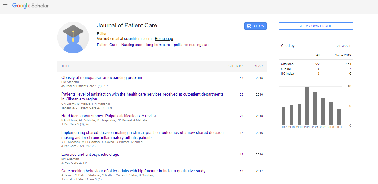
- Google Scholar
Useful Links
Share This Page
Journal Flyer
Open Access Journals
- Agri and Aquaculture
- Biochemistry
- Bioinformatics & Systems Biology
- Business & Management
- Chemistry
- Clinical Sciences
- Engineering
- Food & Nutrition
- General Science
- Genetics & Molecular Biology
- Immunology & Microbiology
- Medical Sciences
- Neuroscience & Psychology
- Nursing & Health Care
- Pharmaceutical Sciences
Commentary - (2023) Volume 9, Issue 1
Care and Monitoring Vital Signs of Patients with Ischemic Stroke
Zhang Jiuan*Received: 02-Jan-2023, Manuscript No. JPC-23-19061; Editor assigned: 05-Jan-2023, Pre QC No. JPC-23-19061 (PQ); Reviewed: 19-Jan-2023, QC No. JPC-23-19061; Revised: 26-Jan-2023, Manuscript No. JPC-23-19061 (R); Published: 02-Feb-2023, DOI: 10.35248/2573-4598.23.9.212
Description
Stroke is a prominent cause of mortality and disability worldwide, and it may be divided into two types: ischemic stroke and hemorrhagic stroke, the latter of which includes intracerebral haemorrhage and subarachnoid haemorrhage. When the blood flow to a portion of the brain is blocked or diminished, brain tissue cannot get oxygen and nutrients, which results in an ischemic stroke [1]. Acute Ischemic Stroke (AIS) can damage either the anterior or posterior circulations, or both. The internal carotid arteries supply oxygenated blood to the brain's anterior circulation while the vertebral arteries merge to form the basilar artery, which feeds into the posterior cerebral and posterior communicating arteries. Because of their proximity to the circle, the anterior and posterior arteries that make up the circle of Willis are referred to as proximal arteries. Anterior circulation strokes involving the internal carotid artery and the Middle Cerebral Artery (MCA) are more prevalent than posterior infarcts [2]. In causes of ischemic stroke, the etiology of ischemic stroke must be identified in order to guide appropriate therapy and secondary prevention. The clinical presentation and appearance of lesions on brain imaging may already reveal information about the underlying cause. To determine the underlying cause of stroke and vascular risk factors, a complete diagnostic work-up is required. There are several etiological classifications for ischemic stroke.
The Acute Stroke Treatment (AST) classification 6 is the most generally used, and it designates five subtypes of ischemic stroke: Large-Artery Atherosclerosis (LAA), Cardiogenic Embolism, Small-Vessel Disease (SVD), stroke of other determined etiology and stroke of unknown etiology. In Cardiogenic Embolism, The cause of 20% of ischemic strokes is cardiogenic embolism. A cerebral artery becomes blocked as a result of a cardiac blood clot that enters the bloodstream and travels to the brain. Further risk factors for cardioembolic strokes include heart failure, Patent Foramen Ovale (PFO), and artificial heart valves. Cardiogenic embolism has been found to result in strokes that are more severe than those caused by other ischemic stroke subtypes. Cardioembolic strokes typically appear as territorial infarcts on imaging, and they can affect more than one artery area or even both sides of the brain [3]. In Large-Artery Atherosclerosis of intracranial or extracranial arteries accounts for about 25% of all ischemic strokes. Inflammation, endothelial dysfunction, and traditional cardiovascular risk factors such as hypertension, diabetes, hypercholesterolemia, and smoking all contribute to the development of atherosclerosis. Strokes induced by LAA are most typically triggered by an artery-to- artery embolism or, less frequently, vascular stenosis leading in hemodynamic infarction. Approximately 20% of ischemic strokes are caused by cardiogenic embolism. A cardiogenic embolism occurs when a cardiac blood clot enters the bloodstream and occludes a cerebral artery. Atrial fibrillation (AF) is the most common cause, with blood clots often forming in the left atrial appendage [4]. Heart failure, Patent Foramen Ovale (PFO), and artificial heart valves are also risk factors for cardioembolic strokes. Cardiogenic embolism has been demonstrated to produce more severe strokes than other ischemic stroke subtypes. Cardioembolic strokes typically manifest as territorial infarcts on imaging and can occur in more than one arterial territory, even bilaterally. In Small-Vessel Disease, Cerebral SVD is one of the three leading causes of ischemic stroke, accounting for approximately 25% of all ischemic strokes. The term SVD refers to a condition that affects the brain's microvasculature. Lipohyalinosis of small perforating arteries feeding deep subcortical tissues and micro-atheroma development at the start of penetrating arteries from major cerebral arteries are the main diseases documented. All stroke patients should be admitted to an acute stroke unit as soon as possible, preferably within 3 hours of the onset of the stroke. Patients should receive stroke nursing care consistent with best practise regardless of the hospital unit to which they are admitted in the absence of a specialised stroke unit. In this case, vital sign monitoring recommendations for acute stroke units also apply to patients admitted to a general hospital unit. The initial and ongoing clinical assessment of a stroke patient after admission to the hospital is critical to improving the patient's long-term outcomes. The sections that follow provide evidence- based guidelines and protocols for nurses to use when assessing patients with acute strokes' overall health status. Following admission to the hospital, the initial nursing assessment of the stroke patient should include evaluating the patient's vital signs, particularly oxygen saturation, blood pressure, and temperature, as well as measuring blood glucose and performing a bedside dysphagia screen/assessment. These assessments apply to all stroke patients, including those who receive reperfusion therapy and thrombectomy. Monitoring these aspects of care is critical for preventing or detecting stroke complications early. A nursing assessment of airway support and breathing is required to determine the need for continued oxygen support. Supplemental oxygen is not recommended for non-hypoxic patients [5]. Supplemental oxygen should be given only if the oxygen saturation is greater than 94% and there are no contraindications. In the first 48 hours after a stroke, nurses should closely monitor the patient's blood pressure. Given the conflicting evidence, it is unclear what blood pressure level should be maintained in patients with acute ischemic stroke to ensure the best outcome. Intensive BP lowering in the acute phase after stroke is not advised. Excellent practise Over the first 24 hours, BP management recommends lowering blood pressure to >220/120 mm Hg. To maintain systemic perfusion levels required to support organ function, hypotension and hypovolemia should be corrected. In hypertensive patients, evidence suggests lowering blood pressure to 185/110 mm Hg before intravenous thrombolysis and keeping it at 180/105 mm Hg for the first 24 hours after treatment. Body temperature, blood glucose, and dysphagia monitoring are considered standard of care for all stroke patients.
References
- Amarenco P, Bogousslavsky J, Caplan LR, Donnan GA, Hennerici MG. Classification of stroke subtypes. Cerebrovascular diseases. Cerebrovasc Dis. 2009;27(5):493-501.
[Crossref] [Google Scholar] [PubMed]
- Sacco RL, Kasner SE, Broderick JP, Caplan LR, Connors JJ, Culebras A, et al. An updated definition of stroke for the 21st century: a statement for healthcare professionals from the American Heart Association/American Stroke Association. Stroke. 2013;44(7):2064-2089.
[Crossref] [Google Scholar] [PubMed]
- Paciaroni M, Caso V, Agnelli G. The concept of ischemic penumbra in acute stroke and therapeutic opportunities. Eur Neurol. 2009;61(6):321-330.
[Crossref] [Google Scholar] [PubMed]
- Balami JS, White PM, McMeekin PJ, Ford GA, Buchan AM. Complications of endovascular treatment for acute ischemic stroke: prevention and management. Int J Stroke. 2018;13(4):348-61.
[Crossref] [Google Scholar] [PubMed]
- Goyal M, Menon BK, van Zwam WH, Dippel DW, Mitchell PJ, Demchuk AM et al. Endovascular thrombectomy after large-vessel ischaemic stroke: a meta-analysis of individual patient data from five randomised trials. Lancet. 2016;387(10029):1723-1731.
[Crossref] [GoogleScholar] [PubMed]
Citation: Jiuan Z (2023) Care and Monitoring Vital Signs of Patients with Ishemic Stroke. J Pat Care. 9:212.
Copyright: © 2023 Jiuan Z. This is an open-access article distributed under the terms of the Creative Commons Attribution License, which permits unrestricted use, distribution, and reproduction in any medium, provided the original author and source are credited.