
Indexed In
- RefSeek
- Hamdard University
- EBSCO A-Z
- Publons
- Euro Pub
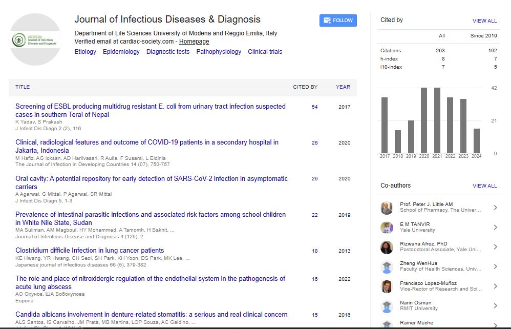
- Google Scholar
Useful Links
Share This Page
Journal Flyer

Open Access Journals
- Agri and Aquaculture
- Biochemistry
- Bioinformatics & Systems Biology
- Business & Management
- Chemistry
- Clinical Sciences
- Engineering
- Food & Nutrition
- General Science
- Genetics & Molecular Biology
- Immunology & Microbiology
- Medical Sciences
- Neuroscience & Psychology
- Nursing & Health Care
- Pharmaceutical Sciences
Short Communication - (2025) Volume 10, Issue 6
Advances in the Diagnosis of Meningitis for Improved Clinical Management and Public Health Outcomes
Peng Brouwer*Received: 29-Oct-2025, Manuscript No. JIDD-25-30663; Editor assigned: 03-Nov-2025, Pre QC No. JIDD-25-30663 (PQ); Reviewed: 17-Nov-2025, QC No. JIDD-25-30663; Revised: 24-Nov-2025, Manuscript No. JIDD-25-30663 (R); Published: 03-Dec-2025, DOI: 10.35248/2576-389X.25.10.367
Description
Meningitis, an inflammation of the meninges surrounding the brain and spinal cord, is a potentially life-threatening condition that requires prompt diagnosis and treatment. The disease can be caused by bacteria, viruses, fungi, parasites, or non-infectious factors such as autoimmune disorders, medications, or malignancies. Bacterial meningitis, caused primarily by Neisseria meningitidis, Streptococcus pneumoniae and Haemophilus influenzae, is associated with high morbidity and mortality if not rapidly identified. Viral meningitis, often caused by enteroviruses, is generally self-limiting but may mimic bacterial forms in the early stages [1-3]. Accurate and timely diagnosis is important for initiating appropriate therapy, preventing complications and implementing public health interventions, including outbreak control and vaccination strategies.
Clinical diagnosis of meningitis begins with recognition of classical symptoms such as fever, headache, neck stiffness and photophobia, nausea, vomiting and altered mental status. In infants and immunocompromised patients, presentation may be atypical, including irritability, lethargy, or seizures. While clinical suspicion is essential, these signs are nonspecific and overlap with other Central Nervous System (CNS) disorders, underscoring the need for laboratory confirmation. Physical examination maneuvers, such as Kernig’s and Brudzinski’s signs, can support suspicion but are not definitive.
Cerebrospinal Fluid (CSF) analysis obtained through lumbar puncture remains the cornerstone of meningitis diagnosis. CSF evaluation includes cell count, glucose, protein, Gram stain, culture and molecular testing. Bacterial meningitis typically presents with elevated white blood cell counts (predominantly neutrophils), decreased glucose and increased protein, whereas viral meningitis shows lymphocytic predominance with normal or slightly elevated protein and normal glucose levels [4]. Gram stain can rapidly identify causative bacteria in many cases, providing immediate guidance for empiric therapy. Culture of CSF, while slower, remains essential for pathogen confirmation, antibiotic susceptibility testing and epidemiologic surveillance.
Molecular diagnostic methods, including Polymerase Chain Reaction (PCR) and real-time PCR, have revolutionized meningitis detection by providing rapid, highly sensitive and specific identification of bacterial, viral and fungal pathogens directly from CSF. PCR is particularly valuable for pathogens that are difficult to culture, such as Listeria monocytogenes, Neisseria meningitidis in partially treated patients and viral agents. Multiplex PCR assays allow simultaneous detection of multiple pathogens, significantly improving diagnostic efficiency, especially during outbreaks or in immunocompromised individuals.
Serologic testing can assist in the diagnosis of certain types of meningitis, particularly those caused by viral infections, autoimmune conditions, or rare pathogens. Detection of pathogen-specific antibodies or antigens in serum or CSF can provide supportive evidence when direct pathogen identification is challenging. Emerging diagnostic approaches, including metagenomic next-generation sequencing, biosensors and rapid antigen tests, hold potential for identifying uncommon or novel pathogens and for use in resource-limited settings [5-7].
Imaging studies, such as Magnetic Resonance Imaging (MRI) or Computed Tomography (CT), are not primary diagnostic tools but can identify complications of meningitis, including abscess formation, hydrocephalus, or cerebral edema. Neuroimaging is also essential before lumbar puncture in cases with elevated intracranial pressure or focal neurologic deficits to reduce the risk of herniation [8].
Rapid and accurate meningitis diagnosis requires integration of clinical assessment, laboratory testing and imaging when indicated. Prompt identification of the causative agent enables targeted antimicrobial therapy, reduces the risk of long-term neurologic sequelae and informs public health measures such as contact tracing, prophylaxis and vaccination campaigns. Early empiric treatment should not be delayed when bacterial meningitis is suspected, while laboratory confirmation guides therapy adjustments and supports epidemiologic monitoring [9,10].
In conclusion, the diagnosis of meningitis relies on a combination of careful clinical evaluation, cerebrospinal fluid analysis, molecular testing and supportive imaging. Advances in PCR and multiplex assays, along with emerging rapid diagnostic technologies, have significantly improved the sensitivity, specificity and speed of pathogen identification. Integration of these approaches ensures timely treatment, improved patient outcomes and effective public health responses. Continued innovation in diagnostic methods is essential to reduce the global burden of meningitis and prevent its severe complications.
References
- Thigpen MC, Whitney CG, Messonnier NE, Zell ER, Lynfield R, Hadler JL, et al. Bacterial meningitis in the United States, 1998-2007. N Engl J Med. 2011;364(21):2016-2025.
[Crossref] [Google Scholar] [PubMed]
- Posadas E, Fisher J. Pediatric bacterial meningitis: an update on early identification and management. Pediatr Emerg Med Pract. 2018;15(11):1-20.
[Crossref] [Google Scholar] [PubMed]
- Ashwal S, Tomasi L, Schneider S, Perkin R, Thompson J. Bacterial meningitis in children: Pathophysiology and treatment. Neurology. 1992;42(4):739-748.
[Crossref] [Google Scholar] [PubMed]
- Van de Beek D, Brouwer MC, Thwaites GE, Tunkel AR. Advances in treatment of bacterial meningitis. Lancet. 2012;380(9854):1693-1702.
[Crossref] [Google Scholar] [PubMed]
- McGill F, Heyderman RS, Panagiotou S, Tunkel AR, Solomon T. Acute bacterial meningitis in adults. Lancet. 2016;388(10063):3036-3047.
- Tunkel AR, Hartman BJ, Kaplan SL, Kaufman BA, Roos KL, Scheld WM, et al. Practice guidelines for the management of bacterial meningitis. Clin Infect Dis. 2004;39(9):1267-1284.
[Crossref] [Google Scholar] [PubMed]
- Sáez-Llorens X, McCracken GH. Bacterial meningitis in children. Lancet. 2003;361(9375):2139-2148.
- Hasbun R, Abrahams J, Jekel J, Quagliarello VJ. Computed tomography of the head before lumbar puncture in adults with suspected meningitis. N Engl J Med. 2001;345(24):1727-1733.
[Crossref] [Google Scholar] [PubMed]
- Granerod J, Crowcroft NS. The epidemiology of acute encephalitis. Neuropsychol Rehabil. 2007;17(4-5):406-428.
[Crossref] [Google Scholar] [PubMed]
- Gundamraj V, Hasbun R. Viral meningitis and encephalitis: An update. Curr Opin Infect Dis. 2023;36(3):177-185.
[Crossref] [Google Scholar] [PubMed]
Citation: Brouwer P (2025). Advances in the Diagnosis of Meningitis for Improved Clinical Management and Public Health Outcomes. J Infect Dis Diagn. 10:367.
Copyright: © 2025 Brouwer P. This is an open-access article distributed under the terms of the Creative Commons Attribution License, which permits unrestricted use, distribution and reproduction in any medium, provided the original author and source are credited.