

Indexed In
- RefSeek
- Hamdard University
- EBSCO A-Z
- Euro Pub
- Google Scholar
Useful Links
Share This Page
Journal Flyer

Open Access Journals
- Agri and Aquaculture
- Biochemistry
- Bioinformatics & Systems Biology
- Business & Management
- Chemistry
- Clinical Sciences
- Engineering
- Food & Nutrition
- General Science
- Genetics & Molecular Biology
- Immunology & Microbiology
- Medical Sciences
- Neuroscience & Psychology
- Nursing & Health Care
- Pharmaceutical Sciences
Case Report - (2026) Volume 11, Issue 1
Acute invasive fungal sinusitis with orbital cellulitis from Cambodian tertiary hospital: a case report
Cheapov Tech1,22Lecturer of medicine faculty, Université des Sciences de la santé and University of Puthisastra, Phn, Cambodia
Received: 30-Jan-2026, Manuscript No. DCRS-26-30965; Editor assigned: 02-Feb-2026, Pre QC No. DCRS-26-30965; Reviewed: 16-Feb-2026, QC No. DCRS-26-30965; Revised: 23-Feb-2026, Manuscript No. DCRS-26-30965; Published: 02-Mar-2026, DOI: 10.35841/2572-5629.26.11.265
Abstract
Background: Acute invasive fungal sinusitis is a fulminant infection. Delay in diagnosis may consequently lead to high mortality notably at poor resources setting like Cambodia.
Objective: We intend to illustrate the diagnostic challenges, aggressive management required, and poor outcomes associated with AIFS at less advanced medical centre.
Outcome: The patient hospitalized around three months after radical full-house endoscopic sinus surgery, orbital exenteration and right hemi maxillectomy to kill the black fungus expansion. The patient’s condition due to the immunocompromising comorbidities, including diabetes mellitus and cardiopathy. This led to the development of severe systemic infection and hospital-acquired pneumonia, ultimately resulting in death around two months. Survival rates for patients affected by this disease remain low in low-resource settings.
Keywords
Mucormycosis; Acute invasive fungal sinusitis; Diabetes mellitus; Multi-disciplinary care
Introduction
Acute Invasive Fungal Sinusitis (AIFS) is a surgical emergency that primarily affect immunocompromised individual notably those with uncontrolled diabetes, blood cancer like hematology malignancy or those undergoing organ transplant. It is highly fatal because the fungi (Mucorales or Aspergillosis) are angioinvasive [1-7]. Geogra- phically, Cambodia located in tropical area associated with consumption of high sugar diet might be a favorable factor for fungus species evolving as an opportunistic infection, likewise in Thailand, Roongrotwattanasiri K, et al., showed six patients with AIFS were well treated with high success rate and the follow up manifested with free mucosa from fungal infection in nasal cavity. In tropical countries like Indonesia, the environment acts as a natural incubator for fungi. The combination of high temperatures (averaging 26°C to 30°C) and high humidity (often above 80%) provides the perfect conditions for environmental molds and yeasts to thrive. For policymakers and clinicians, understanding the "fungal burden" the total number of people affected by serious fungal infections is critical for resource allocation and early diagnosis [2].
Case Presentation
A 58-year-old man presented to the Emergency Department with acute symptoms suggestive of orbital cellulitis, including painful eyelid edema, erythema, fever, and purulent nasal discharge (Figure 1).
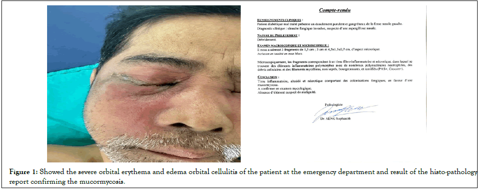
Figure 1: Showed the severe orbital erythema and edema orbital cellulitis of the patient at the emergency department and result of the histo-pathology report confirming the mucormycosis.
He was initially evaluated by an ophthalmologist because of severe orbital manifestations. However, due to overcrowding in the Emergency Department (ED), there was a delay of approximately two days before contrast-enhanced Computed Tomography (CT) was performed and an otolaryngology consultation was obtained.
Nasal endoscopic examination revealed extensive fungal involvement of the nasal cavity, characterized by a pathognomonic black eschar affecting the inferior and middle turbinate’s, with associated obstruction of the nasolacrimal duct and the ostiomeatal complex (Figure 1A).
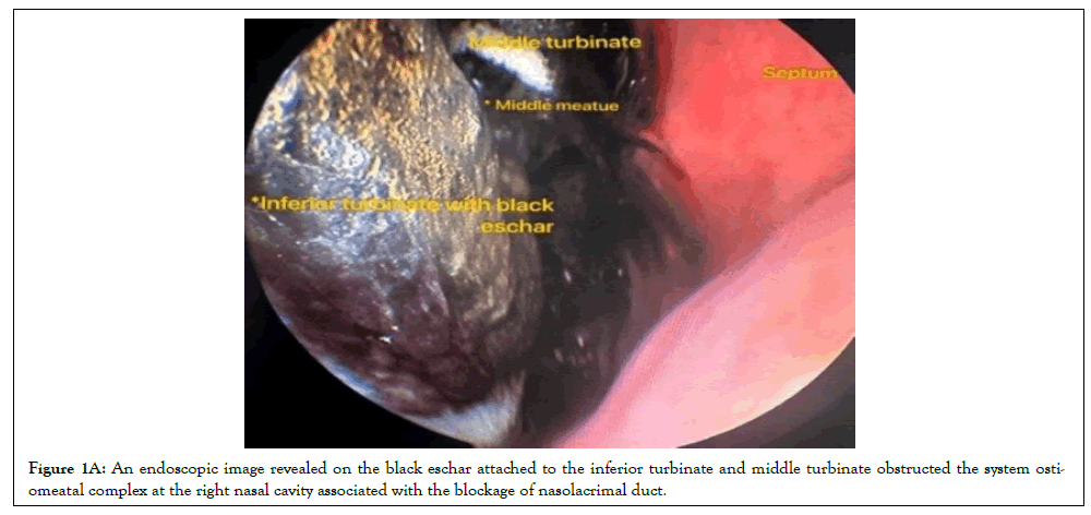
Figure 1A: An endoscopic image revealed on the black eschar attached to the inferior turbinate and middle turbinate obstructed the system ostiomeatal complex at the right nasal cavity associated with the blockage of nasolacrimal duct.
CT imaging of the paranasal sinuses demonstrated an extensive infiltrative process with bilateral sinonasal involvement, including complete opacification of the anterior and posterior ethmoid sinuses with extension into the sphenoid sinus (Figures 2 and 2A).
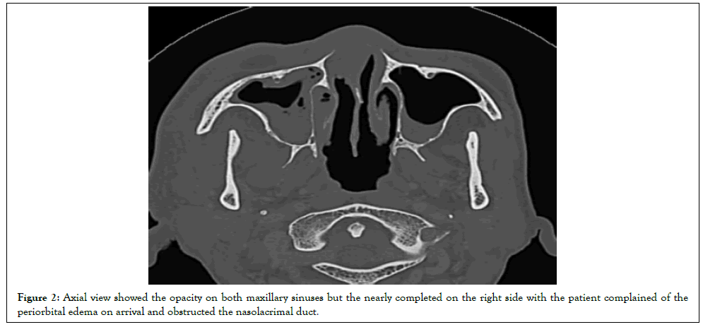
Figure 2: Axial view showed the opacity on both maxillary sinuses but the nearly completed on the right side with the patient complained of the periorbital edema on arrival and obstructed the nasolacrimal duct.
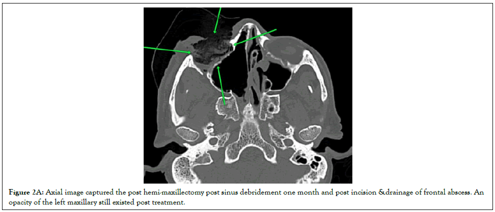
Figure 2A: Axial image captured the post hemi-maxillectomy post sinus debridement one month and post incision &drainage of frontal abscess. An opacity of the left maxillary still existed post treatment.
Magnetic Resonance Imaging (MRI) was not performed due to impaired renal function and the patient’s poor general condition.
Histopathological examination of necrotic nasal tissue revealed characteristic fungal hyphae, confirming the diagnosis of Acute Invasive Fungal Rhinosinusitis (AIFRS).
Results
This case report showed the clinical course of a patient with orbital cellulitis and erythema in the rural area where diagnostic facilities are limited. The delay of diagnosis might consequently lead to high morbidity and mortality. Histopathological examination at hospital could only identify the tissue fragment corresponds to fibroinflammatory and necrotic tissue containing a polymorphous inflammatory infiltrate with numerous neutrophilic polymorphonuclear cells, cellular debris, and non-septate, budding, and branching mycelial filaments (PAS positive, Grocott positive) [8-9]. The patient’s hospital course was complicated by the development of Diabetic Keto Acidosis (DKA). Management was multidisciplinary, involving:
Pharmacotherapy: Intravenous (IV) Meropenem (3g/day) and Vancomycin (2g/day) for secondary bacterial coverage, alongside aggressive glycemic control using IV Actrapid and oral Empagliflozin.
Antifungal therapy: Systemic Amphotericin B was initiated at a dose of 0.5 mg/kg every 24 hours (patient weight 1.75 m and 70 kilograms, however, his creatinine augmented to over 100 mg/dL and blood urea nitrogen reached on 30 mmol/L over 1 week course of drug. Due to there is no actual protocol for anti-fungal in AIFS, the amphotericin B was followed according to the treatment experiences from infectiologist at hospital.
Surgical intervention: The patient underwent urgent radical endoscopic sinus debridement. Due to the angioinvasive nature of the disease, subsequent hemi-maxillectomy was required to excise necrotic maxillary bone [4-7].
Despite aggressive full-house of endoscopic sinus debridement (Draft III) and systemic antifungal therapy, the infection demonstrated unrelenting cephalad progression. One month following the initial procedure, neuroimaging revealed a frontal cerebral abscess, necessitating emergency surgical incision and drainage. Due to extensive ocular involvement, an orbital exenteration was also performed (Figures 3).
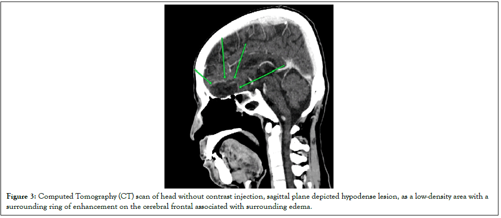
Figure 3: Computed Tomography (CT) scan of head without contrast injection, sagittal plane depicted hypodense lesion, as a low-density area with a surrounding ring of enhancement on the cerebral frontal associated with surrounding edema.
Ultimately, the patient’s clinical course was further complicated by severe hospital-acquired pneumonia; the patient succumbed to this sever respiratory complication, exacerbated by their underlying pulmonary disease, in mid-January. It is critical to note that the outcomes reported here are a function of restricted clinical resources and should not serve as a benchmark for the overall efficacy of AIFS management. These results are not representative of potential outcomes in high-resource settings where superior diagnostic tools and advanced therapeutic modalities allow for more rapid intervention [10,11].
Discussion
Acute Invasive Fungal Sinusitis (AIFS) is a rare yet fulminant condition associated with substantial morbidity and mortality, particularly in immunocompromised individuals. We report a 58-year-old man with poorly controlled diabetes mellitus who developed AIFS complicated by rhino-cerebral mucormycosis. Uncontrolled diabetes markedly increases susceptibility to mucormycosis through impaired neutrophil function, sustained hyperglycemia, and an acidic metabolic milieu that facilitates fungal invasion [6]. Delayed diagnosis remains a critical challenge.
In the case series by Maell C, et al., all diagnoses were delayed, no patient achieved visual recovery beyond counting fingers at 24 months, and mortality was high. Histopathological examination remains the diagnostic cornerstone, demonstrating characteristic fungal hyphae with uniform morphology and diameters of 3-6 μm, whereas mycological cultures are limited by low sensitivity and prolonged processing times. The pronounced angioinvasive nature of mucormycosis results in rapid tissue necrosis, vascular thrombosis, and frequent intracranial extension, thereby complicating management and portending a poor prognosis.
Despite prompt surgical debridement and systemic antifungal therapy, disease progression may be aggressive. The present case highlights the diagnostic complexity of AIFS and the sharply increased mortality risk following cerebral involvement. Reported survival rates are 72.7% at hospital discharge and decline to 57.8% at six months. High-dose antifungal therapy for a minimum of two weeks is recommended; however, first-line treatment failure occurs in up to 50% of cases. Prognosis is influenced by both the site of infection and underlying immunosuppressive conditions, including advanced age, solid-organ transplantation, hematological malignancies, Human Immunodeficiency Virus (HIV) infection, autoimmune disease requiring immunosuppression, and intensive chemotherapy [12].
In Southeast Asia, tropical climates combined with rising metabolic risk factors, particularly poor glycemic control, may contribute to an increasing incidence of opportunistic fungal infections. These factors likely create an environment favorable to aggressive fungal proliferation among susceptible populations. Effective reduction in AIFS-related mortality necessitates a multidisciplinary strategy encompassing early clinical suspicion, rapid histopathological confirmation, and concurrent optimization of metabolic derangements. However, therapeutic standardization remains constrained by disease rarity and the absence of randomized controlled trials, rendering outcome optimization an ongoing clinical challenge [13-19].
Conclusion
In summary, this case report highlights the clinical course and outcome of acute invasive fungal sinusitis in a low-resource setting, emphasizing the aggressive nature of the disease and the significant challenges in its management. The available literature from both high and low resource settings remains limited, particularly in tropical regions such as Indonesia, Thailand, and Cambodia. Indonesia’s warm and humid tropical climate supports a wide range of environmental fungi, underscoring the importance of understanding the national fungal disease burden to inform clinicians and policymakers. As the spectrum of fungal pathogens continues to evolve, the involvement of clinical microbiologists and public health experts is essential to improve early detection, prevention strategies, and patient outcomes in this high-mortality disease. Moreover, the morbidity and mortality risk of not addressing the underlying fungal infection in these patients, these misdiagnoses can lead to unnecessary financial burden and antibiotic resistance. However, this case report does not represent outcomes from advanced-resource settings, where broader diagnostic facilities and advanced therapeutic options may lead to different clinical outcomes.
Declarations
Ethics Approval
This case report was reviewed and formally approved through verbal authorization by the Ethics Committee of Calmette Hospital, Phnom Penh, Cambodia.
Consent to Participate
The patient was fully informed about the case report and provided consent to participate for the benefit of other patients affected by a similar disease.
Consent for Publication
Written informed consent was obtained from the patient for the publication of this case report and any accompanying images.
Data Availability Statement
All data generated or analysed during this study are included in this published article.Competing Interests
The authors declare that they have no competing interests.
Funding
The authors received no financial support for the research, authorship, and/or publication of this article.
Authors Contributions
All authors contributed to the conception of the work, data acquisition, analysis, and interpretation. All authors drafted and critically revised the manuscript and approved the final version.
Acknowledgements
The authors would like to thank the medical and nursing staff of ENT department. Calmette Hospital for their support in the management of this patient.
References
- Roongrotwattanasiri K, Fooanant S, Naksane L. Invasive fungal rhinosinusitis in Maharaj Nakorn Chiang Mai Hospital. J Med Assoc Thai. 2007;90(11):2524-2528.
[Google Scholar] [Pubmed]
- Wahyuningsih R, Adawiyah R, Sjam R, Prihartono J, Ayu Tri Wulandari E, Rozaliyani A, et al. Serious fungal disease incidence and prevalence in Indonesia. Mycoses. 2021;64(10):1203-1212.
[Crossref] [Google Scholar] [Pubmed]
- Luo YT, Zhu CR, He B, Yan AH, Wei HQ. Diagnostic and therapeutic strategies of acute invasive fungal rhinosinusitis. Asian J Surg. 2023;46(1):58-65.
[Crossref] [Google Scholar] [Pubmed]
- Akhondi H, Woldemariam B, Rajasurya V. Fungal sinusitis. 2023. StatPearls Publishing. [Crossref]
[Google Scholar] [Pubmed]
- Deutsch PG, Whittaker J, Prasad S. Invasive and non-invasive fungal rhinosinusitis a review and update of the evidence. Medicina. 2019;55(7):319.
[Crossref] [Google Scholar] [Pubmed]
- Coutel M, Duprez T, Huart C, Wacheul E, Boschi A. Invasive fungal sinusitis with ophthalmological complications: Case series and review of the literature. Neuro-Ophthalmology. 2021;45(3):193-204.
[Crossref] [Google Scholar] [Pubmed]
- Payne SJ, Mitzner R, Kunchala S, Roland L, McGinn JD. Acute invasive fungal rhinosinusitis: A 15-year experience with 41 patients. Otolaryngol Head Neck Surg. 2016;154(4):759-64.
[Crossref] [Google Scholar] [Pubmed]
- Raizada N, Jyotsna VP, Kandasamy D, Xess I, Thakar A, Tandon N. Invasive fungal rhinosinusitis in patients with diabetes. J Infect Dev Ctries. 2018;12(09):787-793.
[Crossref] [Google Scholar] [Pubmed]
- Quang LX, Tam TT, Dang LH, Chen YC, Hung SH, Tai TT, et al. Acute invasive fungal rhinosinusitis in post-COVID-19 patients in Vietnam. J Formos Med Assoc. 2024;123(3):357-365.
[Crossref] [Google Scholar] [Pubmed]
- Xiaodong L, Mei C, Kaijin Z, Li Z, Dandan W, Yi Y, et al. A case study of acute invasive fungal sinusitis complicating central retinal artery occlusion.
- Yağmur AR, Çufalı ÖF, Çolak M, Akbal Çufalı Ş, Kayalı Dinç AS, Yeniçeri A, et al. Invasive fungal rhinosinusitis, clinical manifestations, and prognostic values: As case series audit. Egypt J Otolaryngol. 2023;39(1):189.
- Tran GH, Luong KA, Ngo TP, Bui TM, Luong BA, Vu HA. Invasive fungal rhinosinusitis: The first histopathological study in vietnam. Head Neck Pathol. 2024;18(1):104.
[Crossref] [Google Scholar] [Pubmed]
- Kurokawa M, Kurokawa R, Baba A, Kim J, Tournade C, Mchugh J, et al. Deadly fungi: Invasive fungal rhinosinusitis in the head and neck. Radiographics. 2022:42(7):2075-2094.
[Crossref] [Google Scholar] [Pubmed]
- Jacob NB, Chaney S. Rhino orbito cerebral mucormycosis: A fatal acute invasive fungal infection in uncontrolled diabetes. J Nurse Pract. 2016 Nov 1;12(10):667-74.
- Weissert N, Mengel A, Bader K, Hennersdorf F, Feil K. Angioinvasive rhino-orbital-cerebral mucormycosis in a patient with type 2 diabetes mellitus: A complex and lethal cause of stroke. Diagnostics. 2024;14(19):2246.
[Crossref] [Google Scholar] [Pubmed]
- Chiari F, Guarino P, Caporale CD, Orsini K, Trevisi G, Presutti L, et al. Clinical features, operative management and surgical results of first Draf III procedure, revision Draf III approach and the use of mucosal flaps and stents: A systematic review. Eur Arch Otorhinolaryngol. 2025;282(2):611-622.
[Crossref] [Google Scholar] [Pubmed]
- Chakrabarti A, Denning DW, Ferguson BJ, Ponikau J, Buzina W, Kita H, et al. Fungal rhinosinusitis: A categorization and definitional schema addressing current controversies. Laryngoscope. 2009;119(9):1809-1818.
[Crossref] [Google Scholar] [Pubmed]
- Do H, Sturz J, Crosser B, Ebelhar A, Brown D. Chronic invasive rhino-orbital sinusitis with mucormycosis. ACS Case Rev Surg. 2023;4(1):57â??60.
- Kumar A, Nakra P, Pawar S. Mucormycosis involving maxillary sinus: A case report. SVOA Dentistry. 2021;2(6):263-266.
Citation: Tech C. (2026). Acute Invasive Fungal Sinusitis with Orbital Cellulitis from Cambodian Tertiary Hospital: A Case Report. Diabetes Case Rep.11:265.
Copyright: © 2026 Tech C. This is an open-access article distributed under the terms of the Creative Commons Attribution License, which permits unrestricted use, distribution, and reproduction in any medium, provided the original author and source are credited.