

Indexed In
- Open J Gate
- Academic Keys
- ResearchBible
- China National Knowledge Infrastructure (CNKI)
- Centre for Agriculture and Biosciences International (CABI)
- RefSeek
- Hamdard University
- EBSCO A-Z
- OCLC- WorldCat
- CABI full text
- Publons
- Geneva Foundation for Medical Education and Research
- Google Scholar
Useful Links
Share This Page
Journal Flyer
Open Access Journals
- Agri and Aquaculture
- Biochemistry
- Bioinformatics & Systems Biology
- Business & Management
- Chemistry
- Clinical Sciences
- Engineering
- Food & Nutrition
- General Science
- Genetics & Molecular Biology
- Immunology & Microbiology
- Medical Sciences
- Neuroscience & Psychology
- Nursing & Health Care
- Pharmaceutical Sciences
Case Report - (2026) Volume 15, Issue 2
A Case Series on Ocular Rhinosporidiosis
SL Adile*, Shashank Bais and Nidhi PandeyReceived: 20-Dec-2024, Manuscript No. JTD-26-27873; Editor assigned: 24-Dec-2024, Pre QC No. JTD-26-27873 (PQ); Reviewed: 07-Jan-2025, QC No. JTD-26-27873; Revised: 07-Apr-2026, Manuscript No. JTD-26-27873 (PQ); Published: 14-Apr-2026, DOI: 10.35241/2329-891X.26.15.483
Abstract
Rhinosporidiosis is caused by the pathogen Rhinosporidium sieberi, now thought to be a fungal-like parasite of the eukaryotic group Mesomycetozoea. It is usually a benign disease with slowly growing polypoid lesions in the nose, nasopharynx, or eyes. The clinical features are characteristics of a painless, friable, mostly unilateral polypoid mass guide the diagnosis, whereas the gold standard for diagnosis is histopathologic findings.
Keywords
Rhinosporidiosis; Mesomycetozoea; Rhinosporidium seeberi; Diagnosis
Introduction
Rhinosporidiosis is a chronic granulomatous infective disorder caused by Rhinosporidium seeberi, an aquatic protistan parasite belonging to a clade, Mesomycetozoea. Its first case was described as a nasal polyp by Guillermo Seeber from Buenos Aires in 1900. Rhinosporidium seeberi was previously considered as a fungus, but now it has been classified under Mesomycetozoea. Clinically rhinosporidiosis presents as a polypoid soft tissue mass, often pedunculated. Nose and nasopharynx are most commonly involved in it (more than 70% of cases). Ocular lesions, particularly conjunctiva and lacrimal sac account for about 15% of cases. Rare sites of this disorder include lips, palate, uvula, maxillary antrum, epiglottis, larynx, trachea, bronchus, ear, scalp, vulva, penis, rectum and skin. Rarely disseminated infections are also reported, involving the limbs, trunk and viscera. Brain involvement may lead to fatality.
Case Presentation
Case 1: A 8-year-old female child presented to OPD with a mass protruding from her left lower conjunctiva since 4 months. She had a history of injury with pencil 4 months back since then she noticed a mass growing in her left lower palpebral conjunctiva which gradually progressed to the current state. The mass is pedunculated, sessile and has strawberry like appearance. The mass was about 2 × 3 cm in size. Her visual acuity was 6/6 in both eyes. The patient underwent mass excision, and the mass was sent for histopathological examination which came out to be Rhinosporidiosis [1].
Ocular rhinosporidiosis is an uncommon manifestation of a chronic granulomatous disease caused by Rhinosporidium seeberi, an organism associated with exposure to contaminated stagnant water. The condition is more prevalent in tropical and subtropical regions and predominantly affects individuals from rural backgrounds. While nasal involvement is the most frequently reported form, ocular involvement represents a significant clinical entity due to its distinctive presentation and potential for recurrence.
This case series describes patients diagnosed with ocular rhinosporidiosis who presented to a tertiary care ophthalmology department. The conjunctiva was the most commonly involved ocular structure, particularly the palpebral and bulbar conjunctiva. Clinically, the lesions appeared as fleshy, vascular, polypoidal growths with a tendency to bleed on touch. Multiple tiny whitish spots on the lesion surface, corresponding to sporangia, were observed in several cases and provided an important diagnostic clue [2].
Patients reported symptoms such as persistent redness, irritation, excessive tearing, and foreign body sensation. A detailed clinical examination followed by surgical excision was performed in all cases. Complete removal of the lesion with careful cauterization of the base was emphasized to reduce the likelihood of recurrence. The diagnosis was confirmed through histopathological analysis, which revealed characteristic thick walled sporangia containing numerous endospores embedded within the connective tissue.
Postoperative care included topical antimicrobial and antiinflammatory therapy. Selected patients received oral dapsone as adjunctive treatment to inhibit further sporangial development. Follow-up evaluations demonstrated satisfactory healing in most cases, with minimal complications. Recurrence was observed in a limited number of patients, highlighting the importance of thorough excision and regular monitoring.
This case series emphasizes the need for clinical awareness of ocular rhinosporidiosis in endemic areas and supports prompt surgical management combined with histopathological confirmation to achieve optimal outcomes (Figure 1) [3-6].
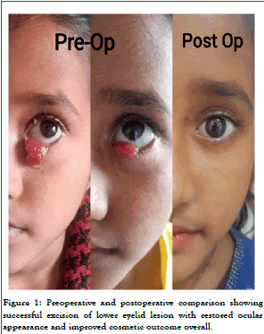
Figure 1: Preoperative and postoperative comparison showing successful excision of lower eyelid lesion with restored ocular appearance and improved cosmetic outcome overall.
Case 1: A 16-year-old male presented to the ophthal OPD with a mass in right lower periorbital region since past 8 months. His visual acuity was 6/6 in both eyes. His anterior segment examination was normal. On examination there was a mass in the right lower periorbital region measuring about 5 × 6 cm. On examination his general condition was good, there was soft non tender diffuse swelling. The remainder of the ocular examination was normal. There was no history of trauma. The patient had a history of I and D done 6 months back in village. The patient had a history of frequent bathing in pond water. The patient underwent mass excision, and the mass was sent for histopathological examination which came out to be Rhinosporidiosis (Figure 2).
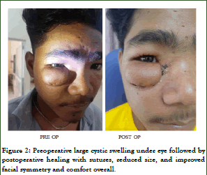
Figure 2: Preoperative large cystic swelling under eye followed by postoperative healing with sutures, reduced size, and improved facial symmetry and comfort overall.
Case 2: A 30-year-old male patient presented to ophthal OPD with complain of mass in right lower periorbital region since past 1 year. His visual acuity is 6/6 in left eye and 6/9 in right eye. On examination the mass was non tender, soft tissue mass measuring 5 × 4 cm. This patient also had a history of I and D done 5 months back in village. Anterior and posterior segment examination was normal. This patient also had a history of bathing in pond water (Figure 3). The mass was excised and sent for histopathological examination which came out to be rhinosporidiosis [7].
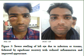
Figure 3: Severe swelling of left eye due to infection or trauma, followed by significant recovery with reduced inflammation and improved appearance.
Results and Discussion
Rhinosporidiosis is a chronic granulomatous and localized infection of the mucus membranes caused by Rhinosporidium seeberi. Although rhinosporidiosis occurs worldwide, it is endemic in Africa and Asia and is most frequent in India, Pakistan, and Sri Lanka (88% of all reported cases). The first case was described as a nasal polyp-from Buenos Aires in 1900, by Guillermo Seeber. Herr, et al. classified this organism as Mesomycetozoa.
The route of transmission is unknown, however it is thought to be acquired by contact with injured epithelium by the causative organism, which usually occurs in an aquatic environment. Those who dive or swim in stagnant water are more likely to get it. This could be the source of infection in our patient, who had previously bathed in pond water [8].
Nose and nasopharynx are the commonest sites involved but ocular lesions, particularly of the conjunctiva and lacrimal sac, account for 15% of cases. Clinically, ocular rhinosporidiosis presents as a freely mobile, pink, friable, richly vascularized polypoid mass that is pedunculated or sessile. The surface is covered with tiny white spots consistent with underlying mature sporangia beneath the epithelium. Diagnosis is mainly clinical but confirmed by histopathology and meticulous excision is the treatment of choice. Recurrences are rare and can be prevented by in toto excision followed by cauterization or cryotherapy at the base of the lesion. In our series, in all d 3 patients’ surgical excisions were done and no recurrences were noted. The potential for recurrence is due to the spillage of endospores on adjacent mucosa [9].
Conclusion
Rhinosporidiosis is a very common infection in India with conjunctival ocular rhinosporidiosis being commonest form of ocular rhinosporidiosis. Its clinical presentations may vary and it is best diagnosed by histopathological examination and use of special stains.
Citation: Adile SL, Bais S, Pandey N (2026) A Case Series on Ocular Rhinosporidiosis. J Trop Dis. 15:483.
Copyright: © 2026 Abebe SL, et al. This is an open-access article distributed under the terms of the Creative Commons Attribution License, which permits unrestricted use, distribution, and reproduction in any medium, provided the original author and source are credited.